Pentadecanoic acidCAS# 1002-84-2 |

Quality Control & MSDS
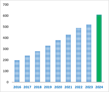
Number of papers citing our products
Chemical structure
3D structure
| Cas No. | 1002-84-2 | SDF | Download SDF |
| PubChem ID | 13849.0 | Appearance | Powder |
| Formula | C15H30O2 | M.Wt | 242.4 |
| Type of Compound | Aliphatics | Storage | Desiccate at -20°C |
| Solubility | Soluble in Chloroform,Dichloromethane,Ethyl Acetate,DMSO,Acetone,etc. | ||
| Chemical Name | pentadecanoic acid | ||
| SMILES | CCCCCCCCCCCCCCC(=O)O | ||
| Standard InChIKey | WQEPLUUGTLDZJY-UHFFFAOYSA-N | ||
| Standard InChI | InChI=1S/C15H30O2/c1-2-3-4-5-6-7-8-9-10-11-12-13-14-15(16)17/h2-14H2,1H3,(H,16,17) | ||
| General tips | For obtaining a higher solubility , please warm the tube at 37 ℃ and shake it in the ultrasonic bath for a while.Stock solution can be stored below -20℃ for several months. We recommend that you prepare and use the solution on the same day. However, if the test schedule requires, the stock solutions can be prepared in advance, and the stock solution must be sealed and stored below -20℃. In general, the stock solution can be kept for several months. Before use, we recommend that you leave the vial at room temperature for at least an hour before opening it. |
||
| About Packaging | 1. The packaging of the product may be reversed during transportation, cause the high purity compounds to adhere to the neck or cap of the vial.Take the vail out of its packaging and shake gently until the compounds fall to the bottom of the vial. 2. For liquid products, please centrifuge at 500xg to gather the liquid to the bottom of the vial. 3. Try to avoid loss or contamination during the experiment. |
||
| Shipping Condition | Packaging according to customer requirements(5mg, 10mg, 20mg and more). Ship via FedEx, DHL, UPS, EMS or other couriers with RT, or blue ice upon request. | ||

Pentadecanoic acid Dilution Calculator
Pentadecanoic acid Molarity Calculator
| 1 mg | 5 mg | 10 mg | 20 mg | 25 mg | |
| 1 mM | 4.1254 mL | 20.6271 mL | 41.2541 mL | 82.5083 mL | 103.1353 mL |
| 5 mM | 0.8251 mL | 4.1254 mL | 8.2508 mL | 16.5017 mL | 20.6271 mL |
| 10 mM | 0.4125 mL | 2.0627 mL | 4.1254 mL | 8.2508 mL | 10.3135 mL |
| 50 mM | 0.0825 mL | 0.4125 mL | 0.8251 mL | 1.6502 mL | 2.0627 mL |
| 100 mM | 0.0413 mL | 0.2063 mL | 0.4125 mL | 0.8251 mL | 1.0314 mL |
| * Note: If you are in the process of experiment, it's necessary to make the dilution ratios of the samples. The dilution data above is only for reference. Normally, it's can get a better solubility within lower of Concentrations. | |||||

Calcutta University

University of Minnesota

University of Maryland School of Medicine

University of Illinois at Chicago

The Ohio State University

University of Zurich

Harvard University

Colorado State University

Auburn University

Yale University

Worcester Polytechnic Institute

Washington State University

Stanford University

University of Leipzig
Universidade da Beira Interior

The Institute of Cancer Research

Heidelberg University

University of Amsterdam

University of Auckland

TsingHua University

The University of Michigan

Miami University

DRURY University

Jilin University

Fudan University

Wuhan University

Sun Yat-sen University

Universite de Paris

Deemed University

Auckland University
The University of Tokyo

Korea University
- Methyl tridecanoate
Catalog No.:BCX1372
CAS No.:1731-88-0
- Tridecanoic acid
Catalog No.:BCX1371
CAS No.:638-53-9
- Octadec-11-enoic acid
Catalog No.:BCX1370
CAS No.:693-72-1
- Cis-11-Eicosenoic acid
Catalog No.:BCX1369
CAS No.:5561-99-9
- Elaidic acid methyl ester
Catalog No.:BCX1368
CAS No.:1937-62-8
- Myristoleic acid
Catalog No.:BCX1367
CAS No.:544-64-9
- Ethyl (2E,4Z)-deca-2,4-dienoate
Catalog No.:BCX1366
CAS No.:3025-30-7
- Morroniaglycone
Catalog No.:BCX1365
CAS No.:1644061-02-8
- Crocetindial
Catalog No.:BCX1364
CAS No.:502-70-5
- Gymnoside VII
Catalog No.:BCX1363
CAS No.:899430-07-0
- Blestriarene B
Catalog No.:BCX1362
CAS No.:127211-03-4
- 6-Benzoylheteratisine
Catalog No.:BCX1361
CAS No.:99759-48-5
- Heptadecanoic acid
Catalog No.:BCX1374
CAS No.:506-12-7
- Methyl heptadecanoate
Catalog No.:BCX1375
CAS No.:1731-92-6
- Nonadecanoic acid
Catalog No.:BCX1376
CAS No.:646-30-0
- Ethyl palmitoleate
Catalog No.:BCX1377
CAS No.:56219-10-4
- Palmitoleic acid
Catalog No.:BCX1378
CAS No.:373-49-9
- Arachidic acid
Catalog No.:BCX1379
CAS No.:506-30-9
- Ethyl Arachidonate
Catalog No.:BCX1380
CAS No.:1808-26-0
- Methyl arachidonate
Catalog No.:BCX1381
CAS No.:2566-89-4
- Ethyl oleate
Catalog No.:BCX1382
CAS No.:111-62-6
- Docosahexaenoic acid ethyl ester
Catalog No.:BCX1383
CAS No.:84494-72-4
- Docosahexaenoic acid methyl ester
Catalog No.:BCX1384
CAS No.:2566-90-7
- Eicosapentaenoic acid ethyl ester
Catalog No.:BCX1385
CAS No.:86227-47-6
Analysis of Fungal Diversity, Physicochemical Properties and Volatile Organic Compounds of Strong-Flavor Daqu from Seven Different Areas.[Pubmed:38672935]
Foods. 2024 Apr 20;13(8):1263.
Strong-flavor Daqu, as a fermentation agent, plays a significant role in shaping the quality of strong-flavor baijius, and fungal species in Daqu are important factors affecting the quality of Daqu. Therefore, we selected strong-flavor Daqu from seven different origins to study the fungal composition and the effects of the fungal composition on the physicochemical properties and volatile organic compounds (VOCs). It was found that the fungal composition influences the physicochemical properties of Daqu. Specifically, there was a positive link between Rhizomucor, Rhizopus, Thermomyces, and liquefying activity and a positive correlation between Aspergillus and fermenting activity. Furthermore, the relationships between esterifying activity and Thermomyces, Rhizomucor, Aspergillus, Pichia, and Saccharomycopsis were found to be positive. The VOCs in Daqu were affected by Aspergillus, Issatchenkia, Pichia, and Thermoascus. Issatchenkia was significantly positively correlated with benzeneethanol as well as Aspergillus and Pentadecanoic acid ethyl ester, ethyl myristate. Pichia and Thermoascus were significantly negatively correlated with benzaldehyde and 2-furaldehyde. This study deepens our understanding of the relationship between VOCs, the physicochemical properties with microbial communities, and reference significance for the production of better-quality strong-flavor Daqu.
The role of volatile organic compounds for assessing characteristics and severity of non-cystic fibrosis bronchiectasis: an observational study.[Pubmed:38633315]
Front Med (Lausanne). 2024 Apr 3;11:1345165.
BACKGROUND: Hypoxic conditions and Pseudomonas aeruginosa (P. aeruginosa) infection are significant factors influencing the prognosis and treatment of patients with bronchiectasis. This study aimed to explore the potential for breath analysis to detect hypoxic conditions and P. aeruginosa infection in bronchiectasis patients by analyzing of volatile organic compounds (VOCs) in exhaled breath condensate (EBC). METHODS: EBC samples were collected from stable bronchiectasis patients and analyzed using solid phase microextraction-gas chromatography-mass spectrometry (SPME-GCMS). The association of VOCs with bronchiectasis patients' phenotypes including hypoxic conditions and P. aeruginosa isolation was analyzed, which may relate to the severity of bronchiectasis disease. RESULTS: Levels of 10-heptadecenoic acid, heptadecanoic acid, longifolene, and decanol in the hypoxia group were higher compared to the normoxia group. Additionally, the levels of 13-octadecenoic acid, octadecenoic acid, phenol, Pentadecanoic acid, and myristic acid were increased in P. aeruginosa (+) group compared to the P. aeruginosa (-) group. Subgroup analysis based on the bronchiectasis severity index (BSI)reveled that the levels of 10-heptadecenoic acid, heptadecanoic acid, decanol, 13-octadecenoic acid, myristic acid, and Pentadecanoic acid were higher in the severe group compared to the moderate group. Multivariate linear regression showed that 10-heptadecenoic acid and age were independent prognostic factors for bronchiectasis patients with hypoxia. Furthermore, octadecenoic acid, phenol and gender were identified as independent prognostic factors for bronchiectasis patients with P. aeruginosa isolation. CONCLUSION: The study provides evidence that specific VOCs in EBC are correlated with the severity of bronchiectasis, and 10-heptadecenoic acid is shown to be a predictive marker for hypoxia condition in bronchiectasis patients.
Comparison of Echocardiography and Myocardial Scintigraphy to Detect Cancer Therapy-Related Cardiovascular Toxicity in Breast Cancer Patients.[Pubmed:38535135]
J Imaging. 2024 Feb 21;10(3):54.
The mortality rate of cancer patients has been decreasing; however, patients often suffer from cardiac disorders due to chemotherapy or other cancer therapies (e.g., cancer-therapy-related cardiovascular toxicity (CVR-CVT)). Therefore, the field of cardio-oncology has drawn more attention in recent years. The first European Society of Cardiology (ESC) guidelines on cardio-oncology was established last year. Echocardiography is the gold standard for the diagnosis of CVR-CVT, but many breast cancer patients are unable to undergo echocardiography due to their surgery wounds or anatomical reasons. We performed a study to evaluate the usefulness of myocardial scintigraphy using Iodine-123 beta-methyl-P-iodophenyl-Pentadecanoic acid ((123)I-BMIPP) in comparison with echocardiography and published the results in the Journal of Imaging last year. This is the secondary analysis following our previous study. A total of 114 breast cancer patients who received chemotherapy within 3 years underwent echocardiography, as well as Thallium ((201)Tl) and (123)I-BMIPP myocardial perfusion and metabolism scintigraphy. The ratio of isotope uptake reduction was scored by Heart Risk View-S software (Nihon Medi-Physics). The scores were then compared with the echocardiography parameters. All the patients' charts and data from January 2022 to November 2023 were reviewed for the secondary analysis. Echocardiogram parameters were obtained from 99 patients (87% of total patients). No correlations were found between the echocardiography parameters and Heart Risk View-S scores of (201)Tl myocardial perfusion scintigraphy, nor those of the BMIPP myocardial metabolism scintigraphy. In total, 8 patients out of 114 (7.0%) died within 22 months, while 3 patients out of 26 CVR-CVT patients (11.5%) died within 22 months. Evaluation by echocardiography was sometimes difficult to perform on breast cancer patients. However, other imaging modalities, including myocardial scintigraphy, cannot serve as alternatives to echocardiography. Cardiac scintigraphy detects circulation disorder or metabolism disorder in the myocardium; therefore, it should be able to reveal myocardial damage to some extent. The mortality rate of breast cancer patients was higher with CVR-CVT. A new modality to detect CVR-CVT besides echocardiography can possibly be anticipated for patients who cannot undergo echocardiography.
A case of cardiac sarcoidosis mimicking acute phase of takotsubo cardiomyopathy evaluated by multimodality cardiac imaging.[Pubmed:38481645]
J Cardiol Cases. 2023 Dec 23;29(3):132-135.
The patient was a 68-year-old woman who experienced loss of consciousness owing to a seizure while walking and bruised her face. Twelve‑lead electrocardiography displayed a complete atrioventricular block. Transthoracic echocardiography displayed hypokinesis from the middle to apex of the myocardium. Emergency coronary angiography displayed no clear stenosis of the coronary arteries, and left ventriculography displayed takotsubo-like abnormal left ventricular wall motion. (99m)Tc-sestamibi/(123)I-beta-methyl iodophenyl Pentadecanoic acid dual single-photon emission computed tomography displayed a perfusion/metabolism mismatch in the left apex, anterior segment, and inferior segment of the myocardium in the acute phase, which improved in the chronic phase. Similar mismatch findings were observed in the ventricular septum, which persisted in the chronic phase. Blood biomarkers of sarcoidosis were positive. Myocardial delayed enhancement was observed in the mid layer of the basal septum and inferior wall on cardiac magnetic resonance imaging. Fluorodeoxyglucose-positron emission tomography displayed signal accumulation in the basal septum. The clinical course of the patient suggested the possibility of cardiac sarcoidosis combined with takotsubo cardiomyopathy. This is a valuable case in which changes over time were detected by multimodality cardiac imaging. LEARNING OBJECTIVE: Cardiac manifestations of both takotsubo cardiomyopathy and sarcoidosis are similar, with both causing abnormal left ventricular wall motion. The co-occurrence of these conditions has been seldomly reported to date. The similarity of the characteristics of each condition poses a challenge in the diagnostic process. The utilization of multimodality cardiac imaging techniques, as demonstrated in the present case, is an effective means of establishing a diagnosis.
Identification of anti-fibrotic and pro-apoptotic bioactive compounds from Ganoderma formosanum and their possible mechanisms in modulating TGF-beta1-induced lung fibrosis.[Pubmed:38458343]
J Ethnopharmacol. 2024 Jun 12;327:118008.
ETHNOPHARMACOLOGICAL RELEVANCE: The Compendium of Materia Medica and the Classic of Materia Medica, the two most prominent records of traditional Chinese medicine, documented the therapeutic benefits of Ganoderma sinense particularly in addressing pulmonary-related ailments. Ganoderma formosanum, an indigenous subspecies of G. sinense from Taiwan, has demonstrated the same therapeutic properties. AIM OF THE STUDY: The aim of this study is to identify bioactive compounds and evaluate the potential of G. formosanum extracts as a novel treatment to alleviate pulmonary fibrosis (PF). Using an in-house drug screening platform, two-stage screening was performed to determine their anti-fibrotic efficacy. METHODS AND MATERIALS: G. formosanum was fractionated into four partitions by solvents of different polarities. To determine their antifibrotic and pro-apoptotic properties, the fractions were analyzed using two TGF-beta1-induced pulmonary fibrosis cell models (NIH-3T3) and human pulmonary fibroblast cell lines, immunoblot, qRT-PCR, and annexin V assays. Subsequently, transcriptomic analysis was conducted to validate the findings and explore possible molecular pathways. The identification of potential bioactive compounds was achieved through UHPLC-MS/MS analysis, while molecular interaction study was investigated by multiple ligands docking and molecular dynamic simulations. RESULTS: The ethyl acetate fraction (EAF) extracted from G. formosanum demonstrated substantial anti-fibrotic and pro-apoptotic effects on TGF-beta1-induced fibrotic models. Moreover, the EAF exhibited no discernible cytotoxicity. Untargeted UHPLC-MS/MS analysis identified potential bioactive compounds in EAF, including stearic acid, palmitic acid, and Pentadecanoic acid. Multiple ligands docking and molecular dynamic simulations further confirmed that those bioactive compounds possess the ability to inhibit TGF-beta receptor 1. CONCLUSION: Potential bioactive compounds in G. formosanum were successfully extracted and identified in the EAF, whose anti-fibrotic and pro-apoptotic properties could potentially modulate pulmonary fibrosis. This finding not only highlights the EAF's potential as a promising therapeutic candidate to treat pulmonary fibrosis, but it also elucidates how Ganoderma confers pulmonary health benefits as described in the ancient texts.
Ephedra alte extracts' GC-MS profiles and antimicrobial activity against multidrug-resistant pathogens (MRSA).[Pubmed:38444505]
Heliyon. 2024 Feb 24;10(5):e27051.
The extracts of E. alte offer promising potential as renewable resources for various chemical derivative products aimed at addressing antibiotic resistance. These extracts exhibited significant activity against methicillin-resistant Staphylococcus aureus (MRSA), a strain known for its resistance to multiple antibiotics. The extracts were found to be effective against several common antibiotics, including Imipenem, Ampicillin, Penicillin G, Oxacillin, and Amoxicillin-clavulanate. GC-MS analysis revealed that the phytoconstituents of E. alte extracts, obtained using both methanol and ethyl acetate, consist of a diverse range of 83 and 160 phytocompounds, respectively. These organic compounds serve as important biochemical precursors for the synthesis of vitamins E and K1, and exhibit antioxidant, antimicrobial, and anti-inflammatory properties in both plants and microorganisms. Notable compounds identified include fatty acids (such as palmitic acid, dodecanoic acid, sebacic acid, Pentadecanoic acid, myristic acid, stearic acid, behenic acid, and linoelaidic acid), phytosterols (Campesterol, beta-sitosterol, Stigmast-5-ene), sugars (D-fructose, Fructofuranans), terpenoids (Phytol, citronellol), and phenolic acids (Protocatechoic acid, shikimic acid). The antimicrobial activity of all E. alte extracts was found to be superior to that of mupirocin and ciprofloxacin, as observed in susceptibility testing against MRSA ATCC 43300 and other pathogenic bacteria and fungi. It is likely that the combined action of the antimicrobial components within the E. alte extract bypasses the mechanisms employed by MRSA to protect itself from antibiotics. Further experiments are needed to investigate the individual effects of each pure compound and their potential synergistic interactions, which may enhance their overall performance.
Fungal-Bacterial Combinations in Plant Health under Stress: Physiological and Biochemical Characteristics of the Filamentous Fungus Serendipita indica and the Actinobacterium Zhihengliuella sp. ISTPL4 under In Vitro Arsenic Stress.[Pubmed:38399809]
Microorganisms. 2024 Feb 17;12(2):405.
Fungal-bacterial combinations have a significant role in increasing and improving plant health under various stress conditions. Metabolites secreted by fungi and bacteria play an important role in this process. Our study emphasizes the significance of secondary metabolites secreted by the fungus Serendipita indica alone and by an actinobacterium Zhihengliuella sp. ISTPL4 under normal growth conditions and arsenic (As) stress condition. Here, we evaluated the arsenic tolerance ability of S. indica alone and in combination with Z. sp. ISTPL4 under in vitro conditions. The growth of S. indica and Z. sp. ISTPL4 was measured in varying concentrations of arsenic and the effect of arsenic on spore size and morphology of S. indica was determined using confocal microscopy and scanning electron microscopy. The metabolomics study indicated that S. indica alone in normal growth conditions and under As stress released Pentadecanoic acid, glycerol tricaprylate, L-proline and cyclo(L-prolyl-L-valine). Similarly, d-Ribose, 2-deoxy-bis(thioheptyl)-dithioacetal were secreted by a combination of S. indica and Z. sp. ISTPL4. Confocal studies revealed that spore size of S. indica decreased by 18% at 1.9 mM and by 15% when in combination with Z. sp. ISTPL4 at a 2.4 mM concentration of As. Arsenic above this concentration resulted in spore degeneration and hyphae fragmentation. Scanning electron microscopy (SEM) results indicated an increased spore size of S. indica in the presence of Z. sp. ISTPL4 (18 +/- 0.75 microm) compared to S. indica alone (14 +/- 0.24 microm) under normal growth conditions. Our study concluded that the suggested combination of microbial consortium can be used to increase sustainable agriculture by combating biotic as well as abiotic stress. This is because the metabolites released by the microbial combination display antifungal and antibacterial properties. The metabolites, besides evading stress, also confer other survival strategies. Therefore, the choice of consortia and combination partners is important and can help in developing strategies for coping with As stress.
Recovery of cardiac metabolic function after high-dose prednisolone in a patient with inflammatory myopathy associated with anti-mitochondrial antibody.[Pubmed:38362584]
J Cardiol Cases. 2023 Nov 11;29(2):85-88.
Myocardial complications in the setting of inflammatory myopathy associated with anti-mitochondrial antibody (AMA) cause various cardiovascular complications. A 64-year-old Japanese man was diagnosed with inflammatory myopathy associated with AMA, and three years after diagnosis, the patient was referred to our hospital with leg edema and dyspnea on exertion. Right ventricular endomyocardial biopsy showed no disease-specific findings, with neither inflammatory cell infiltration nor non-caseating epithelioid cell granuloma, and only mild fibrosis; therefore, we finally diagnosed this patient with cardiac involvement in inflammatory myopathy associated with AMA. (123)I-beta-methyl-p-iodophenyl-Pentadecanoic acid (BMIPP) cardiac scintigraphy showed decreased uptake in wider areas discordant with late gadolinium enhancement (LGE) on cardiovascular magnetic resonance (CMR). One month after starting prednisolone (PSL), the symptoms of congestive heart failure and left ventricular (LV) systolic function had improved. Additionally, BMIPP uptake in the LV myocardium significantly improved compared to that before PSL administration, although decreased BMIPP uptake remained in areas concordant with LGE on CMR. Moreover, it is suggested that recovery of cardiac metabolic function after high-dose PSL administration, which was confirmed through improvement in BMIPP uptake in the LV myocardium, may have led to the improvement in both LV systolic function and heart failure. LEARNING OBJECTIVE: Although the definitive diagnosis of cardiac involvement in inflammatory myopathy associated with anti-mitochondrial antibody is difficult because of the rarity of this condition and no disease-specific findings in imaging and histology, physicians should consider this in patients with cardiac dysfunction and muscle weakness. (123)I-beta-methyl-p-iodophenyl-Pentadecanoic acid scintigraphy should be used to assess cardiac metabolic function and treatment efficacy and should be considered for patient management.
Exploration of bioactive compounds from Olea dioica in Western Ghats of Karnataka using GC-MS.[Pubmed:38344286]
3 Biotech. 2024 Mar;14(3):63.
Bioactive compounds in plants are essential for the formation of novel chemotherapeutic drugs, which have been used in Ayurveda to treat a variety of illnesses. Indian medicinal herbs have been used for thousands of years to treat a variety of illnesses, such as fever, cancer, snake bites, rheumatism, skin problems, and neurodegenerative diseases. GC-MS was used to locate and categorize bioactive components in Olea dioica leaves. The results showed that presence of octanoic acid, methyl ester, decanoic acid, methyl ester, desulphosinigrin, l-gala-l-ido-octose, methyl tetradecanoate, Tetradecanoic acid, 6-benzoxazolesulfonamide, N-(2-hydroxyethyl)-2-methyl-, 10-chloro-5-methoxy-5H-dibenzo[a,d][7]annulene, Pentadecanoic acid, oleic acid, n-hexadecanoic acid, hexanedioic acid, dioctyl ester, and squalene. The methanol extract of Olea dioica was effective against a wide spectrum of pathogenic bacteria at four different concentrations, with the highest activity against Escherichia coli, Klebsiella pneumoniae, Staphylococcus aureus, Xanthomonas campestris, and Salmonella typhimurium. It also showed moderate activity against Agrobacterium tumefaciens, Pseudomonas aeruginosa, Streptomyces pneumonia, and Pseudomonas syringae. The pharmacological properties of O. dioica, as well as their variety and comprehensive phytochemistry, could be exploited as a potent antimicrobial agent for future therapeutics.
[Analysis of contents of different kinds of fatty acids in carotid atherosclerotic plaques].[Pubmed:38281799]
Zhonghua Yi Xue Za Zhi. 2024 Jan 30;104(5):325-331.
Objective: To analyze the contents of different kinds of fatty acids in carotid atherosclerotic plaques. Methods: A total of 24 patients who underwent carotid endarterectomy at the Second Affiliated Hospital of Naval Medical University from October 2021 to September 2022 due to moderate and severe carotid artery stenosis were retrospectively enrolled, including 20 males and 4 females, with a median age[M(Q(1), Q(3))] of 68.5 (63.5, 72.3) years. According to the symptoms of cerebral ischemia, the patients were divided into a symptomatic group (12 cases) and an asymptomatic group (12 cases). Regarding the pathological characteristics, the patients were divided into a stable group (14 cases) and a vulnerable group (10 cases) according to carotid plaque pathology scores. The expression differences of different types of fatty acids in carotid plaques were analyzed by targeted fatty acid metabolomics technology based on ultra-performance liquid chromatography-mass spectrometry (UPLC-ESI-MS/MS) analysis. Results: In the 24 samples, the median amount of fatty acids [M (Q(1), Q(3))] was 1 113 (330, 5 897) ng/g. A total of 13 medium and long-chain fatty acids were detected, including saturated fatty acids, monounsaturated fatty acids and polyunsaturated fatty acids. The content of saturated fatty acids was 584 (290, 9 888) ng/g, accounting for the highest proportion of 51.8%. The content of polyunsaturated fatty acids was 1 444 (393, 4 264) ng/g, accounting for 44.4%. The content of monounsaturated fatty acids was 2 793 (1 558, 3 247) ng/g, accounting for 3.8%. The contents of linoleic acid, alpha-linolenic acid and oleic acid in carotid plaques in the symptomatic group were 1 760 (581, 3 006), 682 (527, 886) and 2 081 (1 358, 2 907) ng/g, respectively, which were lower than those in the asymptomatic group 3 149 (2 226, 4 683), 1 423 (964, 2 270) and 3 178 (2 352, 3 993) ng/g (all P<0.05). The contents of linoleic acid, alpha-linolenic acid and oleic acid in carotid plaques in the vulnerable group were 1 537 (588, 2 921), 649 (477, 850) and 2 081 (1 129, 2 831) ng/g, respectively, which were lower than those in the stable group 3 149 (2 047, 4 416), 1 423 (940, 2 184) and 3 091 (2 201, 3 973) ng/g (all P<0.05). There were no significant differences in the contents of 11, 14-eicosadienoic acid, gamma-linolenic acid, eicosapentaenoic acid, arachidonic acid, erucic acid, margaric acid, Pentadecanoic acid, stearic acid, dodecanoic acid and palmitic acid (all P>0.05). Conclusions: Saturated fatty acids are the main type in carotid plaques. The contents of oleic acid, alpha-linolenic acid and linoleic acid decrease in vulnerable plaques.
Phytochemical analysis, radical scavenging and glioblastoma U87 cells toxicity studies of stem bark of buckthorn (Rhamnus pentapomica R. Parker).[Pubmed:38167318]
BMC Complement Med Ther. 2024 Jan 2;24(1):12.
BACKGROUND: During the past two decades, the correlation between oxidative stress and a variety of serious illnesses such as atherosclerosis, chronic obstructive pulmonary disease (COPD), Alzheimer disease (AD) and cancer has been established. Medicinal plants and their derived phytochemicals have proven efficacy against free radicals and their associated diseases. The current work was aimed to evaluate the phytochemical constituents of Rhamnus pentapomica R. Parker via Gas Chromatography-Mass Spectrometry (GC-MS) and its antioxidant and anti-glioblastoma potentials. METHODS: The bioactive compounds were analysed in Rhamnus pentapomica R. Parker stem bark extracts by GC-MS analysis, and to evaluate their antioxidant and anti-glioblastoma effects following standard procedures. The stem bark was extracted with 80% methanol for 14 days to get crude methanolic extract (Rp.Cme) followed by polarity directed fractionation using solvents including ethyl acetate, chloroform, butanol to get ethyl acetate fraction (Rp.EtAc), chloroform fraction (Rp.Chf) and butanol fraction (Rp.Bt) respectively. Antioxidant assay was performed using DPPH free radicals and cell viability assay against U87 glioblastoma cancer cell lines was performed via MTT assay. RESULTS: In GC-MS analysis, thirty-one compounds were detected in Rp.Cme, 22 in Rp.Chf, 24 in Rp.EtAc and 18 compounds were detected in Rp.Bt. Among the identified compounds in Rp.Cme, 9-Octadecenoic acid (Z)-methyl ester (7.73%), Octasiloxane (5.13%) and Heptasiloxane (5.13%), Hexadecanoic acid, methyl ester (3.76%) and Pentadecanoic acid, 14-methyl-, methyl Ester (3.76%) were highly abundant.. In Rp.Chf, Benzene, 1,3-dimethyl- (3.24%) and in Rp.EtAc Benzene, 1,3-dimethyl-(11.29%) were highly abundant compounds. Antioxidant studies revealed that Rp.Cme and Rp.EtAc exhibit considerable antioxidant potentials with IC(50) values of 153.53 mug/ml and 169.62 mug/ml respectively. Both fractions were also highly effective against glioblastoma cells with IC(50) of 147.64 mug/ml and 76.41ug/ml respectively. CONCLUSION: Phytochemical analysis revealed the presence of important metabolites which might be active against free radicals and glioblastoma cells. Various samples of the plant exhibited considerable antioxidant and anti-glioblastoma potentials warranting further detailed studies.
The effect of long-term moderate exercise on myocardial metabolome in rats.[Pubmed:38149568]
Chin J Physiol. 2023 Nov-Dec;66(6):558-566.
Regular moderate physical exercise is beneficial for the cardiovascular system. Our prior study has demonstrated a long-term moderate exercise (4-week of 60-min 74.0% V̇O(2max) treadmill running) is optimal in protecting from exhaustive exercise-induced cardiac ischemic injury. This study is aimed to investigate the effect of long-term moderate exercise on myocardial metabolome in rats. Thirteen male Sprague-Dawley rats were randomly assigned into the control group (C) and the long-term moderate exercise group (E). The targeted metabolomics of the myocardium was analyzed by ultra-performance liquid chromatography coupled to tandem mass spectrometry (UPLC-MS/MS) system. Results showed that the metabolites categories of bile acids (BAs), fatty acids (FAs), and phenylpropanoic acids were significantly decreased. The biosynthesis of unsaturated FAs pathway was significantly downregulated. The altered metabolites in the E Group included decreased FAs (Pentadecanoic acid, 10Z-heptadecenoic acid, dihomo-gamma-linolenic acid, docosahexaenoic acid, docosapentaenoic acid, and 10Z-nonadecenoic acid), decreased BAs (chenodeoxycholic acid and beta-muricholic acid), decreased organic acids (glycolic acid and 2-hydroxyglutaric acid), decreased carbohydrate (N-acetylneuraminic acid, Neu5Ac), decreased amino acids (alpha-aminobutyric acid and norvaline), decreased phenylpropanoic acids (hydroxyphenyllactic acid), and benzoic acids (4-hydroxybenzoic acid and phthalic acid). The results indicated that long-term moderate exercise has promoted lipids utilization in myocardium while exerted little influence on carbohydrate metabolism and diminished many detrimental metabolites. Notably, decrease of myocardial carbohydrate Neu5Ac after long-term moderate exercise might predict a prospective metabolomics biomarker for cardioprotection. This research has displayed the effect of long-term moderate exercise on myocardial metabolomic profiling in rats and indicated some promising metabolites which can be applied for exercise benefits in future.
Elemental evaluation, nutritional analysis, GC-MS analysis and ameliorative effects of Artocarpus communis J.R.Forst. & G.Forst. seeds' phytoconstituents on metabolic syndrome via in silico approach.[Pubmed:38112300]
J Biomol Struct Dyn. 2023 Dec 19:1-21.
The nutritional as well as beneficial effects of the Artocarpus communis seed on metabolic syndrome complications have not been studied. In this research, the aim was to investigate the nutritional composition and beneficial effects of Artocarpus communis seeds' phytoconstituents on the p53 core, fat mass and obesity-associated (FTO) protein and cytochrome P450 CYP11A1 domains. The elements and phytochemicals in the seed were determined through atomic absorption spectroscopy assay and gas chromatography-mass spectrometry (GC-MS) analysis, respectively. Also, the compounds detected were docked to the p53 core, FTO protein and cytochrome P450 CYP11A1 domains protein. Artocarpus communis seed contains sodium (7.824 +/- 0.0134 ppm), magnesium (10.187 +/- 0.0239 ppm) and iron (1.924 +/- 0.0017), while zinc and cadmium were undetected. Phenolics and flavonoids were the most abundant phytochemicals in the seed. Phytoconstituents, such as Pentadecanoic acid, hexadecanoic acid and methyl ester, possessing different therapeutic effects were identified via GC-MS analysis. In A. communis seed, 3-methyl-4-nitro-5-(1-pyrazolyl) pyrazole and phenanthrene were able to bind more peculiarly and specifically to the p53 core, FTO protein and cytochrome P450 CYP11A1 domains. One of the important processes that were hypothesized for the recovery of metabolic syndrome in affected victims is shown by the molecular dynamics analysis, which shows that the binding of these chemicals to the targeted structure stabilized the proteins. Therefore, Artocarpus communis seeds could be a new strategy for the management of metabolic syndrome.Communicated by Ramaswamy H. Sarma.
GC-MS Fingerprinting Combined with Chemical Pattern-Recognition Analysis Reveals Novel Chemical Markers of the Medicinal Seahorse.[Pubmed:38067553]
Molecules. 2023 Nov 28;28(23):7824.
Seahorse is a valuable marine-animal drug widely used in traditional Chinese medicine (TCM), and which was first documented in the "Ben Cao Jing Ji Zhu" during the Liang Dynasty. Hippocampus kelloggi (HK) is the most common seahorse species in the medicinal material market and is one of the genuine sources of medicinal seahorse documented in the Chinese pharmacopeia. It is mainly cultivated in the Shandong, Fujian, and Guangxi Provinces in China. However, pseudo-HK, represented by Hippocampus ingens (HI) due to its similar appearance and traits, is often found in the market, compromising the safety and efficacy of clinical use. Currently, there is a lack of reliable methods for identifying these species based on their chemical composition. In this study, we employed, for the first time, a strategy combining gas chromatography-mass spectrometry (GC-MS) fingerprints and chemical patterns in order to identify HK and HI; it is also the first metabolomic study to date of HI as to chemical components. The obtained results revealed remarkable similarities in the chemical fingerprints, while significant differences were also observed. By employing hierarchical cluster analysis (HCA) and principal component analysis (PCA), based on the relative contents of their characteristic peaks, all 34 samples were successfully differentiated according to their species of origin, with samples from the same species forming distinct clusters. Moreover, nonadecanoic acid and behenic acid were exclusively detected in HK samples, further distinguishing them from HI samples. Additionally, the relative contents of lauric acid, tetradecanoic acid, Pentadecanoic acid, n-hexadecanoic acid, palmitoleic acid, margaric acid, oleic acid, fenozan acid, eicosapentaenoic acid (EPA), and docosahexaenoic acid (DHA) exhibited significant differences between HK and HI (p < 0.0001), as determined by an unpaired t-test. Orthogonal partial least squares discriminant analysis (OPLS-DA) identified seven components (DHA, EPA, n-hexadecanoic acid, tetradecanoic acid, palmitoleic acid, octadecanoic acid, and margaric acid) with high discriminatory value (VIP value > 1). Thus, nonadecanoic acid, behenic acid, and these seven compounds can be utilized as chemical markers for distinguishing HK from HI. In conclusion, our study successfully developed a combined strategy of GC-MS fingerprinting and chemical pattern recognition for the identification of HK and HI, and we also discovered chemical markers that can directly differentiate between the two species. This study can provide a foundation for the authentication of Hippocampus and holds significant importance for the conservation of wild seahorse resources.