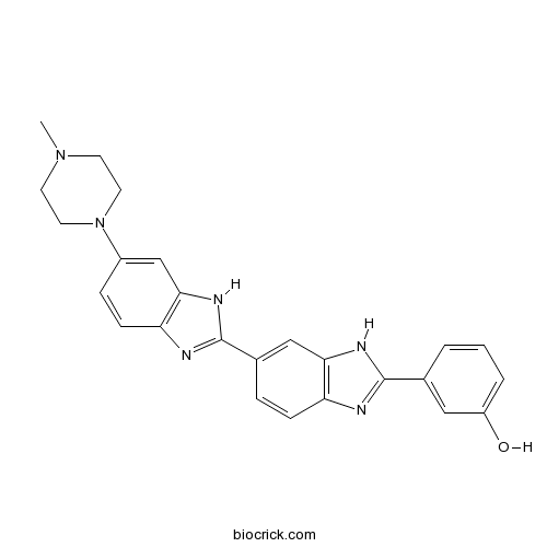
HOE-S 785026Blue fluorescent dyes used CAS# 132869-83-1 |

Quality Control & MSDS
3D structure
Package In Stock
Number of papers citing our products

| Cas No. | 132869-83-1 | SDF | Download SDF |
| PubChem ID | 10410101 | Appearance | Powder |
| Formula | C25H24N6O | M.Wt | 424.5 |
| Type of Compound | N/A | Storage | Desiccate at -20°C |
| Synonyms | meta-Hoechst | ||
| Solubility | DMSO : ≥ 37 mg/mL (87.16 mM); | ||
| Chemical Name | 3-[6-[6-(4-methylpiperazin-1-yl)-1H-benzimidazol-2-yl]-1H-benzimidazol-2-yl]phenol | ||
| SMILES | CN1CCN(CC1)C2=CC3=C(C=C2)N=C(N3)C4=CC5=C(C=C4)N=C(N5)C6=CC(=CC=C6)O | ||
| Standard InChIKey | WOFDIDGQAOQHGY-UHFFFAOYSA-N | ||
| Standard InChI | InChI=1S/C25H24N6O/c1-30-9-11-31(12-10-30)18-6-8-21-23(15-18)29-25(27-21)17-5-7-20-22(14-17)28-24(26-20)16-3-2-4-19(32)13-16/h2-8,13-15,32H,9-12H2,1H3,(H,26,28)(H,27,29) | ||
| General tips | For obtaining a higher solubility , please warm the tube at 37 ℃ and shake it in the ultrasonic bath for a while.Stock solution can be stored below -20℃ for several months. We recommend that you prepare and use the solution on the same day. However, if the test schedule requires, the stock solutions can be prepared in advance, and the stock solution must be sealed and stored below -20℃. In general, the stock solution can be kept for several months. Before use, we recommend that you leave the vial at room temperature for at least an hour before opening it. |
||
| About Packaging | 1. The packaging of the product may be reversed during transportation, cause the high purity compounds to adhere to the neck or cap of the vial.Take the vail out of its packaging and shake gently until the compounds fall to the bottom of the vial. 2. For liquid products, please centrifuge at 500xg to gather the liquid to the bottom of the vial. 3. Try to avoid loss or contamination during the experiment. |
||
| Shipping Condition | Packaging according to customer requirements(5mg, 10mg, 20mg and more). Ship via FedEx, DHL, UPS, EMS or other couriers with RT, or blue ice upon request. | ||
| Description | HOE-S 785026 is a blue fluorescent dyes, which can be used as a cell dye for DNA.In Vitro:Hoechst stains are part of a family of blue fluorescent dyes used to stain DNA. HOES 785026 is a Hoechst stains are part of a family of blue fluorescent dyes used to stain DNA. | |||||

HOE-S 785026 Dilution Calculator

HOE-S 785026 Molarity Calculator
| 1 mg | 5 mg | 10 mg | 20 mg | 25 mg | |
| 1 mM | 2.3557 mL | 11.7786 mL | 23.5571 mL | 47.1143 mL | 58.8928 mL |
| 5 mM | 0.4711 mL | 2.3557 mL | 4.7114 mL | 9.4229 mL | 11.7786 mL |
| 10 mM | 0.2356 mL | 1.1779 mL | 2.3557 mL | 4.7114 mL | 5.8893 mL |
| 50 mM | 0.0471 mL | 0.2356 mL | 0.4711 mL | 0.9423 mL | 1.1779 mL |
| 100 mM | 0.0236 mL | 0.1178 mL | 0.2356 mL | 0.4711 mL | 0.5889 mL |
| * Note: If you are in the process of experiment, it's necessary to make the dilution ratios of the samples. The dilution data above is only for reference. Normally, it's can get a better solubility within lower of Concentrations. | |||||

Calcutta University

University of Minnesota

University of Maryland School of Medicine

University of Illinois at Chicago

The Ohio State University

University of Zurich

Harvard University

Colorado State University

Auburn University

Yale University

Worcester Polytechnic Institute

Washington State University

Stanford University

University of Leipzig

Universidade da Beira Interior

The Institute of Cancer Research

Heidelberg University

University of Amsterdam

University of Auckland

TsingHua University

The University of Michigan

Miami University

DRURY University

Jilin University

Fudan University

Wuhan University

Sun Yat-sen University

Universite de Paris

Deemed University

Auckland University

The University of Tokyo

Korea University
Description: IC50 Value: N/A Hoechst stains are part of a family of blue fluorescent dyes used to stain DNA. These Bis-benzimides were originally developed by Hoechst AG, which numbered all their compounds so that the dye Hoechst 33342 is the 33342nd compound made by the company. There are three related Hoechst stains: Hoechst 33258, Hoechst 33342, and Hoechst 34580. The dyes Hoechst 33258 and Hoechst 33342 are the ones most commonly used and they have similarexcitation/emission spectra. Both dyes are excited by ultraviolet light at around 350 nm, and both emit blue/cyan fluorescent light around anemission maximum at 461 nm. Unbound dye has its maximum fluorescence emission in the 510-540 nm range. Hoechst dyes are soluble in water and in organic solvents such as dimethyl formamide or dimethyl sulfoxide. Concentrations can be achieved of up to 10 mg/mL. Aqueous solutions are stable at 2-6 °C for at least six months when protected from light. For long-term storage the solutions are instead frozen at ≤-20 °C. The dyes bind to the minor groove of double-stranded DNA with a preference for sequences rich in adenine andthymine. Although the dyes can bind to all nucleic acids, AT-rich double-stranded DNA strands enhance fluorescence considerably. Hoechst dyes are cell-permeable and can bind to DNA in live or fixed cells. Therefore, these stains are often called supravital, which means that cells survive a treatment with these compounds. Cells that express specific ATP-binding cassette transporter proteins can also actively transport these stains out of their cytoplasm.
- Lercanidipine hydrochloride
Catalog No.:BCC5238
CAS No.:132866-11-6
- PD 81723
Catalog No.:BCC7032
CAS No.:132861-87-1
- Terchebulin
Catalog No.:BCN3264
CAS No.:132854-40-1
- YS-49
Catalog No.:BCC2067
CAS No.:132836-42-1
- Blonanserin
Catalog No.:BCC3740
CAS No.:132810-10-7
- CP 96345
Catalog No.:BCC7509
CAS No.:132746-60-2
- PyBrOP
Catalog No.:BCC2821
CAS No.:132705-51-2
- [Leu31,Pro34]-Neuropeptide Y (human, rat)
Catalog No.:BCC5722
CAS No.:132699-73-1
- Fmoc-Tle-OH
Catalog No.:BCC2657
CAS No.:132684-60-7
- Senecionine N-oxide
Catalog No.:BCN2130
CAS No.:13268-67-2
- Otophylloside O
Catalog No.:BCN7337
CAS No.:1326583-08-7
- CCMQ
Catalog No.:BCC6984
CAS No.:132623-44-0
- Bis(4-hydroxy-3,5-dimethylphenyl) sulfone
Catalog No.:BCC8884
CAS No.:13288-70-5
- Ramosetron Hydrochloride
Catalog No.:BCC5272
CAS No.:132907-72-3
- 9-Hydroxy-13E-labden-15-oic acid
Catalog No.:BCN6177
CAS No.:132915-47-0
- Rifampin
Catalog No.:BCC4839
CAS No.:13292-46-1
- Macrocarpal A
Catalog No.:BCN6178
CAS No.:132951-90-7
- Mequindox
Catalog No.:BCC9021
CAS No.:13297-17-1
- Fmoc-Lys(Fmoc)-OPfp
Catalog No.:BCC3522
CAS No.:132990-14-8
- (-)-Asarinin
Catalog No.:BCN2290
CAS No.:133-04-0
- Asarinin
Catalog No.:BCN2769
CAS No.:133-05-1
- 3-Indolebutyric acid (IBA)
Catalog No.:BCC6491
CAS No.:133-32-4
- Trichlormethiazide
Catalog No.:BCC4872
CAS No.:133-67-5
- 13-Epijhanol
Catalog No.:BCN4713
CAS No.:133005-15-9
Macro-Raman spectroscopy for bulk composition and homogeneity analysis of multi-component pharmaceutical powders.[Pubmed:28448887]
J Pharm Biomed Anal. 2017 Jul 15;141:180-191.
A new macro-Raman system equipped with a motorized translational sample stage and low-frequency shift capabilities was developed for bulk composition and homogeneity analysis of multi-component pharmaceutical powders. Different sampling methods including single spot and scanning measurement were compared. It was found that increasing sample volumes significantly improved the precision of quantitative composition analysis, especially for poorly mixed powders. The multi-pass cavity of the macro-Raman system increased effective sample volumes by 20 times from the sample volume defined by the collection optics, i.e., from 0.02muL to about 0.4muL. A stochastic model simulating the random sampling process of polydisperse microparticles was used to predict the sampling errors for a specific sample volume. Comparison of fluticasone propionate mass fractions of the commercial products Flixotide((R)) 250 and Seretide((R)) 500 simulated for different sampling volumes with experimentally measured compositions verified that the effective sample volume of a single point macro-Raman measurement in the multi-pass cavity of this instrument was between 0.3muL and 0.5muL. The macro-Raman system was also successfully used for blend uniformity analysis. It was concluded that demixing occurred in the binary mixture of l-leucine and d-mannitol from the observation that the sampling errors indicated by the standard deviations of measured leucine mass fractions increased during mixing, and the standard deviation values were all larger than the theoretical lower limit determined by the simulation. Since sample volume was shown to have a significant impact on measured homogeneity characteristics, it was concluded that powder homogeneity analysis results, i.e., the mean of individual test results and absolute and relative standard deviations, must be presented together with the effective sample volumes of the applied testing techniques for any measurement of powder homogeneity to be fully meaningful.
Modeling nasopharyngeal carcinoma in three dimensions.[Pubmed:28454359]
Oncol Lett. 2017 Apr;13(4):2034-2044.
Nasopharyngeal carcinoma (NPC) is a type of cancer endemic in Asia, including Malaysia, Southern China, Hong Kong and Taiwan. Treatment resistance, particularly in recurring cases, remains a challenge. Thus, studies to develop novel therapeutic agents are important. Potential therapeutic compounds may be effectively examined using two-dimensional (2D) cell culture models, three-dimensional (3D) spheroid models or in vivo animal models. The majority of drug assessments for cancers, including for NPC, are currently performed with 2D cell culture models. This model offers economical and high-throughput screening advantages. However, 2D cell culture models cannot recapitulate the architecture and the microenvironment of a tumor. In vivo models may recapitulate certain architectural and microenvironmental conditions of a tumor, however, these are not feasible for the screening of large numbers of compounds. By contrast, 3D spheroid models may be able to recapitulate a physiological microenvironment not observed in 2D cell culture models, in addition to avoiding the impediments of in vivo animal models. Thus, the 3D spheroid model offers a more representative model for the study of NPC growth, invasion and drug response, which may be cost-effective without forgoing quality.
CD24, CD44 and EpCAM enrich for tumour-initiating cells in a newly established patient-derived xenograft of nasopharyngeal carcinoma.[Pubmed:28959019]
Sci Rep. 2017 Sep 28;7(1):12372.
Subpopulations of nasopharyngeal carcinoma (NPC) contain cells with differential tumourigenic properties. Our study evaluates the tumourigenic potential of CD24, CD44, EpCAM and combination of EpCAM/CD44 cells in NPC. CD44br and EpCAMbr cells enriched for higher S-phase cell content, faster-growing tumourigenic cells leading to tumours with larger volume and higher mitotic figures. Although CD44br and EpCAMbr cells significantly enriched for tumour-initiating cells (TICs), all cells could retain self-renewal property for at least four generations. Compared to CD44 marker alone, EpCAM/CD44dbr marker did not enhance for cells with faster-growing ability or higher TIC frequency. Cells expressing high CD44 or EpCAM had lower KLF4 and p21 in NPC subpopulations. KLF4-overexpressed EpCAMbr cells had slower growth while Kenpaullone inhibition of KLF4 transcription increased in vitro cell proliferation. Compared to non-NPC, NPC specimens had increased expression of EPCAM, of which tumours from advanced stage of NPC had higher expression. Together, our study provides evidence that EpCAM is a potentially important marker in NPC.
Selection of suitable endogenous reference genes for qPCR in kidney and hypothalamus of rats under testosterone influence.[Pubmed:28591185]
PLoS One. 2017 Jun 7;12(6):e0176368.
Real-time quantitative PCR (qPCR) is the most reliable and accurate technique for analyses of gene expression. Endogenous reference genes are being used to normalize qPCR data even though their expression may vary under different conditions and in different tissues. Nonetheless, verification of expression of reference genes in selected studied tissue is essential in order to accurately assess the level of expression of target genes of interest. Therefore, in this study, we attempted to examine six commonly used reference genes in order to identify the gene being expressed most constantly under the influence of testosterone in the kidneys and hypothalamus. The reference genes include glyceraldehyde-3-phosphate dehydrogenase (GAPDH), actin beta (ACTB), beta-2 microglobulin (B2m), hypoxanthine phosphoribosyltransferase 1 (HPRT), peptidylprolylisomerase A (Ppia) and hydroxymethylbilane synthase (Hmbs). The cycle threshold (Ct) value for each gene was determined and data obtained were analyzed using the software programs NormFinder, geNorm, BestKeeper, and rank aggregation. Results showed that Hmbs and Ppia genes were the most stably expressed in the hypothalamus. Meanwhile, in kidneys, Hmbs and GAPDH appeared to be the most constant genes. In conclusion, variations in expression levels of reference genes occur in kidneys and hypothalamus under similar conditions; thus, it is important to verify reference gene levels in these tissues prior to commencing any studies.
Effects of Root Extracts of Eurycoma longifolia Jack on Corpus Cavernosum of Rat.[Pubmed:28226311]
Med Princ Pract. 2017;26(3):258-265.
OBJECTIVE: This study was conducted to investigate the mechanisms of action of Eurycoma longifolia in rat corpus cavernosum. MATERIALS AND METHODS: Tincture of the roots was concentrated to dryness by evaporating the ethanol in vacuo. This ethanolic extract was partitioned into 5 fractions sequentially with hexane, dichloromethane (DCM), ethyl acetate, butanol, and water. The corpus cavernosum relaxant activity of each fraction was investigated. The DCM fraction which showed the highest potency in relaxing phenylephrine-precontracted corpora cavernosa was purified by column chromatography. The effects of the most potent DCM subfraction in relaxing phenylephrine-precontracted corpora cavernosa, DCM-I, on angiotensin I- or angiotensin II-induced contractions in corpora cavernosa were investigated. The effects of DCM-I pretreatment on the responses of phenylephrine-precontracted corpora cavernosa to angiotensin II or bradykinin were also studied. An in vitro assay was conducted to evaluate the effect of DCM-I on angiotensin-converting enzyme activity. RESULTS: Fraction DCM-I decreased the maximal contractions (100%) evoked by angiotensin I and angiotensin II to 30 +/- 14% and 26 +/- 16% (p < 0.001), respectively. In phenylephrine-precontracted corpora cavernosa, DCM-I pretreatment caused angiotensin II to induce 82 +/- 27% relaxation of maximal contraction (p < 0.01) and enhanced (p < 0.001) bradykinin-induced relaxations from 47 +/- 8% to 100 +/- 5%. In vitro, DCM-I was able to reduce (p < 0.001) the maximal angiotensin-converting enzyme activity to 78 +/- 0.24%. CONCLUSION: Fraction DCM-I was able to antagonize angiotensin II-induced contraction to cause corpus cavernosum relaxation via inhibition of angiotensin II type 1 receptor and enhance bradykinin-induced relaxation through inhibition of angiotensin-converting enzyme.


