Datura metel
Datura metel
1. The products in our compound library are selected from thousands of unique natural products; 2. It has the characteristics of diverse structure, diverse sources and wide coverage of activities; 3. Provide information on the activity of products from major journals, patents and research reports around the world, providing theoretical direction and research basis for further research and screening; 4. Free combination according to the type, source, target and disease of natural product; 5. The compound powder is placed in a covered tube and then discharged into a 10 x 10 cryostat; 6. Transport in ice pack or dry ice pack. Please store it at -20 °C as soon as possible after receiving the product, and use it as soon as possible after opening.

Natural products/compounds from Datura metel
- Cat.No. Product Name CAS Number COA
-
BCN1199
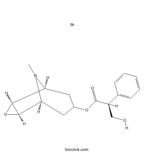
Scopolamine hydrobromide114-49-8
Instructions

-
BCN5903
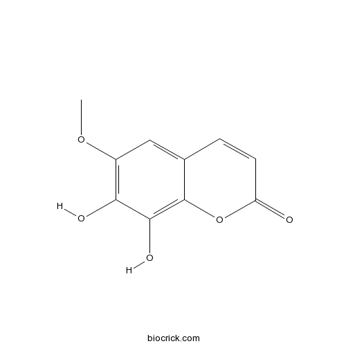
Fraxetin574-84-5
Instructions

-
BCN4213
N-trans-Feruloyltyramine66648-43-9
Instructions

-
BCN4376
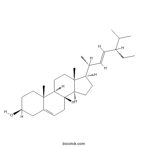
Stigmasterol83-48-7
Instructions

[Chemical constituents from roots of Datura metel].[Pubmed: 29751713]
The chemical constituents from the n-butanol fraction of the 70% ethanol extract of Datura metel roots were separated by silica gel and ODS chromatogram columns as well as preparative HPLC. On the basis of spectral data analysis, their structures were elucidated. Twenty-one compounds were obtained and their structures were identified as citroside A (1), coniferin (2), paeoniflorin (3), (6R,7E,9R)-9-hydroxy-4,7-megastigmadien-3-one 9-O-[α-L-arabin-opyranosyl-(l→6)-β-D-glucopyranoside] (4), (1R,7R,10R,11R)-12-hydroxyl anhuienosol (5), kaurane acid glycoside A (6), ent-2-oxo-15,16-dihydroxypimar-8(14)-en-16-O-β-glucopyranoside (7), ginsenoside Rg₁(8), ginsenoside Re (9), notoginsenosides R₁(10), N-butyl-O-β-D-fructofuranoside (11), salidroside (12), hexyl β-sophoroside (13), 2,6-dimethoxy-4-hydroxyphenol 1-glucoside (14), benzyl-O-β-D-xylopyranoxyl(1→6)-β-D-glucopyranoside (15), (Z)-3-hexenyl-O-α-L-arabinopyranosyl-(1→6)-β-D-glucopyranoside (16), N-[2-(3,4-dihydro-xyphenyl)-2-hydroxyethyl]-3-(4-methoxyphenyl) prop-2-enamide (17), cannabisin D (18), cannabisin E (19), melongenamide B (20), paprazine (21). Compounds 2-17 and 20-21 were isolated from the Solanaceae family for the first time.
Comparisons of the pharmacokinetic and tissue distribution profiles of withanolide B after intragastric administration of the effective part of Datura metel L. in normal and psoriasis guinea pigs.[Pubmed: 29574380]
A simple, highly sensitive ultra-performance liquid chromatography- electrospray ionization-mass spectrometry (LC-ESI-MS) method has been developed to quantify of withanolide B and obakunone (IS) in guinea pig plasma and tissues, and to compare the pharmacokinetics and tissue distribution of withanolide B in normal and psoriasis guinea pigs. After mixing with IS, plasma and tissues were pretreated by protein precipitation with methanol. Chromatographic separation was performed on a C18 column using aqueous (0.1% formic acid) and acetonitrile (0.1% formic acid) solutions at 0.4 mL/min as the mobile phase. The gradient program was selected (0-4.0 min, 2-98% B; 4.0-4.5 min, 98-2% B; and 4.5-5 min, 2% B). Detection was performed on a 4000 QTRAP UPLC-ESI-MS/MS system from AB Sciex in the multiple reaction monitoring (MRM) mode. Withanolide B and obakunone (IS) were monitored under positive ionization conditions. The optimized mass transition ion-pairs (m/z) for quantitation were 455.1/109.4 for withanolide B and 455.1/161.1 for obakunone.
Differential Inhibition of Helicoverpa armigera (Lep.: Noctuidae) Gut Digestive Trypsin by Extracted and Purified Inhibitor of Datura metel (Solanales: Solanaceae).[Pubmed: 29240906]
The cotton bollworm, Helicoverpa armigera Hubner (Lep: Noctuidae), is an economically important pest of numerous major food crops worldwide. Protease inhibitors from plants, expressed constitutively in transgenic crops, have potential for pest management as an alternative to chemical pesticides. In this study, a protease inhibitor was isolated, purified, and characterized from Datura metel L. seeds. The purity of the isolated inhibitor was confirmed by reverse-phase high-performance liquid chromatography, and activity staining showed one major peak and one clear activity band for the protein. Electrophoretic studies following gel filtration and ion-exchange chromatography revealed two and one bands for purified proteins, respectively. Partial biochemical characterizations of the purified inhibitor were determined. Maximum inhibitory activity was observed at 40-45°C (optimal temperature) when tested against gut extracts of fourth to sixth instar H. armigera larvae. Thermo-stability of the trypsin inhibitor against sixth instar larval midgut trypsin was observed up to 50°C when incubated for 30 min and 2 h. Among metal ions tested, Fe2+, Cu2+, and Mn2+ were found to decrease the trypsin inhibitory activity, whereas Hg2+, Mg2+, K+, Zn2+, Na+, Ca2+, and Cd2+ were found to significantly increase the inhibitory effect. This trypsin inhibitor showed competitive inhibition where the apparent value of Michaelis-Menten Km increased, but the value of Vmax remained unchanged.
Novel Microbial Sources of Tropane Alkaloids: First Report of Production by Endophytic Fungi Isolated from Datura metel L.[Pubmed: 29063971]
Eighteen endophytic fungi were isolated from various tissues of Datura metel and genes encoding for putrescine N-methyltransferase (PMT), tropinone reductase 1 (TR1) and hyoscyamine 6β-hydroxylase (H6H) were used as molecular markers for PCR-based screening approach for tropane alkaloids (TAs) producing endophytic fungi. These fungi were identified taxonomically by sequence analysis of the internal transcribed spacer region (ITS1-5.8S-ITS2) and also based on morphological characteristics of the fungal spore as Colletotrichum boninense, Phomopsis sp., Fusarium solani, Colletotrichum incarnatum, Colletotrichum siamense and Colletotrichum gloeosporioides. The production of TAs hyoscyamine and scopolamine by the fungi has been ascertained using chromatography and spectroscopy methods by comparison with the standards. Among the fungi, the highest yields of hyoscyamine (3.9 mg/L) and scopolamine (4.1 mg/L) were found in C. incarnatum culture. This is the first report of endophytic fungi possess the PMT, TR1 and H6H genes and produces TAs. These endophytic fungi have significant potential to be applied in fermentation technology to meet the demands for TAs economically.
Potential of CLSM in studying some modern and fossil palynological objects.[Pubmed: 28940409]
We have tested possibilities and limitations of confocal laser scanning microscopy to study the morphology of pollen and spores and inner structure of sporoderms. As test objects, we used pollen grains of the modern angiosperm Ribes niveum (Grossulariaceae) and Datura metel (Solanaceae), fossil angiosperm pollen grains of Pseudointegricorpus clarireticulatum and Wodehouseia spinata dated to the Late Cretaceous, fossil gymnosperm pollen grains of Cycadopites-type dated to the Middle Jurassic, and fossil megaspores Maexisporites rugulaeferus, M. grosstriletus, and Trileites sp. dated to the Early Triassic. For comparative purpose, we studied the same objects with application of conventional light, scanning electron (to entire pollen grains and spores or to semithin sections of their walls), or transmission electron microscopy. The resolution of confocal microscope is much lower than that of electron microscopes, as are its abilities to reconstruct the surface patterns and inner structure. On the other hand, it can provide information that is unreachable by other microscopical methods. Thus, the structure of endoapertures in angiosperm pollen grains can be directly observed. It is also helpful in studies of asymmetrical pollen and pollen grains bearing various appendages and having complicated exine structure, because rotation of 3-D reconstructions allows one to examine all sides and structures of the pollen grain. The exact location of all visible and concealed structures in the sporoderm can be detected; this information helps to describe the morphology and inner structure of pollen grains and to choose necessary directions of further ultrathin sectioning for a transmission electron microscopical study. In studies of fossil pollen grains that are preserved in clumps and stuck to cuticles, confocal microscope is useful in determining the number of apertures in individual pollen grains. This can be done by means of virtual sections through 3-D reconstructions of pollen grains. Fossil megaspores are too large and too thick-walled objects for a confocal study; however, confocal microscope was able to reveal a degree of compression of fossil megaspores, the presence of a cavity between the outer and inner sporoderm layers, and to get some information about sporoderm inner structure.


