Quisqualis indica
Quisqualis indica
1. The products in our compound library are selected from thousands of unique natural products; 2. It has the characteristics of diverse structure, diverse sources and wide coverage of activities; 3. Provide information on the activity of products from major journals, patents and research reports around the world, providing theoretical direction and research basis for further research and screening; 4. Free combination according to the type, source, target and disease of natural product; 5. The compound powder is placed in a covered tube and then discharged into a 10 x 10 cryostat; 6. Transport in ice pack or dry ice pack. Please store it at -20 °C as soon as possible after receiving the product, and use it as soon as possible after opening.
Natural products/compounds from Quisqualis indica
- Cat.No. Product Name CAS Number COA
-
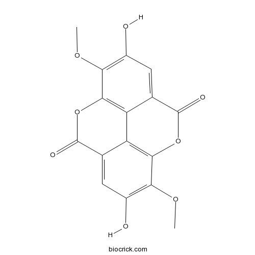
BCN5062
3,8-Di-O-methylellagic acid2239-88-5
Instructions
-
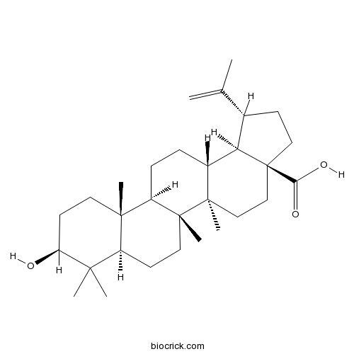
BCN5524
Betulinic acid472-15-1
Instructions
-
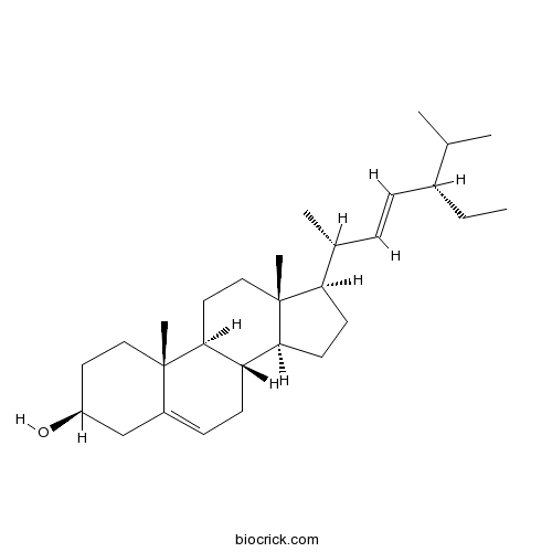
BCN4376
Stigmasterol83-48-7
Instructions
-
BCN4373
Ethyl gallate831-61-8
Instructions

Synthesis and characterization of copper nanoparticles stabilized with Quisqualis indica extract: Evaluation of its cytotoxicity and apoptosis in B16F10 melanoma cells.[Pubmed: 29156527]
Green synthesis of metallic nanoparticles is a cost-effective environment-friendly technique and Quisqualis indica has ethnomedicinal values. With this background in this study, the floral extract of Q. indica was used to fabricate copper nanoparticles (QCuNPs) from copper acetate. Biophysical analysis revealed the formation of spherical, monodisperse, crystalline QCuNPs. Significant cytotoxic potentials of the nanoformulation were determined by MTT and lactate dehydrogenase (LDH) assay on B16F10 melanoma cells. Estimation of GSH and ROS demonstrated that QCuNPs induced melanoma cell death by induction of oxidative stress. Gene transcript analysis showed up-regulation of caspase-dependent as well as caspase-independent (AIF) apoptotic genes in treated cells. Comparative proteomics study mostly showed the abundance of apoptotic and cell cycle arrest proteins in treated samples. The in vivo therapeutic efficacy was studied in mice bearing B16F10 melanoma tumor where a significant decrease in tumor growth was observed in nanoparticles treated animal model. In conclusion, QCuNPs caused cytotoxicity and apoptosis in melanoma cells and its mechanism was established from gene expression and proteomic studies. QCuNPs exhibited potential suppression of B16F10 melanoma cell proliferation and substantial inhibition of tumor growth in animals. As per our information, this is the first study exploring the potential of Q. indica for the formulation of eco-friendly copper nanoparticle which will have great future application in the medicinal field.
Quisqualis indica Improves Benign Prostatic Hyperplasia by Regulating Prostate Cell Proliferation and Apoptosis.[Pubmed: 28943529]
Quisqualis indica (QI) has been used for treating disorders such as stomach pain, constipation, and digestion problem. This study was aimed to evaluate the therapeutic efficacy of QI extract on treating benign prostatic hyperplasia (BPH) in LNCaP human prostate cancer cell line and a testosterone-induced BPH rat model. LNCaP cells were treated with QI plus testosterone propionate (TP), and androgen receptor (AR) and prostate specific antigen (PSA) expression levels were assessed by Western blotting. To induce BPH, the rats were subjected to a daily subcutaneous injection of TP (3 mg/kg) for 4 weeks. The rats in treatment group were orally gavaged with QI (150 mg/kg) together with the TP injection. In-vitro studies showed that TP-induced increases in AR and PSA expression in LNCaP cells were reduced by QI treatment. In BPH-model rats, the prostate weight, testosterone in serum, dihydrotestosterone (DHT) concentration and 5α-reductase type 2 mRNA expression in prostate tissue were significantly reduced following the treatment with QI. TP-induced prostatic hyperplasia and the expression of proliferating cell nuclear antigen (PCNA) and cyclin D1 were significantly attenuated in QI-treated rats. In addition, QI induced apoptosis by up-regulating caspase-3 and -9 activity and decreasing the B-cell lymphoma 2 (Bcl-2)/Bcl-2-associated X protein (Bax) ratio in prostate tissues of BPH rats. Further investigation showed that TP-induced activation of AKT and glycogen synthase kinase 3β (GSK3β) was reduced by QI administration. Therefore, our findings suggest that QI attenuates the BPH state in rats through anti-proliferative and pro-apoptotic activities and might be useful in the clinical treatment of BPH.
Protective effects of Cambodian medicinal plants on tert‑butyl hydroperoxide‑induced hepatotoxicity via Nrf2‑mediated heme oxygenase‑1.[Pubmed: 27959441]
Liver diseases are considered to be primary contributors to morbidity and mortality rates in humans. Oxidative stress is critical in liver injury, and oxidant‑induced liver injury may be caused by toxins, including tert‑butyl hydroperoxide (t‑BHP). The present study investigated the hepatoprotective activities of 64 crude ethanol extracts of Cambodian medicinal plants against t‑BHP‑induced cytotoxicity in human liver‑derived HepG2 cells, and assessed their cytoprotective mechanism pertaining to the expression of heme oxygenase (HO)‑1 and nuclear factor E2‑related factor 2 (Nrf2). Protective effects in HepG2 cells were determined by MTT assay. Protein expression levels of HO‑1 and Nrf2 were determined by western blotting and mRNA expression levels were determined by reverse transcription‑quantitative polymerase chain reaction. Of the 64 extracts, 19 extracts exhibited high hepatoprotective activities: Ampelocissus martini, Bauhinia bracteata, Bombax ceiba, Borassus flabellifer, Cardiospermum halicacabum, Cayratia trifolia, Cinnamomum caryophyllus, Cyperus rotundus, Dasymaschalon lomentaceum, Ficus benjamina, Mangifera duperreana, Morinda citrifolia, Pandanus humilis, Peliosanthes weberi, Phyllanthus emblica, Quisqualis indica, Smilax glabra, Tinospora crispa and Willughbeia cochinchinensis, with half maximal effective concentrations ranging between 59.23 and 157.80 µg/ml. Further investigations revealed that, of these 19 extracts, HO‑1 and Nrf2 were expressed in P. weberi and T. crispa expressed in a dose‑dependent manner. In addition, the activities of reactive oxygen species were suppressed following treatment of these two extracts in t‑BHP‑induced HepG2 cells. These results indicated that, of the 64 Cambodian plants, P. weberi and T. crispa exhibited hepatoprotective effects on t‑BHP‑induced cytotoxicity in HepG2 cells, possibly by the induction of Nrf2‑mediated expression of HO‑1. Taken together, these results suggested that T. crispa or P. weberi may offer potential for therapeutic applications in liver disease characterized by oxidative stress.
One-pot biogenic fabrication of silver nanocrystals using Quisqualis indica: Effectiveness on malaria and Zika virus mosquito vectors, and impact on non-target aquatic organisms.[Pubmed: 27491031]
Currently, mosquito vector control is facing a number of key challenges, including the rapid development of resistance to synthetic pesticides and the recent spread of aggressive arbovirus outbreaks. The biosynthesis of silver nanoparticles (AgNPs) is currently considered an environmental friendly alternative to the employ of pyrethroids, carbamates and microbial agents (e.g. Bacillus thuringiensis var. israelensis), since AgNPs are easy to produce, effective and stable in the aquatic environment. However, their biophysical features showed wide variations according to the botanical agent using for the green synthesis, outlining the importance of screening local floral resources used as reducing and stabilizing agents. In this study, we focused on the biophysical properties and the mosquitocidal action of Quisqualis indica-fabricated AgNPs. AgNPs were characterized using spectroscopic (UV, FTIR, XRD) and microscopic (AFM, SEM, TEM and EDX) techniques. AFM, SEM and TEM confirmed the synthesis of poly-dispersed AgNPs with spherical shape and size ranging from 1 to 30nm. XRD shed light on the crystalline structure of these AgNPs. The acute toxicity of Quisqualis indica extract and AgNPs was evaluated against malaria, arbovirus, and filariasis vectors, Anopheles stephensi, Aedes aegypti and Culex quinquefasciatus, as well as on three important non-target aquatic organisms. The Q. indica leaf extract showed moderate larvicidal effectiveness on Cx. quinquefasciatus (LC50=220.42), Ae. aegypti (LC50=203.63) and An. stephensi (LC50=185.98). Q. indica-fabricated AgNPs showed high toxicity against Cx. quinquefasciatus (LC50=14.63), Ae. aegypti (LC50=13.55) and An. stephensi (LC50=12.52), respectively. Notably, Q. indica-synthesized AgNPs were moderately toxic to non-target aquatic mosquito predators Anisops bouvieri (LC50=653.05μg/mL), Diplonychus indicus (LC50=860.94μg/mL) and Gambusia affinis (LC50=2183.16μg/mL), if compared to the targeted mosquitoes. Overall, the proposed one-pot biogenic fabrication of AgNPs using Q. indica is a low-cost and eco-friendly tool in the fight against Zika virus, malaria and filariasis vectors, with little impact against non-target aquatic mosquito predators.
Pollinator responses to floral colour change, nectar, and scent promote reproductive fitness in Quisqualis indica (Combretaceae).[Pubmed: 27072926]
Floral colour change is visual signals for pollinators to avoid old flowers and increase pollination efficiency. Quisqualis indica flowers change colour from white to pink to red may be associated with a shift from moth to butterfly pollination. To test this hypothesis, we investigated Q. indica populations in Southwest China. Flowers secreted nectar continuously from the evening of anthesis until the following morning, then decreased gradually with floral colour change. The scent compounds in the three floral colour stages were similar; however, the scent composition was different, and the scent emission rate decreased from the white to red stage. Dichogamy in Q. indica prevents self-pollination and interference of male and female functions. Controlled pollinations demonstrated that this species is self-incompatible and needs pollinators for seed production. Different pollinators were attracted in each floral colour stage; mainly moths at night and bees and butterflies during the day. Observations of open-pollinated inflorescences showed that white flowers had a higher fruit set than pink or red flowers, indicating the high contribution of moths to reproductive success. We concluded that the nectar and scent secretion are related to floral colour change in Q. indica, in order to attract different pollinators and promote reproductive fitness.
25-O-acetyl-23,24-dihydro-cucurbitacin F induces cell cycle G2/M arrest and apoptosis in human soft tissue sarcoma cells.[Pubmed: 25701753]
Quisqualis indica is used in traditional Chinese medicine to treat cancer and related syndromes and also known for its anthelminthic effects.
Phytochemistry and pharmacogenomics of natural products derived from traditional Chinese medicine and Chinese materia medica with activity against tumor cells.[Pubmed: 18202018]
The cure from cancer is still not a reality for all patients, which is mainly due to the limitations of chemotherapy (e.g., drug resistance and toxicity). Apart from the high-throughput screening of synthetic chemical libraries, natural products represent attractive alternatives for drug development. We have done a systematic bioactivity-based screening of natural products derived from medicinal plants used in traditional Chinese medicine. Plant extracts with growth-inhibitory activity against tumor cells have been fractionated by chromatographic techniques. We have isolated the bioactive compounds and elucidated the chemical structures by nuclear magnetic resonance and mass spectrometry. By this strategy, we identified 25-O-acetyl-23,24-dihydro-cucurbitacin F as a cytotoxic constituent of Quisqualis indica. Another promising compound identified by this approach was miltirone from Salvia miltiorrhiza. The IC50 values for miltirone of 60 National Cancer Institute cell lines were associated with the microarray-based expression of 9,706 genes. By COMPARE and hierarchical cluster analyses, candidate genes were identified, which significantly predicted sensitivity or resistance of cell lines to miltirone.