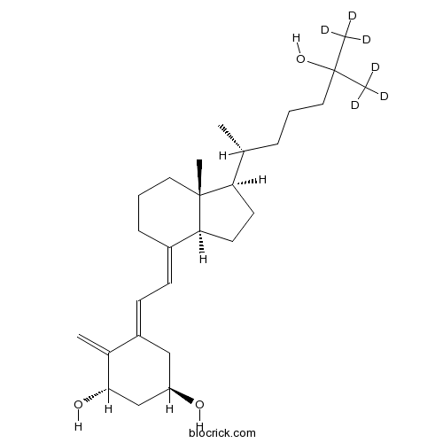
Calcitriol D6CAS# 78782-99-7 |

- 25,26-Dihydroxyvitamin D3
Catalog No.:BCC4201
CAS No.:29261-12-9
- Vitamin D4
Catalog No.:BCC2042
CAS No.:511-28-4
- Impurity B of Calcitriol
Catalog No.:BCC1645
CAS No.:66791-71-7
- Calcifediol-D6
Catalog No.:BCC4075
CAS No.:78782-98-6
Quality Control & MSDS
3D structure
Package In Stock
Number of papers citing our products

| Cas No. | 78782-99-7 | SDF | Download SDF |
| PubChem ID | 57369253 | Appearance | Powder |
| Formula | C27H44O3 | M.Wt | 416.6 |
| Type of Compound | N/A | Storage | Desiccate at -20°C |
| Solubility | Soluble in Chloroform | ||
| Chemical Name | (1R,3S)-5-[2-[(1R,3aS,7aR)-7a-methyl-1-[(2R)-7,7,7-trideuterio-6-hydroxy-6-(trideuteriomethyl)heptan-2-yl]-2,3,3a,5,6,7-hexahydro-1H-inden-4-ylidene]ethylidene]-4-methylidenecyclohexane-1,3-diol | ||
| SMILES | CC(CCCC(C)(C)O)C1CCC2C1(CCCC2=CC=C3CC(CC(C3=C)O)O)C | ||
| Standard InChIKey | GMRQFYUYWCNGIN-OWKVYBDPSA-N | ||
| Standard InChI | InChI=1S/C27H44O3/c1-18(8-6-14-26(3,4)30)23-12-13-24-20(9-7-15-27(23,24)5)10-11-21-16-22(28)17-25(29)19(21)2/h10-11,18,22-25,28-30H,2,6-9,12-17H2,1,3-5H3/t18-,22-,23-,24+,25+,27-/m1/s1/i3D3,4D3 | ||
| General tips | For obtaining a higher solubility , please warm the tube at 37 ℃ and shake it in the ultrasonic bath for a while.Stock solution can be stored below -20℃ for several months. We recommend that you prepare and use the solution on the same day. However, if the test schedule requires, the stock solutions can be prepared in advance, and the stock solution must be sealed and stored below -20℃. In general, the stock solution can be kept for several months. Before use, we recommend that you leave the vial at room temperature for at least an hour before opening it. |
||
| About Packaging | 1. The packaging of the product may be reversed during transportation, cause the high purity compounds to adhere to the neck or cap of the vial.Take the vail out of its packaging and shake gently until the compounds fall to the bottom of the vial. 2. For liquid products, please centrifuge at 500xg to gather the liquid to the bottom of the vial. 3. Try to avoid loss or contamination during the experiment. |
||
| Shipping Condition | Packaging according to customer requirements(5mg, 10mg, 20mg and more). Ship via FedEx, DHL, UPS, EMS or other couriers with RT, or blue ice upon request. | ||
| Description | Calcitriol D6 is the deuterated form of Calcitriol(1,25-Dihydroxyvitamin D3; Rocaltrol ), which is the hormonally active form of vitamin D, Calcitriol is the active metabolite of vitamin D3 that activates the vitamin D receptor (VDR).
IC50 value:
Target: vitamin D receptor
Calcitriol(1,25-Dihydroxyvitamin D3; Rocaltrol ) displays calcemic actions. Calcitriol stimulates intestinal and renal Ca2+ absorption and regulates bone Ca2+ turnover. Calcitriol (1,25-Dihydroxyvitamin D3; Rocaltrol )exhibits antitumor activity; Calcitriol(1,25-Dihydroxyvitamin D3; Rocaltrol ) inhibits in vivo and in vitro cell proliferation in a wide range of cells including breast, prostate, colon, skin and brain carcinomas and myeloid leukemia cells. References: | |||||

Calcitriol D6 Dilution Calculator

Calcitriol D6 Molarity Calculator
| 1 mg | 5 mg | 10 mg | 20 mg | 25 mg | |
| 1 mM | 2.4004 mL | 12.0019 mL | 24.0038 mL | 48.0077 mL | 60.0096 mL |
| 5 mM | 0.4801 mL | 2.4004 mL | 4.8008 mL | 9.6015 mL | 12.0019 mL |
| 10 mM | 0.24 mL | 1.2002 mL | 2.4004 mL | 4.8008 mL | 6.001 mL |
| 50 mM | 0.048 mL | 0.24 mL | 0.4801 mL | 0.9602 mL | 1.2002 mL |
| 100 mM | 0.024 mL | 0.12 mL | 0.24 mL | 0.4801 mL | 0.6001 mL |
| * Note: If you are in the process of experiment, it's necessary to make the dilution ratios of the samples. The dilution data above is only for reference. Normally, it's can get a better solubility within lower of Concentrations. | |||||

Calcutta University

University of Minnesota

University of Maryland School of Medicine

University of Illinois at Chicago

The Ohio State University

University of Zurich

Harvard University

Colorado State University

Auburn University

Yale University

Worcester Polytechnic Institute

Washington State University

Stanford University

University of Leipzig

Universidade da Beira Interior

The Institute of Cancer Research

Heidelberg University

University of Amsterdam

University of Auckland

TsingHua University

The University of Michigan

Miami University

DRURY University

Jilin University

Fudan University

Wuhan University

Sun Yat-sen University

Universite de Paris

Deemed University

Auckland University

The University of Tokyo

Korea University
The biologically active form of vitamin D3. Calcium regulator; vitamin (antirachitic); antihyperparathyroid; antineoplastic; antipsoriatic.
- Calcifediol-D6
Catalog No.:BCC4075
CAS No.:78782-98-6
- Deapi-platycodin D
Catalog No.:BCN2614
CAS No.:78763-58-3
- TC 1698 dihydrochloride
Catalog No.:BCC7394
CAS No.:787587-06-8
- Flumazenil
Catalog No.:BCC1259
CAS No.:78755-81-4
- Shizukanolide C
Catalog No.:BCN6570
CAS No.:78749-47-0
- D-AP4
Catalog No.:BCC6549
CAS No.:78739-01-2
- Ozagrel HCl
Catalog No.:BCC4926
CAS No.:78712-43-3
- Plantagoside
Catalog No.:BCN8077
CAS No.:78708-33-5
- 4,4'-Biphenyldicarboxylic acid
Catalog No.:BCC8655
CAS No.:787-70-2
- Z-D-Asp-OH
Catalog No.:BCC2786
CAS No.:78663-07-7
- Dehydroandrographolidesuccinate
Catalog No.:BCN8359
CAS No.:786593-06-4
- Terbinafine HCl
Catalog No.:BCC4863
CAS No.:78628-80-5
- Zeylenol
Catalog No.:BCC8267
CAS No.:78804-17-8
- 4-Benzyloxycarbonyl-2-piperazinone
Catalog No.:BCC8699
CAS No.:78818-15-2
- Epibrassinolide
Catalog No.:BCC5479
CAS No.:78821-43-9
- Garcinol
Catalog No.:BCC5623
CAS No.:78824-30-3
- Demethylasterriquinone B1
Catalog No.:BCC7189
CAS No.:78860-34-1
- 1-chloro-6-(5-(prop-1-ynyl)thiophen-2-yl)hexa-3,5-diyn-2-ol
Catalog No.:BCN1352
CAS No.:78876-52-5
- 1-chloro-6-(5-ethynylthiophen-2-yl)hexa-3,5-diyn-2-ol
Catalog No.:BCN1351
CAS No.:78876-53-6
- 6-Thio-dG
Catalog No.:BCC6507
CAS No.:789-61-7
- Orobanone
Catalog No.:BCN3562
CAS No.:78916-35-5
- Deacetylnimbinene
Catalog No.:BCN4578
CAS No.:912545-53-0
- Iloprost
Catalog No.:BCC7247
CAS No.:78919-13-8
- Chicanine
Catalog No.:BCN7818
CAS No.:78919-28-5
The vitamin D receptor (VDR) is expressed in skeletal muscle of male mice and modulates 25-hydroxyvitamin D (25OHD) uptake in myofibers.[Pubmed:24949660]
Endocrinology. 2014 Sep;155(9):3227-37.
Vitamin D deficiency is associated with a range of muscle disorders, including myalgia, muscle weakness, and falls. In humans, polymorphisms of the vitamin D receptor (VDR) gene are associated with variations in muscle strength, and in mice, genetic ablation of VDR results in muscle fiber atrophy and motor deficits. However, mechanisms by which VDR regulates muscle function and morphology remain unclear. A crucial question is whether VDR is expressed in skeletal muscle and directly alters muscle physiology. Using PCR, Western blotting, and immunohistochemistry (VDR-D6 antibody), we detected VDR in murine quadriceps muscle. Detection by Western blotting was dependent on the use of hyperosmolar lysis buffer. Levels of VDR in muscle were low compared with duodenum and dropped progressively with age. Two in vitro models, C2C12 and primary myotubes, displayed dose- and time-dependent increases in expression of both VDR and its target gene CYP24A1 after 1,25(OH)2D (1,25 dihydroxyvitamin D) treatment. Primary myotubes also expressed functional CYP27B1 as demonstrated by luciferase reporter studies, supporting an autoregulatory vitamin D-endocrine system in muscle. Myofibers isolated from mice retained tritiated 25-hydroxyvitamin D3, and this increased after 3 hours of pretreatment with 1,25(OH)2D (0.1 nM). No such response was seen in myofibers from VDR knockout mice. In summary, VDR is expressed in skeletal muscle, and vitamin D regulates gene expression and modulates ligand-dependent uptake of 25-hydroxyvitamin D3 in primary myofibers.
Determination of the vitamin D analog EB 1089 (seocalcitol) in human and pig serum using liquid chromatography-tandem mass spectrometry.[Pubmed:10798301]
J Chromatogr B Biomed Sci Appl. 2000 Mar 31;740(1):117-28.
A liquid chromatographic-tandem mass spectrometric assay in human and pig serum has been developed for quantitative analysis of EB 1089 (seocalcitol). EB 1089 is a novel vitamin D analog under development for the treatment of cancer. The analyte was extracted from serum after protein precipitation using an automated solid-phase extraction procedure involving both a reversed-phase and normal-phase procedure on a single C18 cartridge. The analytical chromatography was performed using a Symmetri C8 50x2.1 mm, 3.5 microm column. The mobile phase was a linear gradient from 75% to 99% methanol with a constant concentration of 2 mM ammonium acetate. EB 1089 and the internal standard [d6]-EB 1089 were detected by using MS-MS. The ion source was operated in the positive electrospray ionisation (ESI) mode. The assay is specific, sensitive, and has a capacity of more than 100 samples per day, with a limit of quantitation of 10 pg ml(-1) for a 1.0-ml sample aliquot. It is now used for routine analysis in connection with pharmacokinetic studies in humans and toxicokinetic studies in pigs.
1,25-Dihydroxyvitamin D3 stimulates avian and mammalian cartilage growth in vitro.[Pubmed:3213605]
J Bone Miner Res. 1988 Feb;3(1):87-91.
We addressed the question of whether 1,25-dihydroxyvitamin D3 (1,25-(OH)2D) could directly stimulate cartilage growth in vitro. Pelvic leaflets from chick embryos and scapular growth plates from fetal pigs were organ cultured in serum-free medium in the presence and absence of 1,25-(OH)2D. After 3 days of incubation, 1,25-(OH)2D had increased the pelvic cartilage wet weight 42% and the dry weight 32% above the weight of cartilages incubated in medium alone. 1,25-(OH)2D (10(-9) M-10(-12) M) caused a dose-dependent increase in weight, with maximal increases at 10(-9) M. Furthermore, two deuterized derivatives of 1,25-(OH)2D, 26,27-D6-1,25-(OH)2D3 and 24,26,27-D8-1,25-(OH)2D3, stimulated pelvic cartilage growth in vitro. 26,27-D6-1,25-(OH)2D stimulated increases in growth plate weight above growth plates incubated in medium alone. 26,27-D6-1,25-(OH)2D3 appeared to be potent at lower concentrations than 1,25-(OH)2D on growth plate cartilage. Thus, 1,25-(OH)2D stimulated in vitro growth in two growing cartilage models, the avian pelvic cartilage and the mammalian scapular growth plate cartilage.
Sensitive method for the determination of paricalcitol by liquid chromatography and mass spectrometry and its application to a clinical pharmacokinetic study.[Pubmed:25098404]
Biomed Chromatogr. 2015 Mar;29(3):452-8.
A highly sensitive, specific and rapid LC-ESI-MS/MS method has been developed and validated for the quantification of paricalcitol (PAR) in human plasma (500 muL) using paricalcitol-d6 (PAR-d6 ) as an internal standard (IS) as per regulatory guidelines. A liquid-liquid extraction method was used to extract the analyte and IS from human plasma. Chromatography was achieved on Zorbax SB C18 column using an isocratic mobile phase in a gradient flow. The total chromatographic run time was 6.0 min and the elution of PAR and PAR-d6 occurred at ~2.6 min. A linear response function was established for the range of concentrations 10-500 pg/mL in human plasma. The intra- and inter-day accuracy and precision values for PAR met the acceptance criteria. The validated assay was applied to quantitate PAR concentrations in human plasma following oral administration of 4 microg capsules to humans.


