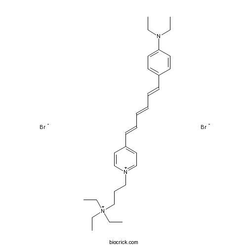
SynaptoRedTM C2CAS# 162112-35-8 |

- StemRegenin 1 (SR1)
Catalog No.:BCC3637
CAS No.:1227633-49-9
Quality Control & MSDS
3D structure
Package In Stock
Number of papers citing our products

| Cas No. | 162112-35-8 | SDF | Download SDF |
| PubChem ID | 2733635 | Appearance | Powder |
| Formula | C30H45Br2N3 | M.Wt | 607.51 |
| Type of Compound | N/A | Storage | Desiccate at -20°C |
| Synonyms | FM4-64 | ||
| Solubility | Soluble to 10 mM in DMSO | ||
| Chemical Name | 3-[4-[6-[4-(diethylamino)phenyl]hexa-1,3,5-trienyl]pyridin-1-ium-1-yl]propyl-triethylazanium;dibromide | ||
| SMILES | CCN(CC)C1=CC=C(C=C1)C=CC=CC=CC2=CC=[N+](C=C2)CCC[N+](CC)(CC)CC.[Br-].[Br-] | ||
| Standard InChIKey | AFVSZGYRRUMOFH-UHFFFAOYSA-L | ||
| Standard InChI | InChI=1S/C30H45N3.2BrH/c1-6-32(7-2)30-20-18-28(19-21-30)16-13-11-12-14-17-29-22-25-31(26-23-29)24-15-27-33(8-3,9-4)10-5;;/h11-14,16-23,25-26H,6-10,15,24,27H2,1-5H3;2*1H/q+2;;/p-2 | ||
| General tips | For obtaining a higher solubility , please warm the tube at 37 ℃ and shake it in the ultrasonic bath for a while.Stock solution can be stored below -20℃ for several months. We recommend that you prepare and use the solution on the same day. However, if the test schedule requires, the stock solutions can be prepared in advance, and the stock solution must be sealed and stored below -20℃. In general, the stock solution can be kept for several months. Before use, we recommend that you leave the vial at room temperature for at least an hour before opening it. |
||
| About Packaging | 1. The packaging of the product may be reversed during transportation, cause the high purity compounds to adhere to the neck or cap of the vial.Take the vail out of its packaging and shake gently until the compounds fall to the bottom of the vial. 2. For liquid products, please centrifuge at 500xg to gather the liquid to the bottom of the vial. 3. Try to avoid loss or contamination during the experiment. |
||
| Shipping Condition | Packaging according to customer requirements(5mg, 10mg, 20mg and more). Ship via FedEx, DHL, UPS, EMS or other couriers with RT, or blue ice upon request. | ||
| Description | Fluorescent dye; stains synaptic vesicles. Becomes fluorescent when incorporated into plasma membrane, used to follow synaptic activity at the synapse or neuromuscular junction. Excitation and emission spectra are ~ 515 and 640 nm respectively. |

SynaptoRedTM C2 Dilution Calculator

SynaptoRedTM C2 Molarity Calculator
| 1 mg | 5 mg | 10 mg | 20 mg | 25 mg | |
| 1 mM | 1.6461 mL | 8.2303 mL | 16.4606 mL | 32.9213 mL | 41.1516 mL |
| 5 mM | 0.3292 mL | 1.6461 mL | 3.2921 mL | 6.5843 mL | 8.2303 mL |
| 10 mM | 0.1646 mL | 0.823 mL | 1.6461 mL | 3.2921 mL | 4.1152 mL |
| 50 mM | 0.0329 mL | 0.1646 mL | 0.3292 mL | 0.6584 mL | 0.823 mL |
| 100 mM | 0.0165 mL | 0.0823 mL | 0.1646 mL | 0.3292 mL | 0.4115 mL |
| * Note: If you are in the process of experiment, it's necessary to make the dilution ratios of the samples. The dilution data above is only for reference. Normally, it's can get a better solubility within lower of Concentrations. | |||||

Calcutta University

University of Minnesota

University of Maryland School of Medicine

University of Illinois at Chicago

The Ohio State University

University of Zurich

Harvard University

Colorado State University

Auburn University

Yale University

Worcester Polytechnic Institute

Washington State University

Stanford University

University of Leipzig

Universidade da Beira Interior

The Institute of Cancer Research

Heidelberg University

University of Amsterdam

University of Auckland

TsingHua University

The University of Michigan

Miami University

DRURY University

Jilin University

Fudan University

Wuhan University

Sun Yat-sen University

Universite de Paris

Deemed University

Auckland University

The University of Tokyo

Korea University
- 4',4'''-Di-O-methylisochamaejasmin
Catalog No.:BCN6849
CAS No.:1620921-68-7
- Dimesna
Catalog No.:BCC1095
CAS No.:16208-51-8
- Myriceric acid C
Catalog No.:BCN1719
CAS No.:162059-94-1
- SC 58125
Catalog No.:BCC5948
CAS No.:162054-19-5
- L-371,257
Catalog No.:BCC7353
CAS No.:162042-44-6
- UNC0379
Catalog No.:BCC8055
CAS No.:1620401-82-2
- Rofecoxib
Catalog No.:BCC4437
CAS No.:162011-90-7
- Bromosporine
Catalog No.:BCC2226
CAS No.:1619994-69-2
- GSK2801
Catalog No.:BCC6498
CAS No.:1619994-68-1
- LY2857785
Catalog No.:BCC8050
CAS No.:1619903-54-6
- Catechin pentaacetate
Catalog No.:BCN1718
CAS No.:16198-01-9
- Esomeprazole Magnesium
Catalog No.:BCC5007
CAS No.:161973-10-0
- RWJ 50271
Catalog No.:BCC7894
CAS No.:162112-37-0
- Flunitrazepam
Catalog No.:BCC6107
CAS No.:1622-62-4
- Melperone hydrochloride
Catalog No.:BCC7385
CAS No.:1622-79-3
- 5'-Deoxy-5-fluoro-N-[(pentyloxy)carbonyl]cytidine 2',3'-diacetate
Catalog No.:BCN1544
CAS No.:162204-20-8
- Dorsmanin A
Catalog No.:BCN4088
CAS No.:162229-27-8
- CI 1020
Catalog No.:BCC7523
CAS No.:162256-50-0
- 7,3'-Dihydroxy-4'-methoxyflavan
Catalog No.:BCN4698
CAS No.:162290-05-3
- Remodelin
Catalog No.:BCC5571
CAS No.:1622921-15-6
- Baccatin X
Catalog No.:BCN7214
CAS No.:1623069-76-0
- Oplopanaxoside C
Catalog No.:BCC8226
CAS No.:162341-29-9
- Baccatin VIII
Catalog No.:BCN7212
CAS No.:1623410-10-5
- Baccatin IX
Catalog No.:BCN7213
CAS No.:1623410-12-7
Temporary Internal Fixation Using C1 Lateral Mass Screw and C2 Pedicle Screw (Goel-Harms Technique) without Bone Grafting for Chronic Atlantoaxial Rotatory Fixation.[Pubmed:28377256]
World Neurosurg. 2017 Jun;102:696.e1-696.e6.
BACKGROUND: The primary treatment strategy for chronic atlantoaxial rotatory fixation (chro-AARF) is traction followed by bracing or application of a halo device. However, to complete these conservative therapies, patient cooperation is mandatory. If conservative therapy fails, surgery is required for reduction and prevention of recurrence. It has been considered that surgery for atlantoaxial rotatory fixation necessitates solid bony fusion. However, once bony fusion is achieved, loss of range of motion is problematic. Here, we report a patient with chro-AARF who was successfully treated with temporary internal fixation using a C1 lateral mass screw and C2 pedicle screw (Goel-Harms technique) without any grafting of bone or use of bone substitute materials. CASE DESCRIPTION: A 9-year-old boy with chro-AARF was referred to our institution. He had a history of pervasive developmental disorders. He did not cooperate for the completion of conservative therapy and could not tolerate this therapy. Therefore, the orthopedic staff and his parents considered surgery. Under general anesthesia, reduction was easily performed. The Goel-Harms screw-rod construct was completed as a temporary internal fixator without any grafting of bone or use of bone substitute materials. After 6 months, the screw-rod construct was removed. Removal of the screw-rod construct was performed easily without complication. There was no ankylosis of the C1-2 joint, and cervical range of motion was maintained 2.8 years after removal of the construct. CONCLUSIONS: When conservative therapy cannot be continued, Goel-Harms surgery as a temporary internal fixator without bone grafting might be a suitable alternative for selected patients with chro-AARF.
A New Technique for the Surgical Treatment of Atlantoaxial Instability: C1 Lateral Mass and C2-3 Transfacet Screwing.[Pubmed:28383091]
Turk Neurosurg. 2017 Mar 9.
AIM: Atlantoaxial instability is a special entity that may be caused by many disorders such as trauma, tumor, arthritis, congenital malformation and infection. Atlantoaxial fixation is needed to provide stability, prevent neurological deficits and correct deformity. The objective of this study is to introduce an alternative technique for the treatment of atlantoaxial instability in patients who have vertebral artery anomaly, anomalous C2 or osteoporosis. MATERIAL AND METHODS: C1-2-3 fixation was performed in a 50-years-old, male patient with atlantoaxial instability due to os odontoideum. C1 lateral masses identified and screw placement was performed. C2 facet joints were identified bilaterally. Superior margin of junction of pedicle and the lamina was used as the entry point and 3.5x22 mm screws were inserted from C2 facet joint to the C3 facet joint in mediolateral and craniocaudal direction under fluoroscopic guidance with caution. The posterior fixation screws are interconnected with two rods. Finally, autologous grafts were placed posterolaterally to encourage the fusion. RESULTS: Patient's complaints relieved after the surgery. C1-C2 instability wasn't seen in the postoperative radiological examinations. CONCLUSION: In the surgical treatment of C1-2 instability, our technique could help to reduce the possibility of vertebral artery injury in patients who have a vertebral artery course anomaly or when it is difficult to place C2 pedicle screws due to anomalous C2 pedicles and osteoporosis. High fusion rate could be achived with this technique due to passing through the four cortical surfaces. No wire or allograft was required. Thus, the instrumentation cost could be reduced.
Molecular Evolutionary Constraints that Determine the Avirulence State of Clostridium botulinum C2 Toxin.[Pubmed:28382496]
J Mol Evol. 2017 Apr;84(4):174-186.
Clostridium botulinum (group-III) is an anaerobic bacterium producing C2 toxin along with botulinum neurotoxins. C2 toxin is belonged to binary toxin A family in bacterial ADP-ribosylation superfamily. A structural and functional diversity of binary toxin A family was inferred from different evolutionary constraints to determine the avirulence state of C2 toxin. Evolutionary genetic analyses revealed evidence of C2 toxin cluster evolution through horizontal gene transfer from the phage or plasmid origins, site-specific insertion by gene divergence, and homologous recombination event. It has also described that residue in conserved NAD-binding core, family-specific domain structure, and functional motifs found to predetermine its virulence state. Any mutational changes in these residues destabilized its structure-function relationship. Avirulent mutants of C2 toxin were screened and selected from a crucial site required for catalytic function of C2I and pore-forming function of C2II. We found coevolved amino acid pairs contributing an essential role in stabilization of its local structural environment. Avirulent toxins selected in this study were evaluated by detecting evolutionary constraints in stability of protein backbone structure, folding and conformational dynamic space, and antigenic peptides. We found 4 avirulent mutants of C2I and 5 mutants of C2II showing more stability in their local structural environment and backbone structure with rapid fold rate, and low conformational flexibility at mutated sites. Since, evolutionary constraints-free mutants with lack of catalytic and pore-forming function suggested as potential immunogenic candidates for treating C. botulinum infected poultry and veterinary animals. Single amino acid substitution in C2 toxin thus provides a major importance to understand its structure-function link, not only of a molecule but also of the pathogenesis.
Three-dimensional dynamic measurements of CH* and C2* concentrations in flame using simultaneous chemiluminescence tomography.[Pubmed:28380735]
Opt Express. 2017 Mar 6;25(5):4640-4654.
The species concentrations of flame chemiluminescence play important role in combustion diagnostics, such as CH* and C2* of hydrocarbon flame, which can provide specific characteristics in combustion control and monitoring. In order to realize both CH* and C2* chemiluminescence intensity detection in propane-air diffusion flame simultaneously, we present three-dimensional dynamic flame detecting method for species concentration determination. Firstly, quantitative flame chemiluminescence multispectral separation technique based on color cameras coupled with double-channel bandpass filters is adopted for dual channel signal division. Next, flame chemiluminescence tomography combining with multi-directional simultaneous capturing is proposed for real time three dimensional observations and detection in flame. Moreover, the proposed technique can quantitatively provide comparison of species intensity between CH* and C2* for further analysis. Considering its credible detecting accuracy and simple requirements, it is believed the proposed technique can be widely used in combustion diagnostics.
Analysis of synaptic vesicle endocytosis in synaptosomes by high-content screening.[Pubmed:22767087]
Nat Protoc. 2012 Jul 5;7(8):1439-55.
Small molecules modulating synaptic vesicle endocytosis (SVE) may ultimately be useful for diseases where pathological neurotransmission is implicated. Only a small number of specific SVE modulators have been identified to date. Slow progress is due to the laborious nature of traditional approaches to study SVE, in which nerve terminals are identified and studied in cultured neurons, typically yielding data from 10-20 synapses per experiment. We provide a protocol for a quantitative, high-throughput method for studying SVE in thousands of nerve terminals. Rat forebrain synaptosomes are attached to 96-well microplates and depolarized; SVE is then quantified by uptake of the dye FM4-64, which is imaged by high-content screening. Synaptosomes that have been frozen and stored can be used in place of fresh synaptosomes, reducing the experimental time and animal numbers required. With a supply of frozen synaptosomes, the assay can be performed within a day, including data analysis.
Vesicular sterols are essential for synaptic vesicle cycling.[Pubmed:21106824]
J Neurosci. 2010 Nov 24;30(47):15856-65.
Synaptic vesicles have a high sterol content, but the importance of vesicular sterols during vesicle recycling is unclear. We used the Drosophila temperature-sensitive dynamin mutant, shibire-ts1, to block endocytosis of recycling synaptic vesicles and to trap them reversibly at the plasma membrane where they were accessible to sterol extraction. Depletion of sterols from trapped vesicles prevented recovery of synaptic transmission after removal of the endocytic block. Measurement of vesicle recycling with synaptopHluorin, FM1-43, and FM4-64 demonstrated impaired membrane retrieval after vesicular sterol depletion. When plasma membrane sterols were extracted before vesicle trapping, no vesicle recycling defects were observed. Ultrastructural analysis indicated accumulation of endosomes and a defect in the formation of synaptic vesicles in synaptic terminals subjected to vesicular sterol depletion. Our results demonstrate the importance of a high vesicular sterol concentration for endocytosis and suggest that vesicular and membrane sterol pools do not readily intermingle during vesicle recycling.
Confocal microscopy of FM4-64 as a tool for analysing endocytosis and vesicle trafficking in living fungal hyphae.[Pubmed:10849201]
J Microsc. 2000 Jun;198(Pt 3):246-59.
Confocal microscopy of amphiphilic styryl dyes has been used to investigate endocytosis and vesicle trafficking in living fungal hyphae. Hyphae were treated with FM4-64, FM1-43 or TMA-DPH, three of the most commonly used membrane-selective dyes reported as markers of endocytosis. All three dyes were rapidly internalized within hyphae. FM4-64 was found best for imaging the dynamic changes in size, morphology and position of the apical vesicle cluster within growing hyphal tips because of its staining pattern, greater photostability and low cytotoxicity. FM4-64 was taken up into both the apical and subapical compartments of living hyphae in a time-dependent manner. The pattern of stain distribution was broadly similar in a range of fungal species tested (Aspergillus nidulans, Botrytis cinerea, Magnaporthe grisea, Neurospora crassa, Phycomyces blakesleeanus, Puccinia graminis, Rhizoctonia solani, Sclerotinia sclerotiorum and Trichoderma viride). With time, FM4-64 was internalized from the plasma membrane appearing in structures corresponding to putative endosomes, the apical vesicle cluster, the vacuolar membrane and mitochondria. These observations are consistent with dye internalization by endocytosis. A speculative model of the vesicle trafficking network within growing hyphae is presented.


