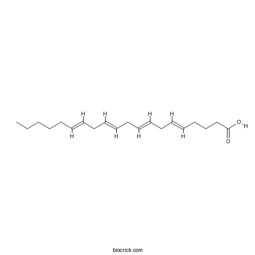
Arachidonic acidEndogenous free fatty acid (in water-soluble emulsion) CAS# 506-32-1 |

Quality Control & MSDS
3D structure
Package In Stock
Number of papers citing our products

| Cas No. | 506-32-1 | SDF | Download SDF |
| PubChem ID | 5312542 | Appearance | Powder |
| Formula | C20H32O2 | M.Wt | 304.47 |
| Type of Compound | Miscellaneous | Storage | Desiccate at -20°C |
| Solubility | Ethanol : ≥ 50 mg/mL (164.22 mM) DMSO : ≥ 50 mg/mL (164.22 mM) H2O : < 0.1 mg/mL (insoluble) *"≥" means soluble, but saturation unknown. | ||
| Chemical Name | (5E,8E,11E,14E)-icosa-5,8,11,14-tetraenoic acid | ||
| SMILES | CCCCCC=CCC=CCC=CCC=CCCCC(=O)O | ||
| Standard InChIKey | YZXBAPSDXZZRGB-CGRWFSSPSA-N | ||
| General tips | For obtaining a higher solubility , please warm the tube at 37 ℃ and shake it in the ultrasonic bath for a while.Stock solution can be stored below -20℃ for several months. We recommend that you prepare and use the solution on the same day. However, if the test schedule requires, the stock solutions can be prepared in advance, and the stock solution must be sealed and stored below -20℃. In general, the stock solution can be kept for several months. Before use, we recommend that you leave the vial at room temperature for at least an hour before opening it. |
||
| About Packaging | 1. The packaging of the product may be reversed during transportation, cause the high purity compounds to adhere to the neck or cap of the vial.Take the vail out of its packaging and shake gently until the compounds fall to the bottom of the vial. 2. For liquid products, please centrifuge at 500xg to gather the liquid to the bottom of the vial. 3. Try to avoid loss or contamination during the experiment. |
||
| Shipping Condition | Packaging according to customer requirements(5mg, 10mg, 20mg and more). Ship via FedEx, DHL, UPS, EMS or other couriers with RT, or blue ice upon request. | ||
| Description | Arachidonic acid is 1 of only 2 unsaturated fatty acids retained in the ovaries of crustaceans and an inhibitor of HR97g, a nuclear receptor expressed in adult ovaries. Arachidonic acid induces retinal arteriolar vasodilation by inhibiting subcellular Ca(2+)-signaling activity in retinal arteriolar myocytes, most likely through a mechanism involving the inhibition of L-type Ca(2+)-channel activity. Arachidonic acid causes an increase in free cytoplasmic calcium concentration ([Ca2+]i) in differentiated skeletal multinucleated myotubes C2C12 and does not induce calcium response in C2C12 myoblasts. |
| Targets | Calcium Channel | Potassium Channel | ROS | Caspase | p38MAPK |
| In vitro | Arachidonic acid activates release of calcium ions from reticulum via ryanodine receptor channels in C2C12 skeletal myotubes.[Pubmed: 24954594]Biochemistry (Mosc). 2014 May;79(5):435-9.
|
| In vivo | Arachidonic acid enhances reproduction in Daphnia magna and mitigates changes in sex ratios induced by pyriproxyfen.[Pubmed: 25393616]Environ Toxicol Chem. 2015 Mar;34(3):527-35.Arachidonic acid is 1 of only 2 unsaturated fatty acids retained in the ovaries of crustaceans and an inhibitor of HR97g, a nuclear receptor expressed in adult ovaries. The authors hypothesized that, as a key fatty acid, Arachidonic acid may be associated with reproduction and potentially environmental sex determination in Daphnia.
Arachidonic acid enhances turnover of the dermal skeleton: studies on zebrafish scales.[Pubmed: 24586706]PLoS One. 2014 Feb 19;9(2):e89347.In fish nutrition, the ratio between omega-3 and omega-6 poly-unsaturated fatty acids influences skeletal development. Supplementation of fish oils with vegetable oils increases the content of omega-6 fatty acids, such as Arachidonic acid in the diet. Arachidonic acid is metabolized by cyclooxygenases to prostaglandin E2, an eicosanoid with effects on bone formation and remodeling. |
| Kinase Assay | Role of ion channels and subcellular Ca2+ signaling in arachidonic acid-induced dilation of pressurized retinal arterioles.[Pubmed: 24699382]Invest Ophthalmol Vis Sci. 2014 May 2;55(5):2893-902.To investigate the mechanisms responsible for the dilatation of rat retinal arterioles in response to Arachidonic acid (AA).
|
| Cell Research | Lymphocyte-derived microparticles induce apoptosis of airway epithelial cells through activation of p38 MAPK and production of arachidonic acid.[Pubmed: 24777398 ]Apoptosis. 2014 Jul;19(7):1113-27.The airway epithelium is critical for the normal integrity and function of the respiratory system. Excessive epithelial cell apoptosis contributes to cell damage and airway inflammation. We previously demonstrated that lymphocyte-derived microparticles (LMPs) induce apoptosis of human bronchial epithelial cells. However, the underlying mechanisms contributing to LMPs-evoked epithelial cell death are largely unknown. |

Arachidonic acid Dilution Calculator

Arachidonic acid Molarity Calculator
| 1 mg | 5 mg | 10 mg | 20 mg | 25 mg | |
| 1 mM | 3.2844 mL | 16.422 mL | 32.844 mL | 65.6879 mL | 82.1099 mL |
| 5 mM | 0.6569 mL | 3.2844 mL | 6.5688 mL | 13.1376 mL | 16.422 mL |
| 10 mM | 0.3284 mL | 1.6422 mL | 3.2844 mL | 6.5688 mL | 8.211 mL |
| 50 mM | 0.0657 mL | 0.3284 mL | 0.6569 mL | 1.3138 mL | 1.6422 mL |
| 100 mM | 0.0328 mL | 0.1642 mL | 0.3284 mL | 0.6569 mL | 0.8211 mL |
| * Note: If you are in the process of experiment, it's necessary to make the dilution ratios of the samples. The dilution data above is only for reference. Normally, it's can get a better solubility within lower of Concentrations. | |||||

Calcutta University

University of Minnesota

University of Maryland School of Medicine

University of Illinois at Chicago

The Ohio State University

University of Zurich

Harvard University

Colorado State University

Auburn University

Yale University

Worcester Polytechnic Institute

Washington State University

Stanford University

University of Leipzig

Universidade da Beira Interior

The Institute of Cancer Research

Heidelberg University

University of Amsterdam

University of Auckland

TsingHua University

The University of Michigan

Miami University

DRURY University

Jilin University

Fudan University

Wuhan University

Sun Yat-sen University

Universite de Paris

Deemed University

Auckland University

The University of Tokyo

Korea University
- Isojacareubin
Catalog No.:BCN6883
CAS No.:50597-93-8
- Columbianadin
Catalog No.:BCN1275
CAS No.:5058-13-9
- Fenspiride HCl
Catalog No.:BCC4659
CAS No.:5053-08-7
- 3-(Carboxymethylamino)propanoic acid
Catalog No.:BCN1791
CAS No.:505-72-6
- Homopiperazine
Catalog No.:BCC8995
CAS No.:505-66-8
- Araneosol
Catalog No.:BCN5613
CAS No.:50461-86-4
- GW441756
Catalog No.:BCC5093
CAS No.:504433-23-2
- Methyl 2alpha-hydroxyhardwickiate
Catalog No.:BCN7595
CAS No.:50428-93-8
- 1,5,6-Trihydroxyxanthone
Catalog No.:BCN7642
CAS No.:5042-03-5
- Isorhamnetin-3-O-beta-D-Glucoside
Catalog No.:BCN1247
CAS No.:5041-82-7
- Isoliquiritin
Catalog No.:BCN5945
CAS No.:5041-81-6
- Juglanin
Catalog No.:BCN6505
CAS No.:5041-67-8
- Nervonic acid
Catalog No.:BCN8374
CAS No.:506-37-6
- Octacosanoic Acid
Catalog No.:BCN5395
CAS No.:506-48-9
- Niranthin
Catalog No.:BCN5614
CAS No.:50656-77-4
- Alkaloid KD1
Catalog No.:BCN1898
CAS No.:50656-87-6
- Alkaloid C
Catalog No.:BCN1897
CAS No.:50656-88-7
- Vandrikidine
Catalog No.:BCN5615
CAS No.:50656-92-3
- Chasmanine
Catalog No.:BCN5409
CAS No.:5066-78-4
- Terfenadine
Catalog No.:BCC3866
CAS No.:50679-08-8
- Boc-Cys(Bzl)-OH
Catalog No.:BCC3376
CAS No.:5068-28-0
- Borneol
Catalog No.:BCN4964
CAS No.:507-70-0
- Pennogenin
Catalog No.:BCN2839
CAS No.:507-89-1
- Vecuronium Bromide
Catalog No.:BCC2498
CAS No.:50700-72-6
Arachidonic acid activates release of calcium ions from reticulum via ryanodine receptor channels in C2C12 skeletal myotubes.[Pubmed:24954594]
Biochemistry (Mosc). 2014 May;79(5):435-9.
Arachidonic acid causes an increase in free cytoplasmic calcium concentration ([Ca2+]i) in differentiated skeletal multinucleated myotubes C2C12 and does not induce calcium response in C2C12 myoblasts. The same reaction of myotubes to Arachidonic acid is observed in Ca2+-free medium. This indicates that Arachidonic acid induces release of calcium ions from intracellular stores. The blocker of ryanodine receptor channels of sarcoplasmic reticulum dantrolene (20 microM) inhibits this effect by 68.7 +/- 6.3% (p < 0.001). The inhibitor of two-pore calcium channels of endolysosomal vesicles trans-NED19 (10 microM) decreases the response to Arachidonic acid by 35.8 +/- 5.4% (p < 0.05). The phospholipase C inhibitor U73122 (10 microM) has no effect. These data indicate the involvement of ryanodine receptor calcium channels of sarcoplasmic reticulum in [Ca2+]i elevation in skeletal myotubes caused by Arachidonic acid and possible participation of two-pore calcium channels from endolysosomal vesicles in this process.
Lymphocyte-derived microparticles induce apoptosis of airway epithelial cells through activation of p38 MAPK and production of arachidonic acid.[Pubmed:24777398]
Apoptosis. 2014 Jul;19(7):1113-27.
The airway epithelium is critical for the normal integrity and function of the respiratory system. Excessive epithelial cell apoptosis contributes to cell damage and airway inflammation. We previously demonstrated that lymphocyte-derived microparticles (LMPs) induce apoptosis of human bronchial epithelial cells. However, the underlying mechanisms contributing to LMPs-evoked epithelial cell death are largely unknown. Here we used bronchial and lung tissue cultures to confirm the pro-apoptotic effects of LMPs. In cell culture experiments, we found that LMPs induced human airway epithelial cell apoptosis with associated increases in caspase-3 activity. In addition, LMPs treatment triggered oxidative stress in epithelial cells by enhancing production of malondialdehyde, superoxide, and reactive oxygen species (ROS), and by inhibiting production of the antioxidant glutathione. Moreover, decreasing cellular ROS with the antioxidant N-acetylcysteine rescued epithelial cell viability. Together, these results demonstrate an important role for oxidative stress in LMPs-induced cell death. In epithelial cells, LMPs treatment induced phosphorylation of p38 MAPK and Arachidonic acid accumulation. Moreover, Arachidonic acid was significantly cytotoxic towards LMPs-treated epithelial cells, whereas inhibition of p38 MAPK was protective against these cytotoxic effects. Similarly, inhibition of Arachidonic acid production led to decreased caspase-3 activity, thus rescuing airway epithelial cells from LMPs-induced cell death. In conclusion, our results show that LMPs induce airway epithelial cell apoptosis by activating p38 MAPK signaling and stimulating production of Arachidonic acid, with consequent increases in oxidative stress and caspase-3 activity. As such, LMPs may be regarded as deleterious markers of epithelial cell damage in respiratory diseases.
Role of ion channels and subcellular Ca2+ signaling in arachidonic acid-induced dilation of pressurized retinal arterioles.[Pubmed:24699382]
Invest Ophthalmol Vis Sci. 2014 May 2;55(5):2893-902.
PURPOSE: To investigate the mechanisms responsible for the dilatation of rat retinal arterioles in response to Arachidonic acid (AA). METHODS: Changes in the diameter of isolated, pressurized rat retinal arterioles were measured in the presence of AA alone and following pre-incubation with pharmacologic agents inhibiting Ca(2+) sparks and oscillations and K(+) channels. Subcellular Ca(2+) signals were recorded in arteriolar myocytes using Fluo-4-based confocal imaging. The effects of AA on membrane currents of retinal arteriolar myocytes were studied using whole-cell perforated patch clamp recording. RESULTS: Arachidonic acid dilated pressurized retinal arterioles under conditions of myogenic tone. Eicosatetraynoic acid (ETYA) exerted a similar effect, but unlike AA, its effects were rapidly reversible. Arachidonic acid-induced dilation was associated with an inhibition of subcellular Ca(2+) signals. Interventions known to block Ca(2+) sparks and oscillations in retinal arterioles caused dilatation and inhibited AA-induced vasodilator responses. Arachidonic acid accelerated the rate of inactivation of the A-type Kv current and the voltage dependence of inactivation was shifted to more negative membrane potentials. It also enhanced voltage-activated and spontaneous large-conductance calcium-activated K(+) (BK) currents, but only at positive membrane potentials. Pharmacologic inhibition of A-type Kv and BK currents failed to block AA-induced vasodilator responses. Arachidonic acid suppressed L-type Ca(2+) currents. CONCLUSIONS: These results suggest that AA induces retinal arteriolar vasodilation by inhibiting subcellular Ca(2+)-signaling activity in retinal arteriolar myocytes, most likely through a mechanism involving the inhibition of L-type Ca(2+)-channel activity. Arachidonic acid actions on K(+) currents are inconsistent with a model in which K(+) channels contribute to the vasodilator effects of AA.
Arachidonic acid enhances reproduction in Daphnia magna and mitigates changes in sex ratios induced by pyriproxyfen.[Pubmed:25393616]
Environ Toxicol Chem. 2015 Mar;34(3):527-35.
Arachidonic acid is 1 of only 2 unsaturated fatty acids retained in the ovaries of crustaceans and an inhibitor of HR97g, a nuclear receptor expressed in adult ovaries. The authors hypothesized that, as a key fatty acid, Arachidonic acid may be associated with reproduction and potentially environmental sex determination in Daphnia. Reproduction assays with Arachidonic acid indicate that it alters female:male sex ratios by increasing female production. This reproductive effect only occurred during a restricted Pseudokirchneriella subcapitata diet. Next, the authors tested whether enriching a poorer algal diet (Chlorella vulgaris) with Arachidonic acid enhances overall reproduction and sex ratios. Arachidonic acid enrichment of a C. vulgaris diet also enhances fecundity at 1.0 microM and 4.0 microM by 30% to 40% in the presence and absence of pyriproxyfen. This indicates that Arachidonic acid is crucial in reproduction regardless of environmental sex determination. Furthermore, the data indicate that P. subcapitata may provide a threshold concentration of Arachidonic acid needed for reproduction. Diet-switch experiments from P. subcapitata to C. vulgaris mitigate some, but not all, of Arachidonic acid's effects when compared with a C. vulgaris-only diet, suggesting that some Arachidonic acid provided by P. subcapitata is retained. In summary, Arachidonic acid supplementation increases reproduction and represses pyriproxyfen-induced environmental sex determination in D. magna in restricted diets. A diet rich in Arachidonic acid may provide protection from some reproductive toxicants such as the juvenile hormone agonist pyriproxyfen. Environ Toxicol Chem 2015;34:527-535. (c) 2014 SETAC.
Arachidonic acid enhances turnover of the dermal skeleton: studies on zebrafish scales.[Pubmed:24586706]
PLoS One. 2014 Feb 19;9(2):e89347.
In fish nutrition, the ratio between omega-3 and omega-6 poly-unsaturated fatty acids influences skeletal development. Supplementation of fish oils with vegetable oils increases the content of omega-6 fatty acids, such as Arachidonic acid in the diet. Arachidonic acid is metabolized by cyclooxygenases to prostaglandin E2, an eicosanoid with effects on bone formation and remodeling. To elucidate effects of poly-unsaturated fatty acids on developing and existing skeletal tissues, zebrafish (Danio rerio) were fed (micro-) diets low and high in Arachidonic acid content. Elasmoid scales, dermal skeletal plates, are ideal to study skeletal metabolism in zebrafish and were exploited in the present study. The fatty acid profile resulting from a high Arachidonic acid diet induced mild but significant increase in matrix resorption in ontogenetic scales of adult zebrafish. Arachidonic acid affected scale regeneration (following removal of ontogenetic scales): mineral deposition was altered and both gene expression and enzymatic matrix metalloproteinase activity changed towards enhanced osteoclastic activity. Arachidonic acid also clearly stimulates matrix metalloproteinase activity in vitro, which implies that resorptive effects of Arachidonic acid are mediated by matrix metalloproteinases. The gene expression profile further suggests that Arachidonic acid increases maturation rate of the regenerating scale; in other words, enhances turnover. The zebrafish scale is an excellent model to study how and which fatty acids affect skeletal formation.
Arachidonic acid promotes glutamate-induced cell death associated with necrosis by 12- lipoxygenase activation in glioma cells.[Pubmed:17400255]
Life Sci. 2007 Apr 24;80(20):1856-64.
Glutamate induced glutathione (GSH) depletion in C6 rat glioma cells, which resulted in cell death. This cell death seemed to be apoptosis through accumulation of reactive oxygen species (ROS) or hydroperoxides representing cytochrome c release from mitochondria and internucleosomal DNA fragmentation. A significant increase of 12-lipoxygenase enzyme activity was observed in the presence of Arachidonic acid (AA) under GSH depletion induced by glutamate. AA promoted the glutamate-induced cell death, which reduced caspase-3 activity and diminished internucleosomal DNA fragmentation. Furthermore, AA reduced intracellular NAD, ATP and membrane potentials, which indicated dysfunction of the mitochondrial membrane. Protease inhibitors such as N-alpha-tosyl-L-phenylalanine chloromethyl ketone (TPCK) and 3, 4-dichloroisocumarin (DCI) but no Ac-DEVD, a caspase inhibitor, suppressed the glutamate-induced cell death. AA reduced the inhibitory effect of TPCK and DCI on the glutamate-induced cell death. These results suggest that AA promotes cell death by inducing necrosis from caspase-3-independent apoptosis. This might occur through lipid peroxidation initiated by ROS or lipid hydroperoxides generated during GSH depletion in C6 cells.
Arachidonic acid metabolism as a potential mediator of cardiac fibrosis associated with inflammation.[Pubmed:17202322]
J Immunol. 2007 Jan 15;178(2):641-6.
An increase in left ventricular collagen (cardiac fibrosis) is a detrimental process that adversely affects heart function. Strong evidence implicates the infiltration of inflammatory cells as a critical part of the process resulting in cardiac fibrosis. Inflammatory cells are capable of releasing Arachidonic acid, which may be further metabolized by cyclooxygenase, lipoxygenase, and cytochrome P450 monooxygenase enzymes to biologically active products, including PGs, leukotrienes, epoxyeicosatrienoic acids, and hydroxyeicosatetraenoic acids. Some of these products have profibrotic properties and may represent a pathway by which inflammatory cells initiate and mediate the development of cardiac fibrosis. In this study, we critically review the current literature on the potential link between this pathway and cardiac fibrosis.
Roles of cPLA2alpha and arachidonic acid in cancer.[Pubmed:17052951]
Biochim Biophys Acta. 2006 Nov;1761(11):1335-43.
Phospholipase A(2)s (PLA(2)s) are key enzymes that catalyze the hydrolysis of membrane phospholipids to release bioactive lipids that play an important role in normal cellular homeostasis. Under certain circumstances, disrupted production of key lipid mediators may adversely impact physiological processes, leading to pathological conditions such as inflammation and cancer. In particular, cytosolic PLA(2)alpha (cPLA(2)alpha) has a high selectivity for liberating Arachidonic acid (AA) that is subsequently metabolized by a panel of downstream enzymes for eicosanoid production. Although concentrations of free AA are maintained at low levels in resting cells, alterations in AA production, often resulting from dysregulation of cPLA(2)alpha activity, are observed in transformed cells. In this review, we summarize recent evidence that cPLA(2)alpha plays a role in the pathogenesis of many human cancers. Much of this evidence has been accumulated from functional studies using cPLA(2)alpha-deficient mice, as well as mechanistic studies in cell culture. We also discuss the potential contribution of cPLA(2)alpha and AA to apoptosis, and the regulatory mechanisms leading to aberrant expression of cPLA(2)alpha.


