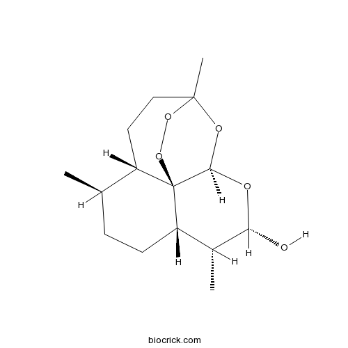
DihydroartemisininCAS# 71939-50-9 |

- alpha-Dihydroartemisinin
Catalog No.:BCN2627
CAS No.:81496-81-3
Quality Control & MSDS
3D structure
Package In Stock
Number of papers citing our products

| Cas No. | 71939-50-9 | SDF | Download SDF |
| PubChem ID | 456410 | Appearance | White cryst. |
| Formula | C15H24O5 | M.Wt | 284.35 |
| Type of Compound | Sesquiterpenes | Storage | Desiccate at -20°C |
| Synonyms | β-Dihydroartemisinin; DHA; Dihydroqinghaosu | ||
| Solubility | DMSO : 41.67 mg/mL (146.54 mM; Need ultrasonic) | ||
| SMILES | CC1CCC2C(C(OC3C24C1CCC(O3)(OO4)C)O)C | ||
| Standard InChIKey | BJDCWCLMFKKGEE-KXTPALSWSA-N | ||
| Standard InChI | InChI=1S/C15H24O5/c1-8-4-5-11-9(2)12(16)17-13-15(11)10(8)6-7-14(3,18-13)19-20-15/h8-13,16H,4-7H2,1-3H3/t8-,9-,10+,11+,12+,13-,14?,15-/m1/s1 | ||
| General tips | For obtaining a higher solubility , please warm the tube at 37 ℃ and shake it in the ultrasonic bath for a while.Stock solution can be stored below -20℃ for several months. We recommend that you prepare and use the solution on the same day. However, if the test schedule requires, the stock solutions can be prepared in advance, and the stock solution must be sealed and stored below -20℃. In general, the stock solution can be kept for several months. Before use, we recommend that you leave the vial at room temperature for at least an hour before opening it. |
||
| About Packaging | 1. The packaging of the product may be reversed during transportation, cause the high purity compounds to adhere to the neck or cap of the vial.Take the vail out of its packaging and shake gently until the compounds fall to the bottom of the vial. 2. For liquid products, please centrifuge at 500xg to gather the liquid to the bottom of the vial. 3. Try to avoid loss or contamination during the experiment. |
||
| Shipping Condition | Packaging according to customer requirements(5mg, 10mg, 20mg and more). Ship via FedEx, DHL, UPS, EMS or other couriers with RT, or blue ice upon request. | ||
| Description | Dihydroartemisinin is widely used as an intermediate in the preparation of other artemisinin-derived antimalarial drugs, recommended as the first-line anti-malarial drug with low toxicity. Dihydroartemisinin has anticancer activity, it inhibited cell proliferation via AKT/GSK3β/cyclinD1/ERK pathway and induced apoptosis is associated with inhibition of Sarco/Endoplasmic reticulum Calcium ATPase activity in colorectal cancer. |
| Targets | mTOR | GLUT | Caspase | ROS | ERK | Akt | GSK-3 | p53 | Antifection | HPV | Autophagy |
| In vitro | Dihydroartemisinin inhibits glucose uptake and cooperates with glycolysis inhibitor to induce apoptosis in non-small cell lung carcinoma cells.[Pubmed: 25799586]PLoS One. 2015 Mar 23;10(3):e0120426.Despite recent advances in the therapy of non-small cell lung cancer (NSCLC), the chemotherapy efficacy against NSCLC is still unsatisfactory. Previous studies show the herbal antimalarial drug Dihydroartemisinin (DHA) displays cytotoxic to multiple human tumors.
Dihydroartemisinin is cytotoxic to papillomavirus-expressing epithelial cells in vitro and in vivo.[Pubmed: 16322232 ]Cancer Res. 2005 Dec 1;65(23):10854-61.Nearly all cervical cancers are etiologically attributable to human papillomavirus (HPV) infection and pharmaceutical treatments targeting HPV-infected cells would be of great medical benefit. Because many neoplastic cells (including cervical cancer cells) overexpress the transferrin receptor to increase their iron uptake, we hypothesized that iron-dependent, antimalarial drugs such as artemisinin might prove useful in treating HPV-infected or transformed cells.
|
| Cell Research | Dihydroartemisinin inhibits cell proliferation via AKT/GSK3β/cyclinD1 pathway and induces apoptosis in A549 lung cancer cells.[Pubmed: 25674233]Dihydroartemisinin inhibits endothelial cell proliferation through the suppression of the ERK signaling pathway.[Pubmed: 25778668]Int J Mol Med. 2015 May;35(5):1381-7.Disrupting tumor angiogenesis serves as an important strategy for cancer therapy. Dihydroartemisinin (DHA), a semi-synthetic derivative of artemisinin, has exhibited potent anti-angiogenic activity. However, the molecular mechanisms underlying this effect have not been fully understood.
The present study aimed to investigate the role of DHA on endothelial cell proliferation, the essential process in angiogenesis. Human umbilical vein endothelial cells (HUVECs) treated with DHA were examined for proliferation, apoptosis and activation of the extracellular signal-regulated kinase (ERK) signaling pathway. Proliferation of HUVECs was inhibited by 20 μM DHA without induction of apoptosis. DHA also reduced the phosphorylation of ERK1/2, and downregulated the mRNA and protein expression of ERK1/2 in HUVECs. In addition, DHA suppressed the transcription and protein expression of ERK1/2 downstream effectors c-Fos and c-Myc. Electrical cell-substrate impedance sensing real-time analysis demonstrated that ERK signaling inhibitor PD98059 comprises the anti-proliferative effects of DHA. Thus, DHA inhibits endothelial cell proliferation by suppressing the ERK signaling pathway.
Int J Clin Exp Pathol. 2014 Dec 1;7(12):8684-91. eCollection 2014.Lung cancer is the most common cause of cancer-related death in the world. The main types of lung cancer are small cell lung carcinoma (SCLC) and non-small-cell lung carcinoma (NSCLC); non small cell lung carcinoma (NSCLC) includes squamous cell carcinoma (SCC), adenocarcinoma and large cell carcinoma, Non small cell lung carcinoma accounts for about 80% of the total lung cancer cases.
|

Dihydroartemisinin Dilution Calculator

Dihydroartemisinin Molarity Calculator
| 1 mg | 5 mg | 10 mg | 20 mg | 25 mg | |
| 1 mM | 3.5168 mL | 17.584 mL | 35.1679 mL | 70.3359 mL | 87.9198 mL |
| 5 mM | 0.7034 mL | 3.5168 mL | 7.0336 mL | 14.0672 mL | 17.584 mL |
| 10 mM | 0.3517 mL | 1.7584 mL | 3.5168 mL | 7.0336 mL | 8.792 mL |
| 50 mM | 0.0703 mL | 0.3517 mL | 0.7034 mL | 1.4067 mL | 1.7584 mL |
| 100 mM | 0.0352 mL | 0.1758 mL | 0.3517 mL | 0.7034 mL | 0.8792 mL |
| * Note: If you are in the process of experiment, it's necessary to make the dilution ratios of the samples. The dilution data above is only for reference. Normally, it's can get a better solubility within lower of Concentrations. | |||||

Calcutta University

University of Minnesota

University of Maryland School of Medicine

University of Illinois at Chicago

The Ohio State University

University of Zurich

Harvard University

Colorado State University

Auburn University

Yale University

Worcester Polytechnic Institute

Washington State University

Stanford University

University of Leipzig

Universidade da Beira Interior

The Institute of Cancer Research

Heidelberg University

University of Amsterdam

University of Auckland

TsingHua University

The University of Michigan

Miami University

DRURY University

Jilin University

Fudan University

Wuhan University

Sun Yat-sen University

Universite de Paris

Deemed University

Auckland University

The University of Tokyo

Korea University
Dihydroartemisinin inhibits NF-κB activity by blocking RelA/p65 translocation to the nucleus. Dihydroartemisinin activates autophagy induction in tumor cells.
In Vitro:Dihydroartemisinin (DHA) is an antimalarial agent. Dihydroartemisinin treatment effectively up-regulates the cytosolic RelA/p65 protein level and down-regulates the nuclear RelA/p65 protein level. Dihydroartemisinin blocks the nuclear translocation of RelA/p65 from the cytosol rather than suppressing RelA/p65 protein synthesis. Dihydroartemisinin induces autophagy in RPMI 8226 cells. Dihydroartemisinin suppresses NF-κB activation in RPMI 8226 cells. The NF-κB Dihydroartemisinin -binding activity is examined by EMSA assay. RPMI 8226 cells are exposed to various concentrations of Dihydroartemisinin (10, 20 and 40 μM) for 12 h, and TNF-α is introduced as a positive control for NF-κB activation. Dihydroartemisinin suppresses NF-κB activation in a dose-dependent manner in contrast with TNF-α[1]. Dihydroartemisinin (DHA) can enhance the anti-tumor effect of photodynamic therapy (PDT) on esophageal cancer cells, and cell viability is investigated using the MTT assay. Eca109 and Ec9706 cells are treated with Dihydroartemisinin (80 μM), PDT (25 and 20 J/cm2, respectively) or their combination. Single treatment with Dihydroartemisinin or PDT causes a 37±5% or 34±6% reduction in viability in Eca109 cells and a 33±7% or 34±6% reduction in Ec9706 cells, respectively. However, when PDT is combined with Dihydroartemisinin, the cell viability is reduced 59±6% or 61±7% in the cell lines, respectively[2].
In Vivo:Single oral doses of Dihydroartemisinin (at 200, 300, 400 or 600 mg/kg), given once on each of day 6-8 post-infection, reduce total-worm burdens by 69.2%-90.6% and female-worm burdens by 62.2%-92.2%, depending on dosage in the first experiment. Similar treatments given on day 34-36 post-infection reduce total-worm burdens by 73.9%-85.5% and female-worm burdens by 83.8%-95.3%[3].
References:
[1]. Hu W, et al. Dihydroartemisinin induces autophagy by suppressing NF-κB activation. Cancer Lett. 2014 Feb 28;343(2):239-48.
[2]. Li YJ, et al. Dihydroartemisinin accentuates the anti-tumor effects of photodynamic therapy via inactivation of NF-κB in Eca109 and Ec9706 esophageal cancer cells. Cell Physiol Biochem. 2014;33(5):1527-36.
[3]. Li HJ, et al. Dihydroartemisinin-praziquantel combinations and multiple doses of dihydroartemisinin in the treatment of Schistosoma japonicum in experimentally infected mice. Ann Trop Med Parasitol. 2011 Jun;105(4):329-33.
- Sarmentosin
Catalog No.:BCN4275
CAS No.:71933-54-5
- ProINDY
Catalog No.:BCC6350
CAS No.:719277-30-2
- S-Adenosyl-L-methionine tosylate
Catalog No.:BCN2230
CAS No.:71914-80-2
- 2-Amino-5-chlorobenzophenone
Catalog No.:BCC8535
CAS No.:719-59-5
- PHA-793887
Catalog No.:BCC2521
CAS No.:718630-59-2
- Camaldulenic acid
Catalog No.:BCN3928
CAS No.:71850-15-2
- Borrelidin
Catalog No.:BCC7964
CAS No.:7184-60-3
- Sativan
Catalog No.:BCN6815
CAS No.:71831-00-0
- Ivermectin B1a
Catalog No.:BCC9005
CAS No.:71827-03-7
- Demethoxyencecalinol
Catalog No.:BCN7765
CAS No.:71822-00-9
- Z-Thr-ol
Catalog No.:BCC2574
CAS No.:71811-27-3
- H-D-Ala-NH2.HCl
Catalog No.:BCC3197
CAS No.:71810-97-4
- Leiocarposide
Catalog No.:BCC8196
CAS No.:71953-77-0
- Artemether
Catalog No.:BCN5973
CAS No.:71963-77-4
- Fmoc-Asp(OtBu)-OH
Catalog No.:BCC3469
CAS No.:71989-14-5
- Fmoc-Asn-OH
Catalog No.:BCC3079
CAS No.:71989-16-7
- Fmoc-Glu(OtBu)-OH
Catalog No.:BCC3494
CAS No.:71989-18-9
- Fmoc-Gln-OH
Catalog No.:BCC3483
CAS No.:71989-20-3
- Fmoc-Ile-OH
Catalog No.:BCC3505
CAS No.:71989-23-6
- Fmoc-Lys(Boc)-OH
Catalog No.:BCC3516
CAS No.:71989-26-9
- Fmoc-Met-OH
Catalog No.:BCC3528
CAS No.:71989-28-1
- Fmoc-Pro-OH
Catalog No.:BCC3538
CAS No.:71989-31-6
- Fmoc-Ser(tBu)-OH
Catalog No.:BCC3544
CAS No.:71989-33-8
- Fmoc-Thr(tBu)-OH
Catalog No.:BCC3552
CAS No.:71989-35-0
Dihydroartemisinin inhibits glucose uptake and cooperates with glycolysis inhibitor to induce apoptosis in non-small cell lung carcinoma cells.[Pubmed:25799586]
PLoS One. 2015 Mar 23;10(3):e0120426.
Despite recent advances in the therapy of non-small cell lung cancer (NSCLC), the chemotherapy efficacy against NSCLC is still unsatisfactory. Previous studies show the herbal antimalarial drug Dihydroartemisinin (DHA) displays cytotoxic to multiple human tumors. Here, we showed that DHA decreased cell viability and colony formation, induced apoptosis in A549 and PC-9 cells. Additionally, we first revealed DHA inhibited glucose uptake in NSCLC cells. Moreover, glycolytic metabolism was attenuated by DHA, including inhibition of ATP and lactate production. Consequently, we demonstrated that the phosphorylated forms of both S6 ribosomal protein and mechanistic target of rapamycin (mTOR), and GLUT1 levels were abrogated by DHA treatment in NSCLC cells. Furthermore, the upregulation of mTOR activation by high expressed Rheb increased the level of glycolytic metabolism and cell viability inhibited by DHA. These results suggested that DHA-suppressed glycolytic metabolism might be associated with mTOR activation and GLUT1 expression. Besides, we showed GLUT1 overexpression significantly attenuated DHA-triggered NSCLC cells apoptosis. Notably, DHA synergized with 2-Deoxy-D-glucose (2DG, a glycolysis inhibitor) to reduce cell viability and increase cell apoptosis in A549 and PC-9 cells. However, the combination of the two compounds displayed minimal toxicity to WI-38 cells, a normal lung fibroblast cell line. More importantly, 2DG synergistically potentiated DHA-induced activation of caspase-9, -8 and -3, as well as the levels of both cytochrome c and AIF of cytoplasm. However, 2DG failed to increase the reactive oxygen species (ROS) levels elicited by DHA. Overall, the data shown above indicated DHA plus 2DG induced apoptosis was involved in both extrinsic and intrinsic apoptosis pathways in NSCLC cells.
Dihydroartemisinin inhibits endothelial cell proliferation through the suppression of the ERK signaling pathway.[Pubmed:25778668]
Int J Mol Med. 2015 May;35(5):1381-7.
Disrupting tumor angiogenesis serves as an important strategy for cancer therapy. Dihydroartemisinin (DHA), a semi-synthetic derivative of artemisinin, has exhibited potent anti-angiogenic activity. However, the molecular mechanisms underlying this effect have not been fully understood. The present study aimed to investigate the role of DHA on endothelial cell proliferation, the essential process in angiogenesis. Human umbilical vein endothelial cells (HUVECs) treated with DHA were examined for proliferation, apoptosis and activation of the extracellular signal-regulated kinase (ERK) signaling pathway. Proliferation of HUVECs was inhibited by 20 microM DHA without induction of apoptosis. DHA also reduced the phosphorylation of ERK1/2, and downregulated the mRNA and protein expression of ERK1/2 in HUVECs. In addition, DHA suppressed the transcription and protein expression of ERK1/2 downstream effectors c-Fos and c-Myc. Electrical cell-substrate impedance sensing real-time analysis demonstrated that ERK signaling inhibitor PD98059 comprises the anti-proliferative effects of DHA. Thus, DHA inhibits endothelial cell proliferation by suppressing the ERK signaling pathway. The present study strengthened the potential of DHA as an angiogenesis inhibitor for cancer treatment.
Dihydroartemisinin is cytotoxic to papillomavirus-expressing epithelial cells in vitro and in vivo.[Pubmed:16322232]
Cancer Res. 2005 Dec 1;65(23):10854-61.
Nearly all cervical cancers are etiologically attributable to human papillomavirus (HPV) infection and pharmaceutical treatments targeting HPV-infected cells would be of great medical benefit. Because many neoplastic cells (including cervical cancer cells) overexpress the transferrin receptor to increase their iron uptake, we hypothesized that iron-dependent, antimalarial drugs such as artemisinin might prove useful in treating HPV-infected or transformed cells. We tested three different artemisinin compounds and found that Dihydroartemisinin (DHA) and artesunate displayed strong cytotoxic effects on HPV-immortalized and transformed cervical cells in vitro with little effect on normal cervical epithelial cells. DHA-induced cell death involved activation of the mitochondrial caspase pathway with resultant apoptosis. Apoptosis was p53 independent and was not the consequence of drug-induced reductions in viral oncogene expression. Due to its selective cytotoxicity, hydrophobicity, and known ability to penetrate epithelial surfaces, we postulated that DHA might be useful for the topical treatment of mucosal papillomavirus lesions. To test this hypothesis, we applied DHA to the oral mucosa of dogs that had been challenged with the canine oral papillomavirus. Although applied only intermittently, DHA strongly inhibited viral-induced tumor formation. Interestingly, the DHA-treated, tumor-negative dogs developed antibodies against the viral L1 capsid protein, suggesting that DHA had inhibited tumor growth but not early rounds of papillomavirus replication. These findings indicate that DHA and other artemisinin derivatives may be useful for the topical treatment of epithelial papillomavirus lesions, including those that have progressed to the neoplastic state.
Dihydroartemisinin-Induced Apoptosis is Associated with Inhibition of Sarco/Endoplasmic Reticulum Calcium ATPase Activity in Colorectal Cancer.[Pubmed:25701954]
Cell Biochem Biophys. 2015 Sep;73(1):137-45.
Dihydroartemisinin (DHA) is a promising anti-cancer compound capable of inhibiting proliferation and inducing apoptosis of various cancer cells, including colorectal cancer. However, the molecular mechanisms have not been well understood. This study aimed to explore the underlying mechanism of DHA-induced apoptosis in HCT-116 cells. Cell counting kit-8 assay and flow cytometry analysis confirmed that DHA inhibited proliferation, arrested cell cycle at G0/G1 phase, and enhanced apoptosis in HCT-116 cells. Fluo-3/AM-stained flow cytometry assay revealed that the intracellular Ca(2+) concentration of HCT-116 cells was increased significantly after DHA treatment. Meanwhile, the activity of sarco/endoplasmic reticulum calcium ATPase (SERCA) was appeared to be reduced in a dose-dependent manner. We further detected the upregulated expression of CAAT/enhancer binding protein homologous protein (CHOP) in DHA-treated HCT-116 cells. Conversely, silencing CHOP resulted in a decrease of DHA-induced apoptosis. In addition, the expression of Bax in cytoplasm was elevated significantly along with the sharply decline of Bcl-2 expression in DHA-treated HCT-116 cells. Moreover, the distributions of Bid on mitochondria were increased, accompanied by the activation of caspase-3 in the presence of DHA. Overall, our data indicated that DHA triggered endoplasmic reticulum (ER) stress through inhibiting SERCA activity to release intracellular Ca(2+) from ER, the upregulated expression of CHOP activated mitochondrial apoptosis pathway to induce apoptosis of HCT-116 cells. Therefore, our findings provide a theoretical foundation for DHA as a potential candidate in treatment of colorectal cancer.


