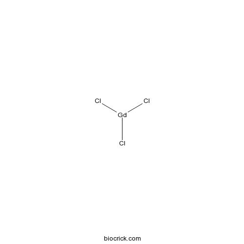
Gadolinium chlorideTRPML3 blocker; stretch-activated calcium channel blocker CAS# 10138-52-0 |

- Etifoxine
Catalog No.:BCC1560
CAS No.:21715-46-8
- Etomidate
Catalog No.:BCC1150
CAS No.:33125-97-2
- Acamprosate calcium
Catalog No.:BCC1327
CAS No.:77337-73-6
- Flumazenil
Catalog No.:BCC1259
CAS No.:78755-81-4
Quality Control & MSDS
3D structure
Package In Stock
Number of papers citing our products

| Cas No. | 10138-52-0 | SDF | Download SDF |
| PubChem ID | 61486 | Appearance | Powder |
| Formula | GdCl3 | M.Wt | 263.61 |
| Type of Compound | N/A | Storage | Desiccate at -20°C |
| Solubility | Soluble to 100 mM in water | ||
| Chemical Name | trichlorogadolinium | ||
| SMILES | Cl[Gd](Cl)Cl | ||
| Standard InChIKey | MEANOSLIBWSCIT-UHFFFAOYSA-K | ||
| Standard InChI | InChI=1S/3ClH.Gd/h3*1H;/q;;;+3/p-3 | ||
| General tips | For obtaining a higher solubility , please warm the tube at 37 ℃ and shake it in the ultrasonic bath for a while.Stock solution can be stored below -20℃ for several months. We recommend that you prepare and use the solution on the same day. However, if the test schedule requires, the stock solutions can be prepared in advance, and the stock solution must be sealed and stored below -20℃. In general, the stock solution can be kept for several months. Before use, we recommend that you leave the vial at room temperature for at least an hour before opening it. |
||
| About Packaging | 1. The packaging of the product may be reversed during transportation, cause the high purity compounds to adhere to the neck or cap of the vial.Take the vail out of its packaging and shake gently until the compounds fall to the bottom of the vial. 2. For liquid products, please centrifuge at 500xg to gather the liquid to the bottom of the vial. 3. Try to avoid loss or contamination during the experiment. |
||
| Shipping Condition | Packaging according to customer requirements(5mg, 10mg, 20mg and more). Ship via FedEx, DHL, UPS, EMS or other couriers with RT, or blue ice upon request. | ||
| Description | Calcium-sensing receptor (CaSR) agonist; induces NLRP3 inflammasome activation in bone marrow-derived macrophages. Blocks stretch-activated calcium channels; inhibits increase in intracellular calcium ion concentration in hypotonic-stimulated pulmonary artery smooth muscle cells. Has been demonstrated to block TRPML channel currents and other TRP channels. |

Gadolinium chloride Dilution Calculator

Gadolinium chloride Molarity Calculator
| 1 mg | 5 mg | 10 mg | 20 mg | 25 mg | |
| 1 mM | 3.7935 mL | 18.9674 mL | 37.9348 mL | 75.8697 mL | 94.8371 mL |
| 5 mM | 0.7587 mL | 3.7935 mL | 7.587 mL | 15.1739 mL | 18.9674 mL |
| 10 mM | 0.3793 mL | 1.8967 mL | 3.7935 mL | 7.587 mL | 9.4837 mL |
| 50 mM | 0.0759 mL | 0.3793 mL | 0.7587 mL | 1.5174 mL | 1.8967 mL |
| 100 mM | 0.0379 mL | 0.1897 mL | 0.3793 mL | 0.7587 mL | 0.9484 mL |
| * Note: If you are in the process of experiment, it's necessary to make the dilution ratios of the samples. The dilution data above is only for reference. Normally, it's can get a better solubility within lower of Concentrations. | |||||

Calcutta University

University of Minnesota

University of Maryland School of Medicine

University of Illinois at Chicago

The Ohio State University

University of Zurich

Harvard University

Colorado State University

Auburn University

Yale University

Worcester Polytechnic Institute

Washington State University

Stanford University

University of Leipzig

Universidade da Beira Interior

The Institute of Cancer Research

Heidelberg University

University of Amsterdam

University of Auckland

TsingHua University

The University of Michigan

Miami University

DRURY University

Jilin University

Fudan University

Wuhan University

Sun Yat-sen University

Universite de Paris

Deemed University

Auckland University

The University of Tokyo

Korea University
- Formosanol
Catalog No.:BCN5826
CAS No.:101312-79-2
- PF-04691502
Catalog No.:BCC3837
CAS No.:1013101-36-4
- Zacopride hydrochloride
Catalog No.:BCC7178
CAS No.:101303-98-4
- Noreugenin
Catalog No.:BCN5827
CAS No.:1013-69-0
- PETCM
Catalog No.:BCC2360
CAS No.:10129-56-3
- 11-Chloro-2,3-dihydro-2-methyl-1H- dibenz[2,3:6,7]oxepino[4,5-c]pyrrol-1-one
Catalog No.:BCC8431
CAS No.:1012884-46-6
- Phenserine
Catalog No.:BCC7529
CAS No.:101246-66-6
- Kushenol M
Catalog No.:BCN3310
CAS No.:101236-51-5
- Kushenol L
Catalog No.:BCN3309
CAS No.:101236-50-4
- Kushenol K
Catalog No.:BCN3448
CAS No.:101236-49-1
- IRAK inhibitor 3
Catalog No.:BCC1656
CAS No.:1012343-93-9
- Picrasidine Q
Catalog No.:BCN3182
CAS No.:101219-61-8
- CP 376395 hydrochloride
Catalog No.:BCC7604
CAS No.:1013933-37-3
- VTP-27999 2,2,2-trifluoroacetate
Catalog No.:BCC2049
CAS No.:1013937-63-7
- GSK 0660
Catalog No.:BCC7688
CAS No.:1014691-61-2
- 5''-O-Syringoylkelampayoside A
Catalog No.:BCN4798
CAS No.:1014974-98-1
- Methyl salvionolate A
Catalog No.:BCN3475
CAS No.:1015171-69-3
- Latifolin
Catalog No.:BCN7778
CAS No.:10154-42-4
- Fmoc-D-Pro-OH
Catalog No.:BCC3540
CAS No.:101555-62-8
- Yadanzioside K
Catalog No.:BCN6714
CAS No.:101559-98-2
- Yadanzioside M
Catalog No.:BCN6712
CAS No.:101559-99-3
- trans-2,3-Dihydro-3-ethoxyeuparin
Catalog No.:BCN6923
CAS No.:1015698-14-2
- GSK 4716
Catalog No.:BCC7557
CAS No.:101574-65-6
- 6-O-Nicotinoylbarbatin C
Catalog No.:BCN5828
CAS No.:1015776-92-7
Combination of sorafenib and gadolinium chloride (GdCl3) attenuates dimethylnitrosamine(DMN)-induced liver fibrosis in rats.[Pubmed:26572488]
BMC Gastroenterol. 2015 Nov 16;15:159.
BACKGROUND/AIMS: Liver sinusoidal endothelial cells (SECs), hepatic stellate cells (HSCs) and Kupffer cells (KCs) are involved in the development of liver fibrosis and represent a potential therapeutic target. The therapeutic effects on liver fibrosis of sorafenib, a multiple tyrosine kinase inhibitor, and Gadolinium chloride (GdCl3), which depletes KCs, were evaluated in rats. METHODS: Liver fibrosis was induced in rats with dimethylnitrosamine, and the effects of sorafenib and/or GdCl3 in these rats were monitored. Interactions among ECs, HSCs and KCs were assessed by laser confocal microscopy. RESULTS: The combination of sorafenib and GdCl3, but not each agent alone, attenuated liver fibrosis and significantly reduced liver function and hydroxyproline (Hyp). Sorafenib significantly inhibited the expression of angiogenesis-associated cell markers and cytokines, including CD31, von Willebrand factor (vWF), and vascular endothelial growth factor, whereas GdCl3 suppressed macrophage-related cell markers and cytokines, including CD68, tumor necrosis factor-alpha, interleukin-1beta, and CCL2. Laser confocal microscopy showed that sorafenib inhibited vWF expression and GdCl3 reduced CD68 staining. Sorafenib plus GdCl3 suppressed the interactions of HSCs, ECs and KCs. CONCLUSION: Sorafenib plus GdCl3 can suppress collagen accumulation, suggesting that this combination may be a potential therapeutic strategy in the treatment of liver fibrosis.
Pretreatment with low-dose gadolinium chloride attenuates myocardial ischemia/reperfusion injury in rats.[Pubmed:26948086]
Acta Pharmacol Sin. 2016 Apr;37(4):453-62.
AIM: We have shown that low-dose Gadolinium chloride (GdCl3) abolishes arachidonic acid (AA)-induced increase of cytoplasmic Ca(2+), which is known to play a crucial role in myocardial ischemia/reperfusion (I/R) injury. The present study sought to determine whether low-dose GdCl3 pretreatment protected rat myocardium against I/R injury in vitro and in vivo. METHODS: Cultured neonatal rat ventricular myocytes (NRVMs) were treated with GdCl3 or nifedipine, followed by exposure to anoxia/reoxygenation (A/R). Cell apoptosis was detected; the levels of related signaling molecules were assessed. SD rats were intravenously injected with GdCl3 or nifedipine. Thirty min after the administration the rats were subjected to LAD coronary artery ligation followed by reperfusion. Infarction size, the release of serum myocardial injury markers and AA were measured; cell apoptosis and related molecules were assessed. RESULTS: In A/R-treated NRVMs, pretreatment with GdCl3 (2.5, 5, 10 mumol/L) dose-dependently inhibited caspase-3 activation, death receptor-related molecules DR5/Fas/FADD/caspase-8 expression, cytochrome c release, AA release and sustained cytoplasmic Ca(2+) increases induced by exogenous AA. In I/R-treated rats, pre-administration of GdCl3 (10 mg/kg) significantly reduced the infarct size, and the serum levels of CK-MB, cardiac troponin-I, LDH and AA. Pre-administration of GdCl3 also significantly decreased the number of apoptotic cells, caspase-3 activity, death receptor-related molecules (DR5/Fas/FADD) expression and cytochrome c release in heart tissues. The positive control drug nifedipine produced comparable cardioprotective effects in vitro and in vivo. CONCLUSION: Pretreatment with low-dose GdCl3 significantly attenuates I/R-induced myocardial apoptosis in rats by suppressing activation of both death receptor and mitochondria-mediated pathways.
Gadolinium chloride elicits apoptosis in human osteosarcoma U-2 OS cells through extrinsic signaling, intrinsic pathway and endoplasmic reticulum stress.[Pubmed:27748868]
Oncol Rep. 2016 Dec;36(6):3421-3426.
Gadolinium (Gd) compounds are important as magnetic resonance imaging (MRI) contrast agents, and are potential anticancer agents. However, no report has shown the effect of Gadolinium chloride (GdCl3) on osteosarcoma in vitro. The present study investigated the apoptotic mechanism of GdCl3 on human osteosarcoma U-2 OS cells. Our results indicated that GdCl3 significantly reduced cell viability of U-2 OS cells in a concentration-dependent manner. GdCl3 led to apoptotic cell shrinkage and DNA fragmentation in U-2 OS cells as revealed by morphologic changes and TUNEL staining. Colorimetric assay analyses also showed that activities of caspase-3, caspase-8, caspase-9 and caspase-4 occurred in GdCl3-treated U-2 OS cells. Pretreatment of cells with pan-caspase inhibitor (Z-VAD-FMK) and specific inhibitors of caspase-3/-8/-9 significantly reduced cell death caused by GdCl3. The increase of cytoplasmic Ca2+ level, ROS production and the decrease of mitochondria membrane potential (DeltaPsim) were observed by flow cytometric analysis in U-2 OS cells after GdCl3 exposure. Western blot analyses demonstrated that the levels of Fas, FasL, cytochrome c, Apaf-1, GADD153 and GRP78 were upregulated in GdCl3-treated U-2 OS cells. In conclusion, death receptor, mitochondria-dependent and endoplasmic reticulum (ER) stress pathways contribute to GdCl3-induced apoptosis in U-2 OS cells. GdCl3 might have potential to be used in treatment of osteosarcoma patients.
Protective effect of low dose gadolinium chloride against isoproterenol-induced myocardial injury in rat.[Pubmed:26089194]
Apoptosis. 2015 Sep;20(9):1164-75.
Acute myocardial injury remains a leading cause of morbidity and mortality worldwide, and large amount of released arachidonic acid (AA) is found to be related to cardiomyocyte apoptosis and necrosis. Previous study suggested that GdCl3 completely abolished AA-induced Ca(2+) response. Thus, this study aims to investigate possible cardioprotection effect of GdCl3 on isoproterenol (ISO)-induced myocardial injury and its underlying mechanism(s). Rats that were randomly allocated to five groups: control, GdCl3, ISO, ISO + GdCl3, and ISO + verapamil. Serum levels of AA and cardiac markers, infarct area, and cell apoptosis in heart were measured by ELISA assay, TTC and TUNEL staining, respectively. Chemical interaction between AA and GdCl3 was evaluated by mass and UV spectrometry. The expressions and translocations of death receptor related molecules into lipid rafts were detected in neonatal rat ventricular myocytes by Western blots. Compared with ISO-administered rats, GdCl3 significantly ameliorated the myocardium injury, demonstrated by restoring serum cardiac troponin I, lactate dehydrogenase, creatine kinase MB and AA to near normal levels, and decreasing infarct area and cell apoptosis. In addition, an activation of AA-Fas pathway was found in ISO-induced myocardial injury, which was abrogated by GdCl3. Furthermore, AA induced cell apoptosis through clustering and activating death receptor related molecules TNFR1, Fas and FADD in lipid rafts, a process significantly prevented by the pretreatment with GdCl3. Finally, GdCl3 at the molar ratio of 1/3 (GdCl3/AA) was mostly effective in abolishing AA-induced Ca(2+) response and cell apoptosis, because an obvious change in the chemical identity of AA was obtained by GdCl3 according to this molar ratio. In conclusion, this study demonstrates for the first time that GdCl3 protects myocardium against ISO-induced cell apoptosis through, at least partly, serving as a scavenger of AA, therefore abolishing its downstream activation of the death receptor regulated apoptosis pathway.
The stretch-activated channel blocker Gd(3+) reduces palytoxin toxicity in primary cultures of skeletal muscle cells.[Pubmed:22900474]
Chem Res Toxicol. 2012 Sep 17;25(9):1912-20.
Palytoxin (PLTX) is one of the most toxic seafood contaminants ever isolated. Reports of human food-borne poisoning ascribed to PLTX suggest skeletal muscle as a primary target site. Primary cultures of mouse skeletal muscle cells were used to study the relationship between Ca(2+) response triggered by PLTX and the development of myotoxic insult. Ca(2+) imaging experiments revealed that PLTX causes a transitory intracellular Ca(2+) response (transient phase) followed by a slower and more sustained Ca(2+) increase (long-lasting phase). The transient phase is due to Ca(2+) release from intracellular stores and entry through voltage-dependent channels and the Na(+)/Ca(2+) exchanger (reverse mode). The long-lasting phase is due to a massive and prolonged Ca(2+) influx from the extracellular compartment. Sulforhodamine B assay revealed that the long-lasting phase is the one responsible for the toxicity in skeletal muscle cells. Our data analyzed, for the first time, pathways of PLTX-induced Ca(2+) entry and their correlation with PLTX-induced toxicity in skeletal muscle cells. The cellular morphology changes induced by PLTX and the sensitivity to gadolinium suggest a role for stretch-activated channels.
The calcium-sensing receptor regulates the NLRP3 inflammasome through Ca2+ and cAMP.[Pubmed:23143333]
Nature. 2012 Dec 6;492(7427):123-7.
Mutations in the gene encoding NLRP3 cause a spectrum of autoinflammatory diseases known as cryopyrin-associated periodic syndromes (CAPS). NLRP3 is a key component of one of several distinct cytoplasmic multiprotein complexes (inflammasomes) that mediate the maturation of the proinflammatory cytokine interleukin-1beta (IL-1beta) by activating caspase-1. Although several models for inflammasome activation, such as K(+) efflux, generation of reactive oxygen species and lysosomal destabilization, have been proposed, the precise molecular mechanism of NLRP3 inflammasome activation, as well as the mechanism by which CAPS-associated mutations activate NLRP3, remain to be elucidated. Here we show that the murine calcium-sensing receptor (CASR) activates the NLRP3 inflammasome, mediated by increased intracellular Ca(2+) and decreased cellular cyclic AMP (cAMP). Ca(2+) or other CASR agonists activate the NLRP3 inflammasome in the absence of exogenous ATP, whereas knockdown of CASR reduces inflammasome activation in response to known NLRP3 activators. CASR activates the NLRP3 inflammasome through phospholipase C, which catalyses inositol-1,4,5-trisphosphate production and thereby induces release of Ca(2+) from endoplasmic reticulum stores. The increased cytoplasmic Ca(2+) promotes the assembly of inflammasome components, and intracellular Ca(2+) is required for spontaneous inflammasome activity in cells from patients with CAPS. CASR stimulation also results in reduced intracellular cAMP, which independently activates the NLRP3 inflammasome. cAMP binds to NLRP3 directly to inhibit inflammasome assembly, and downregulation of cAMP relieves this inhibition. The binding affinity of cAMP for CAPS-associated mutant NLRP3 is substantially lower than for wild-type NLRP3, and the uncontrolled mature IL-1beta production from CAPS patients' peripheral blood mononuclear cells is attenuated by increasing cAMP. Taken together, these findings indicate that Ca(2+) and cAMP are two key molecular regulators of the NLRP3 inflammasome that have critical roles in the molecular pathogenesis of CAPS.
Calcium signaling in live cells on elastic gels under mechanical vibration at subcellular levels.[Pubmed:22053183]
PLoS One. 2011;6(10):e26181.
A new device was designed to generate a localized mechanical vibration of flexible gels where human umbilical vein endothelial cells (HUVECs) were cultured to mechanically stimulate these cells at subcellular locations. A Fluorescence Resonance Energy Transfer (FRET)-based calcium biosensor (an improved Cameleon) was used to monitor the spatiotemporal distribution of intracellular calcium concentrations in the cells upon this mechanical stimulation. A clear increase in intracellular calcium concentrations over the whole cell body (global) can be observed in the majority of cells under mechanical stimulation. The chelation of extracellular calcium with EGTA or the blockage of stretch-activated calcium channels on the plasma membrane with streptomycin or Gadolinium chloride significantly inhibited the calcium responses upon mechanical stimulation. Thapsigargin, an endoplasmic reticulum (ER) calcium pump inhibitor, or U73122, a phospholipase C (PLC) inhibitor, resulted in mainly local calcium responses occurring at regions close to the stimulation site. The disruption of actin filaments with cytochalasin D or inhibition of actomyosin contractility with ML-7 also inhibited the global calcium responses. Therefore, the global calcium response in HUVEC depends on the influx of calcium through membrane stretch-activated channels, followed by the release of inositol trisphosphate (IP3) via PLC activation to trigger the ER calcium release. Our newly developed mechanical stimulation device can also provide a powerful tool for the study of molecular mechanism by which cells perceive the mechanical cues at subcellular levels.
The varitint-waddler (Va) deafness mutation in TRPML3 generates constitutive, inward rectifying currents and causes cell degeneration.[Pubmed:18162548]
Proc Natl Acad Sci U S A. 2008 Jan 8;105(1):353-8.
Varitint-waddler (Va and Va(J)) mice are deaf and have vestibular impairment, with inner ear defects that include the degeneration and loss of sensory hair cells. The semidominant Va mutation results in an alanine-to-proline substitution at residue 419 (A419P) of the presumed ion channel TRPML3. Another allele, Va(J), has the A419P mutation in addition to an I362T mutation. We found that hair cells, marginal cells of stria vascularis, and other cells lining the cochlear and vestibular endolymphatic compartments express TRPML3. When heterologously expressed in LLC-PK1-CL4 epithelial cells, a culture model for hair cells, TRPML3 accumulated in lysosomes and in espin-enlarged microvilli that resemble stereocilia. We also demonstrated that wild-type TRPML3 forms channels that are blocked by Gd(3+), have a conductance of 50-70 pS and, like many other TRP channels, open at very positive potentials and thus rectify outwardly. In addition to this outward current, TRPML3(419P) and (I362T+A419P) generated a constitutive inwardly rectifying current that suggests a sensitivity to hyperpolarizing negative potentials and that depolarized the cells. Cells expressing TRPML3(A419P) or (I362T+A419P), but not wild-type TRPML3, died and were extruded from the epithelium in a manner reminiscent of degenerating hair cells in Va mice. The increased open probability of TRPML3(A419P) and (I362T+A419P) at physiological potentials likely underlies hair cell degeneration and deafness in Va and Va(J) mice.


