SphingomyelinCAS# 6254-89-3 |

Quality Control & MSDS
3D structure
Package In Stock

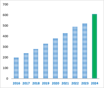
Number of papers citing our products
| Cas No. | 6254-89-3 | SDF | Download SDF |
| PubChem ID | 9939941.0 | Appearance | Powder |
| Formula | C39H79N2O6P | M.Wt | 703.03 |
| Type of Compound | N/A | Storage | Desiccate at -20°C |
| Synonyms | N-palmitoyl-D-erythro-sphingosylphosphorylcholine | ||
| Solubility | Soluble in Chloroform,Dichloromethane,Ethyl Acetate,DMSO,Acetone,etc. | ||
| Chemical Name | [(E,2S,3R)-2-(hexadecanoylamino)-3-hydroxyoctadec-4-enyl] 2-(trimethylazaniumyl)ethyl phosphate | ||
| SMILES | CCCCCCCCCCCCCCCC(=O)NC(COP(=O)([O-])OCC[N+](C)(C)C)C(C=CCCCCCCCCCCCCC)O | ||
| Standard InChIKey | RWKUXQNLWDTSLO-GWQJGLRPSA-N | ||
| Standard InChI | InChI=1S/C39H79N2O6P/c1-6-8-10-12-14-16-18-20-22-24-26-28-30-32-38(42)37(36-47-48(44,45)46-35-34-41(3,4)5)40-39(43)33-31-29-27-25-23-21-19-17-15-13-11-9-7-2/h30,32,37-38,42H,6-29,31,33-36H2,1-5H3,(H-,40,43,44,45)/b32-30+/t37-,38+/m0/s1 | ||
| General tips | For obtaining a higher solubility , please warm the tube at 37 ℃ and shake it in the ultrasonic bath for a while.Stock solution can be stored below -20℃ for several months. We recommend that you prepare and use the solution on the same day. However, if the test schedule requires, the stock solutions can be prepared in advance, and the stock solution must be sealed and stored below -20℃. In general, the stock solution can be kept for several months. Before use, we recommend that you leave the vial at room temperature for at least an hour before opening it. |
||
| About Packaging | 1. The packaging of the product may be reversed during transportation, cause the high purity compounds to adhere to the neck or cap of the vial.Take the vail out of its packaging and shake gently until the compounds fall to the bottom of the vial. 2. For liquid products, please centrifuge at 500xg to gather the liquid to the bottom of the vial. 3. Try to avoid loss or contamination during the experiment. |
||
| Shipping Condition | Packaging according to customer requirements(5mg, 10mg, 20mg and more). Ship via FedEx, DHL, UPS, EMS or other couriers with RT, or blue ice upon request. | ||

Sphingomyelin Dilution Calculator
Sphingomyelin Molarity Calculator
| 1 mg | 5 mg | 10 mg | 20 mg | 25 mg | |
| 1 mM | 1.4224 mL | 7.1121 mL | 14.2241 mL | 28.4483 mL | 35.5604 mL |
| 5 mM | 0.2845 mL | 1.4224 mL | 2.8448 mL | 5.6897 mL | 7.1121 mL |
| 10 mM | 0.1422 mL | 0.7112 mL | 1.4224 mL | 2.8448 mL | 3.556 mL |
| 50 mM | 0.0284 mL | 0.1422 mL | 0.2845 mL | 0.569 mL | 0.7112 mL |
| 100 mM | 0.0142 mL | 0.0711 mL | 0.1422 mL | 0.2845 mL | 0.3556 mL |
| * Note: If you are in the process of experiment, it's necessary to make the dilution ratios of the samples. The dilution data above is only for reference. Normally, it's can get a better solubility within lower of Concentrations. | |||||

Calcutta University

University of Minnesota

University of Maryland School of Medicine

University of Illinois at Chicago

The Ohio State University

University of Zurich

Harvard University

Colorado State University

Auburn University

Yale University

Worcester Polytechnic Institute

Washington State University

Stanford University

University of Leipzig
Universidade da Beira Interior
The Institute of Cancer Research

Heidelberg University

University of Amsterdam

University of Auckland

TsingHua University

The University of Michigan

Miami University

DRURY University
Jilin University

Fudan University

Wuhan University

Sun Yat-sen University

Universite de Paris

Deemed University

Auckland University

The University of Tokyo

Korea University
- 22,23-Dihydroergosterol
Catalog No.:BCX1118
CAS No.:516-79-0
- Inflacoumarin A
Catalog No.:BCX1117
CAS No.:158446-33-4
- Strigolactone
Catalog No.:BCX1116
CAS No.:76974-79-3
- Steviol-13-O-Glucoside
Catalog No.:BCX1115
CAS No.:60129-60-4
- Nifedipine impurity B
Catalog No.:BCX1114
CAS No.:50428-14-3
- Irisolidone 7-O-glucoside
Catalog No.:BCX1113
CAS No.:126308-74-5
- Maytansinol
Catalog No.:BCX1112
CAS No.:57103-68-1
- Maltooctaose
Catalog No.:BCX1111
CAS No.:66567-45-1
- Benzoyltropein
Catalog No.:BCX1110
CAS No.:19145-60-9
- Hyperforin acetate
Catalog No.:BCX1109
CAS No.:68324-06-1
- 4-(Acetyloxy)benzeneethanol
Catalog No.:BCX1108
CAS No.:60037-43-6
- Disodium uridine diphosphoglucose
Catalog No.:BCX1107
CAS No.:28053-08-9
- Abyssinone II
Catalog No.:BCX1120
CAS No.:77263-08-2
- Orchioside B
Catalog No.:BCX1121
CAS No.:851780-22-8
- Beta-Tomatine
Catalog No.:BCX1122
CAS No.:17406-46-1
- Monensin B
Catalog No.:BCX1123
CAS No.:30485-16-6
- Jaligonic acid B
Catalog No.:BCX1124
CAS No.:2375176-78-4
- Zymosterol
Catalog No.:BCX1125
CAS No.:128-33-6
- (-)-Epitaxifolin
Catalog No.:BCX1126
CAS No.:114761-89-6
- 2,3,4,6-tetraacetate Salidroside
Catalog No.:BCX1127
CAS No.:28251-63-0
- Salidroside pentaacetate
Catalog No.:BCX1128
CAS No.:39032-08-1
- Agigenin
Catalog No.:BCX1129
CAS No.:55332-76-8
- Ostruthine
Catalog No.:BCX1130
CAS No.:148-83-4
- L-Guluronic Acid Sodium Salt
Catalog No.:BCX1131
CAS No.:15769-56-9
Expanding the CHARMM36 United Atom Chain Model for the Inclusion of Sphingolipids.[Pubmed:38688001]
J Phys Chem B. 2024 Apr 30.
The inclusion of accurate yet computationally inexpensive lipid force fields (FF) is pertinent for the study of lipids and lipid-containing systems using molecular dynamics (MD). Within the past decade, the implementation and further expansion of a united atom (UA) FF for lipids have been developed in the CHARMM family of FFs. The most recent version of the UA presented more accurate descriptions of lipid properties for several phospholipids with saturated and monounsaturated chains, termed C36UAr. However, the original C36UAr model lacks parameters for an important class of lipids, such as sphingolipids. The focus of this article is to broaden the scope of the C36UAr chain model to incorporate these lipids. In this study, two common sphingolipids, N-palmitoyl Sphingomyelin and N-stearoyl Sphingomyelin are converted to a UA-chain representation and simulated to investigate the accuracy and speed over the all-atom FF model for sphingolipids. Improvements were found among multiple parameters, for example, in the surface area per lipid (SA/lip) and hydrogen order parameters, over the all-atom simulations of these Sphingomyelins in C36, while as much as halving the simulation time for simulations of the same setup otherwise. Thus, the accuracy and efficiency found in this study are consistent with those found in the C36UAr model for phospholipids and expand the application of C36UAr to a wider array of membrane models to better match that in vivo.
Cholesterol Changes Interfacial Water Alignment in Model Cell Membranes.[Pubmed:38687869]
J Am Chem Soc. 2024 Apr 30.
The nanoscopic layer of water that directly hydrates biological membranes plays a critical role in maintaining the cell structure, regulating biochemical processes, and managing intermolecular interactions at the membrane interface. Therefore, comprehending the membrane structure, including its hydration, is essential for understanding the chemistry of life. While cholesterol is a fundamental lipid molecule in mammalian cells, influencing both the structure and dynamics of cell membranes, its impact on the structure of interfacial water has remained unknown. We used surface-specific vibrational sum-frequency generation spectroscopy to study the effect of cholesterol on the structure and hydration of monolayers of the lipids 1,2-dipalmitoyl-sn-glycero-3-phosphocholine (DPPC), 1,2-dioleoyl-sn-glycero-3-phosphocholine (DOPC), and egg Sphingomyelin (SM). We found that for the unsaturated lipid DOPC, cholesterol intercalates in the membrane without significantly changing the orientation of the lipid tails and the orientation of the water molecules hydrating the headgroups of DOPC. In contrast, for the saturated lipids DPPC and SM, the addition of cholesterol leads to clearly enhanced packing and ordering of the hydrophobic tails. It is also observed that the orientation of the water hydrating the lipid headgroups is enhanced upon the addition of cholesterol. These results are important because the orientation of interfacial water molecules influences the cell membranes' dipole potential and the strength and specificity of interactions between cell membranes and peripheral proteins and other biomolecules. The lipid nature-dependent role of cholesterol in altering the arrangement of interfacial water molecules offers a fresh perspective on domain-selective cellular processes, such as protein binding.
Characterization of lipids and volatile compounds in boiled donkey meat by lipidomics and volatilomics.[Pubmed:38685881]
J Food Sci. 2024 Apr 30.
Lipids are crucial substances for the formation and retention of volatile compounds (VOCs). The lipid and VOC profiles of boiled donkey meat were investigated by lipidomics and volatilomics. In total, 4277 lipids belonging to 39 subclasses were identified, comprising 26.93% triglycerides (TGs), 15.74% phosphatidylcholins (PCs), and 9.40% phosphatidylethanolamines. The relative percentage of TG in the meat significantly decreases (p < 0.001) from 0 to 40 min, after which there is no significant change, whereas PCs, Sphingomyelins, and methyl phosphatidylcholines (MePCs) show the opposite trend. TG(16:1_18:1_18:2) and TG(16:0_16:1_18:2) appear to be key lipids for retaining VOCs in boiled donkey meat. Furthermore, PC(18:3e_16:0) and MePC(31:0e) were found to be potential markers for discriminating donkey meat. A total of 83 VOCs were detected, including 25.30% aldehydes, 18.07% hydrocarbons, 14.46% ketones, and 13.25% alcohols. Eleven characteristic VOCs with relative odor activity values >1 were identified as the predominant flavor compounds in boiled donkey meat, mainly hexanal and 1-octen-3-ol. Of the 258 differential lipids, 72 of them, especially polyunsaturated-fatty acid-rich lipids, are the main contributors to the formation of VOCs. Together, the key lipids for retention and formation of VOCs in donkey meat were revealed, providing a theoretical basis for VOC regulation.
Lipidomic changes occurring in platelets during extended cold storage.[Pubmed:38679572]
Transfus Med. 2024 Apr 28.
OBJECTIVES: Cold storage is being implemented as an alternative to conventional room-temperature storage for extending the shelf-life of platelet components beyond 5-7 days. The aim of this study was to characterise the lipid profile of platelets stored under standard room-temperature or cold (refrigerated) conditions. METHODS: Matched apheresis derived platelet components in 60% PAS-E/40% plasma (n = 8) were stored at room-temperature (20-24 degrees C with agitation) or in the cold (2-6 degrees C without agitation). Platelets were sampled on day 1, 5 and 14. The lipidome was assessed by ultra-pressure liquid chromatography ion mobility quadrupole time of flight mass spectrometry (UPLC IMS QToF). Changes in bioactive lipid mediators were measured by ELISA. RESULTS: The total phospholipid and sphingolipid content of the platelets and supernatant were 44 544 +/- 2915 mug/mL and 38 990 +/- 10 880 mug/mL, respectively, and was similar over 14 days, regardless of storage temperature. The proportion of the procoagulant lipids, phosphatidylserine (PS) and phosphatidylethanolamine (PE), increased by 2.7% and 12.2%, respectively, during extended cold storage. Cold storage for 14 days increased Sphingomyelin (SM) by 4.1% and decreased ceramide by 1.6% compared to day 1. Further, lysophosphatidylcholine (LPC) species remained unchanged during cold storage for 14 days. The concentration of 12- and 15-hydroxyeicosatetraenoic acid (HETE) were lower in the supernatant of cold-stored platelets than room-temperature controls stored for 14 days. CONCLUSION: The lipid profile of platelets was relatively unchanged during storage for 5 days, regardless of temperature. However, during extended cold storage (14 days) the proportion of the procoagulant lipids, PS and PE, increased, while LPC and bioactive lipids were stable.
The sphingolipids change in exosomes from cancer patients and association between exosome release and sphingolipids level based on a pseudotargeted lipidomics method.[Pubmed:38677835]
Anal Chim Acta. 2024 May 29;1305:342527.
The lipid based ESCRT-independent mechanism, which contributes to MVB formation, is one of the crucial procedures in exosome biogenesis. n-SMase is a key lipid metabolism enzyme in this mechanism and can induce the hydrolysis of Sphingomyelins (SMs) to ceramides (Cers), thereby promoting the formation of ILVs inside MVBs. Therefore, the regulation of n-SMase can realize the alteration in exosome release. According to the fact that cancer-associated cells have a tendency to release more exosomes than healthy cells, lipid extracts in exosomes from healthy volunteers, HCC and ICC patients were analyzed by a novel pseudotargeted lipidomics method focused on sphingolipids (SLs) to explore whether cancer-related features regulate the release of exosomes through the above pathway. Multivariate analysis based on the SLs expression could distinguish three groups well indicated that the SLs expression among the three groups were different. In cancer groups, two species of critical Cers were up-regulated, denoted as Cer (d18:1_16:0) and Cer (d18:1_18:0), while 55 kinds of SLs were down-regulated, including 40 species of SMs, such as SM (d18:1_16:0), SM (d18:1_18:1) and SM (d18:1_24:0). Meanwhile, several species of SM/Cer exhibited significant down-regulation. This substantial enhancement of the SMs hydrolysis to Cers process during exosome biogenesis suggested that cancer-related features may potentially promote an increase in exosome release through ESCRT-independent mechanism. Moreover, differential SLs have a capability of becoming potential biomarkers for disease diagnosis and classification with an AUC value of 0.9884 or 0.9806 for the comparison between healthy group and HCC or ICC groups, respectively. In addition, an association analysis conducted on the cell lines showed that changes in the SM/Cer contents in cells and their exosomes were negatively correlated with the levels of released exosomes, implied the regulation of exosome release levels can be achieved by modulating n-SMase and subsequent SL expression.
Lipid responses to perfluorooctane sulfonate exposure for multiple rat organs.[Pubmed:38669874]
Ecotoxicol Environ Saf. 2024 Apr 25;277:116368.
Perfluorooctane sulfonate (PFOS) is a persistent chemical that has long been a threat to human health. However, the molecular effects of PFOS on various organs are not well studied. In this study, male Sprague-Dawley rats were treated with various doses of PFOS through gavage for 21 days. Subsequently, the liver, lung, heart, kidney, pancreas, testis, and serum of the rats were harvested for lipid analysis. We applied a focusing lipidomic analytical strategy to identify key lipid responses of phosphorylcholine-containing lipids, including phosphatidylcholines and Sphingomyelins. Partial least squares discriminant analysis revealed that the organs most influenced by PFOS exposure were the liver, kidney, and testis. Changes in the lipid profiles of the rats indicated that after exposure, levels of diacyl-phosphatidylcholines and 22:6-containing phosphatidylcholines in the liver, kidney, and testis of the rats decreased, whereas the level of 20:3-containing phosphatidylcholines increased. Furthermore, levels of polyunsaturated fatty acids-containing plasmenylcholines decreased. Changes in Sphingomyelin levels indicated organ-dependent responses. Decreased levels of Sphingomyelins in the liver, nonmonotonic dose responses in the kidney, and irregular responses in the testis after PFOS exposure are observed. These lipid responses may be associated with alterations pertaining to phosphatidylcholine synthesis, fatty acid metabolism, membrane properties, and oxidative stress in the liver, kidney, and testis. Lipid responses in the liver could have contributed to the observed increase in liver to body weight ratios. The findings suggest potential toxicity and possible mechanisms associated with PFOS in multiple organs.
Phospholipid analyses of rabbit ocular surface tissues.[Pubmed:38663719]
Exp Eye Res. 2024 Apr 23;243:109911.
The tissues of the integument covering the ocular surface comprise a mucus membrane functioning as a protective physical barrier and has the ability to mount a defensive inflammatory response. Since lipid metabolism has a role in both of these functions, we studied normal membrane phospholipids (PL) of the cornea and bulbar conjunctiva to (1) determine baseline PL profiles of these tissues, (2) compare and contrast these individual PL metabolite profiles as well as groups of metabolites, and (3) describe pathway-specific metabolic interrelations among these tissues. Corneal and conjunctival tissue samples were isolated from rabbit eyes (n = 30) and extracted with chloroform-methanol using a modified Folch procedure. (31)P nuclear magnetic resonance spectroscopy was used to qualitatively and quantitatively measure tissue PL profiles. The cornea and conjunctiva, respectively, have the following PL composition (mole % of total detected phospholipid): phosphatidylglycerol (PG) -, 0.4; lysophosphatidylethanolamine 1.2, -; phosphatidic acid -, 0.4; diPG (cardiolipin) 2.1, 3.5; unknown PL at the chemical shift of 0.13 delta 1.5, 0.9; ethanolamine plasmalogen 11.2, 13.0; phosphatidylethanolamine 11.5, 12.8; phosphatidylserine 8.9, 10.1; Sphingomyelin 10.2, 10.7; lysophosphatidylcholine 0.9, 1.4; phosphatidylinositol 5.3, 5.3; phosphatidylcholine (PC) plasmalogen or alkylacylPC 2.2, 1.9; PC 45.1, 40.0. In addition, 28 PL metabolic indices were calculated from these data, which permitted pathway-specific lipid analyses. This study (1) establishes PL profiles of the two ocular tissues of the integument that cover the surface of the eye, (2) compares and contrasts indices comprised of ratios and combinations of PL, and (3) describes pathway-specific metabolic interrelations among these tissues to serve as baselines for studies involving the distribution of tissue phospholipids.
Mechanics of biomimetic free-standing lipid membranes: insights into the elasticity of complex lipid compositions.[Pubmed:38655466]
RSC Adv. 2024 Apr 22;14(19):13044-13052.
The creation of free-standing lipid membranes has been so far of remarkable interest to investigate processes occurring in the cell membrane since its unsupported part enables studies in which it is important to maintain cell-like physicochemical properties of the lipid bilayer, that nonetheless depend on its molecular composition. In this study, we prepare pore-spanning membranes that mimic the composition of plasma membranes and perform force spectroscopy indentation measurements to unravel mechanistic insights depending on lipid composition. We show that this approach is highly effective for studying the mechanical properties of such membranes. Furthermore, we identify a direct influence of cholesterol and Sphingomyelin on the elasticity of the bilayer and adhesion between the two leaflets. Eventually, we explore the possibilities of imaging in the unsupported membrane regions. For this purpose, we investigate the adsorption and movement of a peripheral protein, the fibroblast growth factor 2, on the complex membrane.
Systematic investigation of genetically determined plasma and urinary metabolites to discover potential interventional targets for colorectal cancer.[Pubmed:38648753]
J Natl Cancer Inst. 2024 Apr 22:djae089.
BACKGROUND: We aimed to identify plasma and urinary metabolites related to colorectal cancer (CRC) risk and elucidate their mediator role in the associations between modifiable risk factors and CRC. METHODS: Metabolite quantitative trait loci were derived from two published metabolomics genome-wide association studies (GWASs), and summary-level data were extracted for 651 plasma metabolites and 208 urinary metabolites. Genetic associations with CRC were obtained from a large-scale GWAS meta-analysis (100,204 cases; 154,587 controls) and the FinnGen cohort (4,957 cases; 304,197 controls). Mendelian randomization (MR) and colocalization analyses were performed to evaluate the causal roles of metabolites in CRC. Druggability evaluation was employed to prioritize potential therapeutic targets. Multivariable MR and mediation estimation were conducted to elucidate the mediating effects of metabolites on the associations between modifiable risk factors and CRC. RESULTS: The study identified 30 plasma metabolites and four urinary metabolites for CRC. Plasma Sphingomyelin and urinary lactose, which were positively associated with CRC risk, could be modulated by drug interventions (ie, Olipudase alfa, Tilactase). Thirteen modifiable risk factors were associated with nine metabolites and eight of these modifiable risk factors were associated with CRC risk. These nine metabolites mediated the effect of modifiable risk factors (Actinobacteria, BMI, waist-hip ratio, fasting insulin, smoking initiation) on CRC. CONCLUSION: This study identified key metabolite biomarkers associated with CRC and elucidated their mediator roles in the associations between modifiable risk factors and CRC. These findings provide new insights into the etiology and potential therapeutic targets for CRC and the etiological pathways of modifiable environmental factors with CRC.
Development and Validation of UPLC-MS/MS Analysis for Sphingolipids Isolated from Velvet Antlers of Cervus elaphus.[Pubmed:38645377]
ACS Omega. 2024 Apr 1;9(15):17229-17237.
Deer velvet antlers, known as tonics, have created a large market as dietary supplements and have been consumed worldwide. Despite the high consumption of velvet antlers as dietary supplements, analytical methods for their identification and standardization remain limited. Quantitative analysis for gangliosides, considered quality indexes for velvet antlers, was developed to indirectly analyze the sialic acid obtained from chemical degradation. Owing to the complex and time-consuming chemical derivatization of gangliosides, a simple and rapid quality evaluation method for velvet antlers must be developed. For the first time, this study reports the isolation and structural elucidation of two new Sphingomyelins (1 and 2), two known Sphingomyelins (3 and 4), and four ceramides (5-8) as chemical markers from the velvet antlers of Cervus elaphus. To expedite and simplify the quality control of velvet antlers, advanced quantitative analysis of sphingolipids has been developed using ultra-performance liquid chromatography-mass spectroscopy.
Jian Gan powder ameliorates immunological liver injury in mice by modulating the gut microbiota and metabolic profiles.[Pubmed:38641655]
Eur J Med Res. 2024 Apr 20;29(1):240.
BACKGROUND: Immunological liver injury (ILI) is a common liver disease associated with the microbiota-gut-liver axis. Jian Gan powder (JGP) exhibits both protective and therapeutic effects on hepatitis virus-induced ILI in the clinic. However, the underlying mechanisms remain elusive. The aim of this study is to investigate the hepatoprotective effects and associated mechanisms of JGP in the context of gut microbiota, utilizing a mouse model of ILI. METHODS: The mouse model was established employing Bacillus Calmette-Guerin (BCG) plus lipopolysaccharide (LPS). Following treatment with JGP (7.5, 15, or 30 g/kg), serum, liver, and fresh fecal samples were analyzed. 16S rRNA gene sequencing and untargeted metabolomics profiling were performed to assess the role of JGP on the gut microbiota and its metabolites. RESULTS: JGP treatment markedly reduced serum IFN-gamma, IL-6, IL-22, and hepatic p-STAT3 (phosphorylated transducer and activator of transcription-3) expression. In contrast, JGP increased the percentage of proliferating cell nuclear antigen-positive liver cells in treated mice. Fecal 16S rRNA gene sequencing revealed that JGP treatment restored the levels of Alloprevotella, Burkholderia-Caballeronia-Paraburkholderia, Muribaculum, Streptococcus, and Stenotrophomonas. Additionally, metabolomics analysis of fecal samples showed that JGP restored the levels of allylestrenol, eplerenone, phosphatidylethanolamine (PE) (P-20:0/0:0), Sphingomyelin (SM) d27:1, soyasapogenol C, chrysin, and soyasaponin I. CONCLUSIONS: JGP intervention improves ILI by restoring gut microbiota and modifying its metabolic profiles. These results provide a novel insight into the mechanism of JGP in treating ILI and the scientific basis to support its clinical application.
How ceramides affect the development of colon cancer: from normal colon to carcinoma.[Pubmed:38635059]
Pflugers Arch. 2024 Apr 18.
The integrity of the colon and the development of colon cancer depend on the sphingolipid balance in colon epithelial cells. In this review, we summarize the current knowledge on how ceramides and their complex derivatives influence normal colon development and colon cancer development. Ceramides, glucosylceramides and Sphingomyelin are essential membrane components and, due to their biophysical properties, can influence the activation of membrane proteins, affecting protein-protein interactions and downstream signalling pathways. Here, we review the cellular mechanisms known to be affected by ceramides and their effects on colon development. We also describe which ceramides are deregulated during colorectal carcinogenesis, the molecular mechanisms involved in ceramide deregulation and how this affects carcinogenesis. Finally, we review new methods that are now state of the art for studying lipid-protein interactions in the physiological environment.
Dehydration of Lipid Membranes Drives Redistribution of Cholesterol Between Lateral Domains.[Pubmed:38634827]
J Phys Chem Lett. 2024 Apr 25;15(16):4515-4522.
Cholesterol-rich lipid rafts are found to facilitate membrane fusion, central to processes like viral entry, fertilization, and neurotransmitter release. While the fusion process involves local, transient membrane dehydration, the impact of reduced hydration on cholesterol's structural organization in biological membranes remains unclear. Here, we employ confocal fluorescence microscopy and atomistic molecular dynamics simulations to investigate cholesterol behavior in phase-separated lipid bilayers under controlled hydration. We unveiled that dehydration prompts cholesterol release from raft-like domains into the surrounding fluid phase. Unsaturated phospholipids undergo more significant dehydration-induced structural changes and lose more hydrogen bonds with water than Sphingomyelin. The results suggest that cholesterol redistribution is driven by the equalization of biophysical properties between phases and the need to satisfy lipid hydrogen bonds. This underscores the role of cholesterol-phospholipid-water interplay in governing cholesterol affinity for a specific lipid type, providing a new perspective on the regulatory role of cell membrane heterogeneity during membrane fusion.
Integrated lipid metabolomics and proteomics analysis reveal the pathogenesis of polycystic ovary syndrome.[Pubmed:38632610]
J Transl Med. 2024 Apr 17;22(1):364.
BACKGROUND: Polycystic ovary syndrome (PCOS) is an endocrinological and metabolic disorder that can lead to female infertility. Lipid metabolomics and proteomics are the new disciplines in systems biology aimed to discover metabolic pathway changes in diseases and diagnosis of biomarkers. This study aims to reveal the features of PCOS to explore its pathogenesis at the protein and metabolic level. METHODS: We collected follicular fluid samples and granulosa cells of women with PCOS and normal women who underwent in vitro fertilization(IVF) and embryo transfer were recruited. The samples were for the lipidomic study and the proteomic study based on the latest metabolomics and proteomics research platform. RESULTS: Lipid metabolomic analysis revealed abnormal metabolism of glycerides, glycerophospholipids, and Sphingomyelin in the FF of PCOS. Differential lipids were strongly linked with the rate of high-quality embryos. In total, 144 differentially expressed proteins were screened in ovarian granulosa cells in women with PCOS compared to controls. Go functional enrichment analysis showed that differential proteins were associated with blood coagulation and lead to follicular development disorders. CONCLUSION: The results showed that the differential lipid metabolites and proteins in PCOS were closely related to follicle quality,which can be potential biomarkers for oocyte maturation and ART outcomes.
Unlocking liver health: Can tackling myosteatosis spark remission in metabolic dysfunction-associated steatotic liver disease?[Pubmed:38623714]
Liver Int. 2024 Apr 16.
Myosteatosis is highly prevalent in metabolic dysfunction-associated steatotic liver disease (MASLD) and could reciprocally impact liver function. Decreasing muscle fat could be indirectly hepatoprotective in MASLD. We conducted a review to identify interventions reducing myosteatosis and their impact on liver function. Non-pharmacological interventions included diet (caloric restriction or lipid enrichment), bariatric surgery and physical activity. Caloric restriction in humans achieving a mean weight loss of 3% only reduces muscle fat. Lipid-enriched diet increases liver fat in human with no impact on muscle fat, except Sphingomyelin-enriched diet which reduces both lipid contents exclusively in pre-clinical studies. Bariatric surgery, hybrid training (resistance exercise and electric stimulation) or whole-body vibration in human decrease both liver and muscle fat. Physical activity impacts both phenotypes by reducing local and systemic inflammation, enhancing insulin sensitivity and modulating the expression of key mediators of the muscle-liver-adipose tissue axis. The combination of diet and physical activity acts synergistically in liver, muscle and white adipose tissue, and further decrease muscle and liver fat. Several pharmacological interventions (patchouli alcohol, KBP-089, 2,4-dinitrophenol methyl ether, adipoRon and atglistatin) and food supplementation (vitamin D or resveratrol) improve liver and muscle phenotypes in pre-clinical studies by increasing fatty acid oxidation and anti-inflammatory properties. These interventions are effective in reducing myosteatosis in MASLD while addressing the liver disease itself. This review supports that disturbances in inter-organ crosstalk are key pathophysiological mechanisms involved in MASLD and myosteatosis pathogenesis. Focusing on the skeletal muscle might offer new therapeutic strategies to treat MASLD by modulating the interactions between liver and muscles.