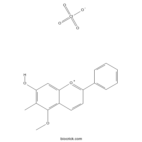
Dracorhodin perchlorateCAS# 125536-25-6 |

Quality Control & MSDS
3D structure
Package In Stock
Number of papers citing our products

| Cas No. | 125536-25-6 | SDF | Download SDF |
| PubChem ID | 74787691 | Appearance | Orange powder |
| Formula | C17H15ClO7 | M.Wt | 366.75 |
| Type of Compound | Flavonoids | Storage | Desiccate at -20°C |
| Solubility | Soluble in methan | ||
| Chemical Name | 5-methoxy-6-methyl-2-phenylchromenylium-7-ol;perchlorate | ||
| SMILES | CC1=C(C=C2C(=C1OC)C=CC(=[O+]2)C3=CC=CC=C3)O.[O-]Cl(=O)(=O)=O | ||
| Standard InChIKey | KRTYZFUODYMZPG-UHFFFAOYSA-N | ||
| Standard InChI | InChI=1S/C17H14O3.ClHO4/c1-11-14(18)10-16-13(17(11)19-2)8-9-15(20-16)12-6-4-3-5-7-12;2-1(3,4)5/h3-10H,1-2H3;(H,2,3,4,5) | ||
| General tips | For obtaining a higher solubility , please warm the tube at 37 ℃ and shake it in the ultrasonic bath for a while.Stock solution can be stored below -20℃ for several months. We recommend that you prepare and use the solution on the same day. However, if the test schedule requires, the stock solutions can be prepared in advance, and the stock solution must be sealed and stored below -20℃. In general, the stock solution can be kept for several months. Before use, we recommend that you leave the vial at room temperature for at least an hour before opening it. |
||
| About Packaging | 1. The packaging of the product may be reversed during transportation, cause the high purity compounds to adhere to the neck or cap of the vial.Take the vail out of its packaging and shake gently until the compounds fall to the bottom of the vial. 2. For liquid products, please centrifuge at 500xg to gather the liquid to the bottom of the vial. 3. Try to avoid loss or contamination during the experiment. |
||
| Shipping Condition | Packaging according to customer requirements(5mg, 10mg, 20mg and more). Ship via FedEx, DHL, UPS, EMS or other couriers with RT, or blue ice upon request. | ||
| Description | Dracorhodin perchlorate inhibits cell growth, and induces apoptosis in fibroblasts in a dose-and time-dependent manner, arresting cell cycle at G1 phase, may as a candidate for anti-breast cancer. Dracorhodin perchlorate can inhibit high glucose-induced serum and glucocorticoid induced protein kinase 1 (SGK1) and fibronectin(FN) expression in human mesangial cells, and this may be part of the mechanism of preventing and treating renal fibrosis of DN. |
| Targets | Caspase | PI3K | Akt | NF-kB | p53 | Bcl-2/Bax | p21 | TNF-α | PARP | MMP(e.g.TIMP) | P450 (e.g. CYP17) |
| In vitro | Dracorhodin perchlorate induces apoptosis in primary fibroblasts from human skin hypertrophic scars via participation of caspase-3.[Pubmed: 24525335]Eur J Pharmacol. 2014 Apr 5;728:82-92.Hypertrophic scar (HS) is an abnormally proliferative disorder characterized by excessive proliferation of fibroblasts and redundant deposition of extracellular matrix. An unbalance between fibroblast proliferation and apoptosis has been assumed to play an important role in HS formation. Dracorhodin perchlorate inhibit high glucose induce serum and glucocorticoid induced protein kinase 1 and fibronectin expression in human mesangial cells.[Pubmed: 20931854]Zhongguo Zhong Yao Za Zhi. 2010 Aug;35(15):1996-2000.To investigate the effect of Dracorhodin perchlorate (DP) on inhibiting high glucose-induced serum and glucocorticoid induced protein kinase 1 (SGK1) and fibronectin (FN) expression in human mesangial cells (HMC), and its mechanism of prevention and treatment on renal fibrosis in diabetic nephropathy (DN) .
Dracorhodin perchlorate suppresses proliferation and induces apoptosis in human prostate cancer cell line PC-3.[Pubmed: 21505988]J Huazhong Univ Sci Technolog Med Sci. 2011 Apr;31(2):215-9.
|
| Cell Research | Dracorhodin perchlorate induced human breast cancer MCF-7 apoptosis through mitochondrial pathways.[Pubmed: 23869191 ]Dracorhodin perchlorate inhibits PI3K/Akt and NF-κB activation, up-regulates the expression of p53, and enhances apoptosis.[Pubmed: 22711363]Apoptosis. 2012 Oct;17(10):1104-19.Dracorhodin perchlorate has been recently shown to induce apoptotic cell death in cancer cells. However, the molecular mechanisms underlying these effects are unknown in human gastric tumor cells. Int J Med Sci. 2013 Jul 7;10(9):1149-56.Dracorhodin perchlorate (DP) was a synthetic analogue of the antimicrobial anthocyanin red pigment dracorhodin. It was reported that DP could induce apoptosis in human prostate cancer, human gastric tumor cells and human melanoma, but the cytotoxic effect of DP on human breast cancer was not investigated. This study would investigate whether DP was a candidate chemical of anti-human breast cancer.
|

Dracorhodin perchlorate Dilution Calculator

Dracorhodin perchlorate Molarity Calculator
| 1 mg | 5 mg | 10 mg | 20 mg | 25 mg | |
| 1 mM | 2.7267 mL | 13.6333 mL | 27.2665 mL | 54.5331 mL | 68.1663 mL |
| 5 mM | 0.5453 mL | 2.7267 mL | 5.4533 mL | 10.9066 mL | 13.6333 mL |
| 10 mM | 0.2727 mL | 1.3633 mL | 2.7267 mL | 5.4533 mL | 6.8166 mL |
| 50 mM | 0.0545 mL | 0.2727 mL | 0.5453 mL | 1.0907 mL | 1.3633 mL |
| 100 mM | 0.0273 mL | 0.1363 mL | 0.2727 mL | 0.5453 mL | 0.6817 mL |
| * Note: If you are in the process of experiment, it's necessary to make the dilution ratios of the samples. The dilution data above is only for reference. Normally, it's can get a better solubility within lower of Concentrations. | |||||

Calcutta University

University of Minnesota

University of Maryland School of Medicine

University of Illinois at Chicago

The Ohio State University

University of Zurich

Harvard University

Colorado State University

Auburn University

Yale University

Worcester Polytechnic Institute

Washington State University

Stanford University

University of Leipzig

Universidade da Beira Interior

The Institute of Cancer Research

Heidelberg University

University of Amsterdam

University of Auckland

TsingHua University

The University of Michigan

Miami University

DRURY University

Jilin University

Fudan University

Wuhan University

Sun Yat-sen University

Universite de Paris

Deemed University

Auckland University

The University of Tokyo

Korea University
- Testosterone phenylpropionate
Catalog No.:BCC9171
CAS No.:1255-49-8
- SR 8278
Catalog No.:BCC6191
CAS No.:1254944-66-5
- Sibutramine hydrochloride monohydrate
Catalog No.:BCC5251
CAS No.:125494-59-9
- Saclofen
Catalog No.:BCC6580
CAS No.:125464-42-8
- LY2874455
Catalog No.:BCC1723
CAS No.:1254473-64-7
- RQ-00203078
Catalog No.:BCC6419
CAS No.:1254205-52-1
- Acetate gossypol
Catalog No.:BCN5354
CAS No.:12542-36-8
- TCN 238
Catalog No.:BCC7901
CAS No.:125404-04-8
- Cedrelone
Catalog No.:BCN6135
CAS No.:1254-85-9
- 6-Acetonyl-N-methyl-dihydrodecarine
Catalog No.:BCN6134
CAS No.:1253740-09-8
- AbK
Catalog No.:BCC8011
CAS No.:1253643-88-7
- NMS-E973
Catalog No.:BCC5335
CAS No.:1253584-84-7
- 3',5,5',7-Tetrahydroxy-4',6-dimethoxyflavone
Catalog No.:BCN6136
CAS No.:125537-92-0
- LY 233053
Catalog No.:BCC5771
CAS No.:125546-04-5
- PF-4708671
Catalog No.:BCC5031
CAS No.:1255517-76-0
- UNC0638
Catalog No.:BCC1135
CAS No.:1255580-76-7
- MnTMPyP Pentachloride
Catalog No.:BCC6532
CAS No.:125565-45-9
- Rotigotine hydrochloride
Catalog No.:BCC1908
CAS No.:125572-93-2
- [Leu31,Pro34]-Neuropeptide Y (porcine)
Catalog No.:BCC5716
CAS No.:125580-28-1
- TRX818
Catalog No.:BCC6458
CAS No.:1256037-58-7
- AI-10-49
Catalog No.:BCC3973
CAS No.:1256094-72-0
- RuBi-Nicotine
Catalog No.:BCC7793
CAS No.:1256362-30-7
- Ledipasvir
Catalog No.:BCC1696
CAS No.:1256388-51-8
- CH5424802
Catalog No.:BCC3749
CAS No.:1256580-46-7
Dracorhodin perchlorate induced human breast cancer MCF-7 apoptosis through mitochondrial pathways.[Pubmed:23869191]
Int J Med Sci. 2013 Jul 7;10(9):1149-56.
OBJECTIVE: Dracorhodin perchlorate (DP) was a synthetic analogue of the antimicrobial anthocyanin red pigment dracorhodin. It was reported that DP could induce apoptosis in human prostate cancer, human gastric tumor cells and human melanoma, but the cytotoxic effect of DP on human breast cancer was not investigated. This study would investigate whether DP was a candidate chemical of anti-human breast cancer. METHODS: The MTT assay reflected the number of viable cells through measuring the activity of cellular enzymes. Phase contrast microscopy visualized cell morphology. Fluorescence microscopy detected nuclear fragmentation after Hoechst 33258 staining. Flowcytometric analysis of Annexin V-PI staining and Rodamine 123 staining was used to detect cell apoptosis and mitochondrial membrane potential (MMP). Real time PCR detected mRNA level. Western blot examined protein expression. RESULTS: DP dose and time-dependently inhibited the growth of MCF-7 cells. DP inhibited MCF-7 cell growth through apoptosis. DP regulated the expression of Bcl-2 and Bax, which were mitochondrial pathway proteins, to decrease MMP, and DP promoted the transcription of Bax and inhibited Bcl-2. Apoptosis-inducing factor (AIF) and cytochrome c which localized in mitochondrial in physiological condition were released into cytoplasm when MMP was decreased. DP activated caspase-9, which was the downstream of mitochondrial pathway. Therefore DP decreased MMP to release AIF and cytochrome c into cytoplasm, further activating caspase 9, lastly led to apoptosis. CONCLUSION: Therefore DP was a candidate for anti-breast cancer, DP induced apoptosis of MCF-7 through mitochondrial pathway.
Dracorhodin perchlorate suppresses proliferation and induces apoptosis in human prostate cancer cell line PC-3.[Pubmed:21505988]
J Huazhong Univ Sci Technolog Med Sci. 2011 Apr;31(2):215.
The growth inhibition and pro-apoptosis effects of Dracorhodin perchlorate on human prostate cancer PC-3 cell line were examined. After administration of 10-80 mumol/L Dracorhodin perchlorate for 12-48 h, cell viability of PC-3 cells was measured by MTT colorimetry. Cell proliferation ability was detected by colony formation assay. Cellular apoptosis was inspected by acridine orange-ethidium bromide fluorescent staining, Hoechst 33258 fluorescent staining, and flow cytometry (FCM) with annexin V-FITC/propidium iodide dual staining. The results showed that Dracorhodin perchlorate inhibited the growth of PC-3 in a dose- and time-dependent manner. IC50 of Dracorhodin perchlorate on PC-3 cells at 24 h was 40.18 mumol/L. Cell clone formation rate was decreased by 86% after treatment with 20 mumol/L of Dracorhodin perchlorate. Some cells presented the characteristic apoptotic changes. The cellular apoptotic rates induced by 10-40 mumol/L Dracorhodin perchlorate for 24 h were 8.43% to 47.71% respectively. It was concluded that Dracorhodin perchlorate significantly inhibited the growth of PC-3 cells by suppressing proliferation and inducing apoptosis of the cells.
Dracorhodin perchlorate inhibits PI3K/Akt and NF-kappaB activation, up-regulates the expression of p53, and enhances apoptosis.[Pubmed:22711363]
Apoptosis. 2012 Oct;17(10):1104-19.
Dracorhodin perchlorate has been recently shown to induce apoptotic cell death in cancer cells. However, the molecular mechanisms underlying these effects are unknown in human gastric tumor cells. In this study, effects of Dracorhodin perchlorate on cell viability, cell cycle, and apoptosis were investigated in SGC-7901 cells. The results showed that Dracorhodin perchlorate induced cellular and DNA morphological changes and decreased the viability of SGC-7901 cells. Dracorhodin perchlorate-mediated cell cycle arrest was associated with a marked decrease in protein levels of phosphorylated retinoblastoma and E2F1. Dracorhodin perchlorate-induced apoptosis is mediated via upregulation of p53, inhibiting the activation of PI3K/Akt, and NF-kappaB, thereby decreasing the expression of the anti-apoptotic proteins, Bcl-2 and Bcl-XL. Interestingly, we also found that Dracorhodin perchlorate significantly suppressed the IGF-1-induced phosphorylation of Akt in the stably expressing EGFP-Akt recombinant CHO-hIR cells and inhibited TNF-induced NF-kappaB transcriptional activity in the NF-kappaBp65-EGFP recombinant U2OS cells, indicating that inhibition of PI3K/Akt and NF-kappaB may provide a molecular basis for the ability of Dracorhodin perchlorate to induce apoptosis. Dracorhodin perchlorate induced up-regulation of p53, thereby resulting in the activation of its downstream targets p21 and Bax following the dissipation of mitochondrial membrane potential and activation of caspase-3 and its substrate, PARP. Moreover, Dracorhodin perchlorate dramatically enhanced the wortmannin- and TNF-induced apoptosis in SGC-7901 cells. These results reveal functional interplay among the PI3K/Akt, p53 and NF-kappaB pathways that are frequently deregulated in cancer and suggest that their simultaneous targeting by Dracorhodin perchlorate could result in efficacious and selective killing of cancer cells.
Dracorhodin perchlorate induces apoptosis in primary fibroblasts from human skin hypertrophic scars via participation of caspase-3.[Pubmed:24525335]
Eur J Pharmacol. 2014 Apr 5;728:82-92.
Hypertrophic scar (HS) is an abnormally proliferative disorder characterized by excessive proliferation of fibroblasts and redundant deposition of extracellular matrix. An unbalance between fibroblast proliferation and apoptosis has been assumed to play an important role in HS formation. To explore the regulative effects of Dracorhodin perchlorate (Dp), one of the derivants of dracorhodin that is a major constituent in the traditional Chinese medicine, on primary fibroblasts from human skin hypertrophic scars, 3-[4,5-dimethylthiazol-2-yl]-2,5-diphenyltetrazolium bromide (MTT) assay and flow cytometric analysis were respectively used to evaluate the inhibitory effect of Dp on the cells and to determine cell cycle distribution. Additionally, cellular apoptosis was separately detected with Hoechst 33258 staining and terminal deoxynucleotidyl transferase (TdT)-mediated dUTP nick end labeling (TUNEL) assay. The expression levels of caspase-3 mRNA and protein were respectively measured with reverse transcription-polymerase chain reaction and western blot analysis, and caspase-3 activity were determined using a colorimetric assay kit. The results showed that Dp significantly inhibited cell growth, and induced apoptosis in fibroblasts in a dose-and time-dependent manner, arresting cell cycle at G1 phase. Additionally, Dp slightly up-regulated caspase-3 mRNA expression in fibroblasts, but significantly down-regulated caspase-3 protein expression in a dose- and time-dependent manner, and concurrently elevated caspase-3 activity. Taken together, these data indicated that Dp could effectively inhibit cell proliferation, and induced cell cycle arrest and apoptosis in fibroblasts, at least partially via modulation of caspase-3 expression and its activity, which suggests that Dp is an effective and potential candidate to develop for HS treatment.
[Dracorhodin perchlorate inhibit high glucose induce serum and glucocorticoid induced protein kinase 1 and fibronectin expression in human mesangial cells].[Pubmed:20931854]
Zhongguo Zhong Yao Za Zhi. 2010 Aug;35(15):1996-2000.
OBJECTIVE: To investigate the effect of Dracorhodin perchlorate (DP) on inhibiting high glucose-induced serum and glucocorticoid induced protein kinase 1 (SGK1) and fibronectin (FN) expression in human mesangial cells (HMC), and its mechanism of prevention and treatment on renal fibrosis in diabetic nephropathy (DN) . METHOD: The HMC were divided into normal glucose group (NG group, 5.5 mmol x L(-1) D-glucose), normal glucose +low DP group (NG + LDP group, 5.5 mmol x L(-1) D-glucose +7.5 micromol x L(-1) DP), normal glucose +high DP group (NG + HDP group, 5.5 mmol x L(-1) D-glucose + 15 micromol x L(-1) DP), high glucose group (HG group,25 mmol x L(-1) D-glucose), high glucose +low DP group (HG + LDP group, 25 mmol x L(-1) D-glucose + 7.5 micromol x L(-1) DP)and high glucose +high DP group (HG +HDP group, 25 mmol x L(-1) D-glucose + 15 micromol x L(-1) DP). Each group was examined at 24 hours. The levels of SGK1 and FN mRNA was detected by real-time fluorescence quantitative PCR,and the expression of SGK1 and FN protein was detected by Western blot or indirect immunofluorescence. RESULT: A basal level of SGK1 and FN in HMC were detected in NG group, and the level of SGK1 and FN mRNA and protein were not evidently different compared to that of NG group adding 7.5 micromol x L(-1) DP for 24 hours. On the other hand, the levels of SGK1 and FN mRNA and protein were obviously decreased by adding 15 micromol x L(-1) DP for 24 hours. Compared to NG group, the levels of SGK1 and FN mRNA and protein were increased in HG group after stimulating for 24 hours (P < 0.01). Compared to HG group, the level of SGK1 and FN mRNA and protein were evidently reduced in HG + LDP and HG + HDP groups by adding 7.5 micromol x L(-1) DP and 15 micromol x L(-1) DP for 24 hours (P < 0.01). CONCLUSION: Dracorhodin perchlorate can inhibit high glucose-induced serum and glucocorticoid induced protein kinase 1 (SGK1) and fibronectin(FN) expression in human mesangial cells, and this may be part of the mechanism of preventing and treating renal fibrosis of DN.


