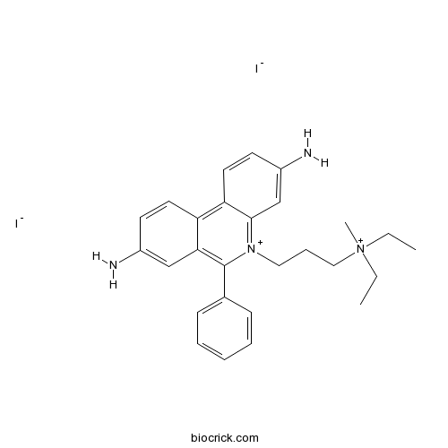
Propidium iodideCAS# 25535-16-4 |

- Pregnenolone
Catalog No.:BCN6255
CAS No.:145-13-1
- Adrenosterone
Catalog No.:BCC4061
CAS No.:382-45-6
- Epiandrosterone
Catalog No.:BCC4481
CAS No.:481-29-8
- Cortisone acetate
Catalog No.:BCC4771
CAS No.:50-04-4
- Deoxycorticosterone acetate
Catalog No.:BCC4655
CAS No.:56-47-3
Quality Control & MSDS
3D structure
Package In Stock
Number of papers citing our products

| Cas No. | 25535-16-4 | SDF | Download SDF |
| PubChem ID | 104981 | Appearance | Powder |
| Formula | C27H34I2N4 | M.Wt | 668.39 |
| Type of Compound | N/A | Storage | Desiccate at -20°C |
| Synonyms | PI | ||
| Solubility | Soluble to 5 mM in water with sonication | ||
| Chemical Name | 3-(3,8-diamino-6-phenylphenanthridin-5-ium-5-yl)propyl-diethyl-methylazanium;diiodide | ||
| SMILES | CC[N+](C)(CC)CCC[N+]1=C2C=C(C=CC2=C3C=CC(=CC3=C1C4=CC=CC=C4)N)N.[I-].[I-] | ||
| Standard InChIKey | XJMOSONTPMZWPB-UHFFFAOYSA-M | ||
| Standard InChI | InChI=1S/C27H33N4.2HI/c1-4-31(3,5-2)17-9-16-30-26-19-22(29)13-15-24(26)23-14-12-21(28)18-25(23)27(30)20-10-7-6-8-11-20;;/h6-8,10-15,18-19,29H,4-5,9,16-17,28H2,1-3H3;2*1H/q+1;;/p-1 | ||
| General tips | For obtaining a higher solubility , please warm the tube at 37 ℃ and shake it in the ultrasonic bath for a while.Stock solution can be stored below -20℃ for several months. We recommend that you prepare and use the solution on the same day. However, if the test schedule requires, the stock solutions can be prepared in advance, and the stock solution must be sealed and stored below -20℃. In general, the stock solution can be kept for several months. Before use, we recommend that you leave the vial at room temperature for at least an hour before opening it. |
||
| About Packaging | 1. The packaging of the product may be reversed during transportation, cause the high purity compounds to adhere to the neck or cap of the vial.Take the vail out of its packaging and shake gently until the compounds fall to the bottom of the vial. 2. For liquid products, please centrifuge at 500xg to gather the liquid to the bottom of the vial. 3. Try to avoid loss or contamination during the experiment. |
||
| Shipping Condition | Packaging according to customer requirements(5mg, 10mg, 20mg and more). Ship via FedEx, DHL, UPS, EMS or other couriers with RT, or blue ice upon request. | ||
| Description | Red-fluorescent DNA stain. Membrane impermeant to live cells; differentiates between live and dead cells in cell populations. |

Propidium iodide Dilution Calculator

Propidium iodide Molarity Calculator
| 1 mg | 5 mg | 10 mg | 20 mg | 25 mg | |
| 1 mM | 1.4961 mL | 7.4807 mL | 14.9613 mL | 29.9226 mL | 37.4033 mL |
| 5 mM | 0.2992 mL | 1.4961 mL | 2.9923 mL | 5.9845 mL | 7.4807 mL |
| 10 mM | 0.1496 mL | 0.7481 mL | 1.4961 mL | 2.9923 mL | 3.7403 mL |
| 50 mM | 0.0299 mL | 0.1496 mL | 0.2992 mL | 0.5985 mL | 0.7481 mL |
| 100 mM | 0.015 mL | 0.0748 mL | 0.1496 mL | 0.2992 mL | 0.374 mL |
| * Note: If you are in the process of experiment, it's necessary to make the dilution ratios of the samples. The dilution data above is only for reference. Normally, it's can get a better solubility within lower of Concentrations. | |||||

Calcutta University

University of Minnesota

University of Maryland School of Medicine

University of Illinois at Chicago

The Ohio State University

University of Zurich

Harvard University

Colorado State University

Auburn University

Yale University

Worcester Polytechnic Institute

Washington State University

Stanford University

University of Leipzig

Universidade da Beira Interior

The Institute of Cancer Research

Heidelberg University

University of Amsterdam

University of Auckland

TsingHua University

The University of Michigan

Miami University

DRURY University

Jilin University

Fudan University

Wuhan University

Sun Yat-sen University

Universite de Paris

Deemed University

Auckland University

The University of Tokyo

Korea University
Propidium iodide is a red-fluorescent dye that can be used to stain cells.
In Vitro:Propidium iodide is a cell-membrane impermeable dye with characteristic excitation maximum at 535 nm and emission maximum at 617 nm which intercalates with nucleic acids with a stoichiometry of one dye per 4-5 base pairs with little sequence preference. Propidium iodide has evidenced of having no toxic effects on neurons, being today’s most common marker for membrane integrity and cell viability when applied prior to fixation (pre-fixation propidium iodide staining method). The pre-fixation staining has been widely used for quantitative assessments of neuronal cell decline in models of acute neurodegeneration, visualized as intensely labeled PI+-pycnotic nuclei of degenerating neurons [1]. Propidium iodide cannot cross the membrane of live cells, making it useful to measure the percentage of apoptotic cells by flow-cytometric analysis. The flow cytometric data shows an excellent correlation with the results obtained with both electrophoretic and colorimetric methods. This new rapid, simple and reproducible method proves useful for assessing apoptosis of specific cell populations in heterogeneous tissues such as bone marrow, thymus and lymph nodes[2].
References:
[1]. Hezel M, et al. Propidium iodide staining: a new application in fluorescence microscopy for analysis of cytoarchitecture in adult and developing rodent brain. Micron. 2012 Oct;43(10):1031-8.
[2]. A rapid and simple method for measuring thymocyte apoptosis by propidium iodidestaining and flow cytometry. J Immunol Methods. 1991 Jun 3;139(2):271-9.
- Mayumbine
Catalog No.:BCN5123
CAS No.:25532-45-0
- Z-Ile-Glu-Pro-Phe-Ome
Catalog No.:BCC5526
CAS No.:255257-97-4
- Isoferulic acid
Catalog No.:BCN5122
CAS No.:25522-33-2
- Ibotenic acid
Catalog No.:BCC6591
CAS No.:2552-55-8
- Bruceine A
Catalog No.:BCC5311
CAS No.:25514-31-2
- Bruceine C
Catalog No.:BCN8000
CAS No.:25514-30-1
- Bruceine B
Catalog No.:BCN7615
CAS No.:25514-29-8
- 1-Acetyl-4-piperidinecarboxylic acid
Catalog No.:BCC8447
CAS No.:25503-90-6
- 4-Phenylbutan-2-one
Catalog No.:BCN3808
CAS No.:2550-26-7
- 3-Epioleanolic acid
Catalog No.:BCN3050
CAS No.:25499-90-5
- Tasquinimod
Catalog No.:BCC1987
CAS No.:254964-60-8
- 4-Allyloxy-2-hydroxybenzophenone
Catalog No.:BCC8675
CAS No.:2549-87-3
- 1-(4-Hydroxybenzoyl)glucose
Catalog No.:BCN6900
CAS No.:25545-07-7
- 7-Methoxy-4-methylcoumarin
Catalog No.:BCN6540
CAS No.:2555-28-4
- Efetaal
Catalog No.:BCN8494
CAS No.:2556-10-7
- 1,6,7-Trihydroxyxanthone
Catalog No.:BCN5124
CAS No.:25577-04-2
- Delta-Tocotrienol
Catalog No.:BCN6696
CAS No.:25612-59-3
- 7beta-Acetoxytaxuspine C
Catalog No.:BCN7219
CAS No.:256347-91-8
- BAY 41-2272
Catalog No.:BCC7932
CAS No.:256376-24-6
- SEW 2871
Catalog No.:BCC7312
CAS No.:256414-75-2
- Schleicheol 1
Catalog No.:BCN4661
CAS No.:256445-66-6
- Schleicheol 2
Catalog No.:BCN5125
CAS No.:256445-68-8
- Ac2-12
Catalog No.:BCC5826
CAS No.:256447-08-2
- Preisocalamendiol
Catalog No.:BCN5126
CAS No.:25645-19-6
Xylem Characterization Using Improved Pseudo-Schiff Propidium Iodide Staining of Whole Mount Samples and Confocal Laser-Scanning Microscopy.[Pubmed:28050834]
Methods Mol Biol. 2017;1544:127-132.
An improved pseudo-Schiff Propidium iodide staining technique well suited for, but not limited to, the visualization of xylem cell walls in whole mount samples is presented. The pseudo-Schiff reaction results in covalent binding of the fluorescent dye Propidium iodide to cell walls. This stable linkage permits the use of clearing agents after staining, which is itself improved following pretreatment of the plant tissue. A subsequent acid alcohol washing step eliminates unbound Propidium iodide to reduce background fluorescence. The method can be used for characterizing xylem cell structure in different organs and species without the need for tissue sectioning.
Measuring the DNA Content of Cells in Apoptosis and at Different Cell-Cycle Stages by Propidium Iodide Staining and Flow Cytometry.[Pubmed:27698234]
Cold Spring Harb Protoc. 2016 Oct 3;2016(10). pii: 2016/10/pdb.prot087247.
All cells are created from preexisting cells. This involves complete duplication of the parent cell to create two daughter cells by a process known as the cell cycle. For this process to be successful, the DNA of the parent cell must be faithfully replicated so that each daughter cell receives a full copy of the genetic information. During the cell cycle, the DNA content of the parent cell increases as new DNA is synthesized (S phase). When there are two full copies of the DNA (G2/M phase), the cell splits to form two new cells (G0/G1 phase). As such, cells in different stages of the cell cycle have different DNA contents. The cell cycle is tightly regulated to safeguard the integrity of the cell and any cell that is defective or unable to complete the cell cycle is programmed to die by apoptosis. When this occurs, the DNA is fragmented into oligonucleosomal-sized fragments that are disposed of when the dead cell is removed by phagocytosis. Consequently apoptotic cells have reduced DNA content compared with living cells. This can be measured by staining cells with Propidium iodide (PI), a fluorescent molecule that intercalates with DNA at a specific ratio. The level of PI fluorescence in a cell is, therefore, directly proportional to the DNA content of that cell. This protocol describes the use of PI staining to determine the percentage of cells in each phase of the cell cycle and the percentage of apoptotic cells in a sample.
Quantitation of Apoptosis and Necrosis by Annexin V Binding, Propidium Iodide Uptake, and Flow Cytometry.[Pubmed:27803250]
Cold Spring Harb Protoc. 2016 Nov 1;2016(11). pii: 2016/11/pdb.prot087288.
The surface of healthy cells is composed of lipids that are asymmetrically distributed on the inner and outer leaflet of the plasma membrane. One of these lipids, phosphatidylserine (PS), is normally restricted to the inner leaflet of the plasma membrane and is, therefore, only exposed to the cell cytoplasm. However, during apoptosis lipid asymmetry is lost and PS becomes exposed on the outer leaflet of the plasma membrane. Annexin V, a 36-kDa calcium-binding protein, binds to PS; therefore, fluorescently labeled Annexin V can be used to detect PS that is exposed on the outside of apoptotic cells. Annexin V can also stain necrotic cells because these cells have ruptured membranes that permit Annexin V to access the entire plasma membrane. However, apoptotic cells can be distinguished from necrotic cells by co-staining with Propidium iodide (PI) because PI enters necrotic cells but is excluded from apoptotic cells. This protocol describes Annexin V binding and PI uptake followed by flow cytometry to detect and quantify apoptotic and necrotic cells.
A pipeline for developing and testing staining protocols for flow cytometry, demonstrated with SYBR Green I and propidium iodide viability staining.[Pubmed:27810378]
J Microbiol Methods. 2016 Dec;131:172-180.
The increasing use of flow cytometry (FCM) for analyses of environmental samples has resulted in a large variety of staining protocols with varying results and limited comparability. Viability assessment with FCM is in this context of particular interest because incorrect staining could severely affect the outcome/interpretation of the results. Here we propose a pipeline for the development and optimization of staining protocols for environmental samples, demonstrated with the common viability dye combination of SYBR Green I (SG) and Propidium iodide (PI). Optimization steps included the assessment of dye solvents, determination of suitable PI concentration, and determining the optimal staining temperature and staining time. We demonstrated that dimethyl sulfoxide (DMSO) could impair membrane integrity, when used for SGPI stock solution preparation, and TRIS buffer was chosen as an alternative. Moreover we selected 6muM as optimal PI final concentration: less than 3muM resulted in incomplete staining of damaged cells, while concentrations higher that 12muM resulted in false PI-positive staining of intact cells. Low temperatures (25 degrees C) resulted in a slow reaction and did not enable the staining of all bacteria, while high temperatures (44 degrees C) caused damage to cells and false PI-positive results. Hence, 35 degrees C was selected as optimal staining temperature. We further showed that a minimum of 15min were necessary to obtain stable staining results. Moreover, we showed that addition of EDTA resulted in 1-39% more PI-positive results compared to an EDTA-free sample, and argue that insufficient evidence currently exist in favor of adding EDTA to all samples in general. Altogether, the data clearly shows the need to be careful, precise and reproducible when staining cells for flow cytometric analyses, and the need to assess and optimize staining protocols with both viable and non-viable bacteria.
p53-dependent and p53-independent anticancer effects of different histone deacetylase inhibitors.[Pubmed:24281001]
Br J Cancer. 2014 Feb 4;110(3):656-67.
BACKGROUND: Histone deacetylase inhibitors (HDACi) are promising antineoplastic agents, but their precise mechanisms of actions are not well understood. In particular, the relevance of p53 for HDACi-induced effects has not been fully elucidated. We investigated the anticancer effects of four structurally distinct HDACi, vorinostat, entinostat, apicidin and valproic acid, using isogenic HCT-116 colon cancer cell lines differing in p53 status. METHODS: Effects were assessed by MTT assay, flow-cytometric analyses of Propidium iodide uptake, mitochondrial depolarisation and cell-cycle distribution, as well as by gene expression profiling. RESULTS: Vorinostat was equally effective in p53 wild-type and null cells, whereas entinostat was less effective in p53 null cells. Histone deacetylase inhibitors treatment suppressed the expression of MDM2 and increased the abundance of p53. Combination treatments showed that vorinostat enhanced the cytotoxic activity of TRAIL and bortezomib, independent of the cellular p53 status. Investigations into the effects of an inhibitor of the sirtuin class of HDAC, tenovin-1, revealed that tenovin-1-mediated cell death hinged on p53. CONCLUSION: These results demonstrate that vorinostat activates p53, but does not require p53 for inducing its anticancer action. Yet they also demonstrate that entinostat-induced cytotoxic effects partially depend on p53, indicating that different HDACi have a different requirement for p53.
SIRT4 protein suppresses tumor formation in genetic models of Myc-induced B cell lymphoma.[Pubmed:24368766]
J Biol Chem. 2014 Feb 14;289(7):4135-44.
Glutamine metabolism plays an essential role for growth and proliferation of many cancer cells by providing metabolites for the maintenance of mitochondrial functions and macromolecular synthesis. Aberrant activation of the transcription factor c-Myc, e.g. caused by t(8;14) chromosomal translocation commonly found in Burkitt lymphoma, is a key driver of cellular glutamine metabolism in many tumors, highlighting the need to identify molecular mechanisms that can suppress glutamine usage in these cancers. Recently, the mitochondrial sirtuin SIRT4 has been reported to function as a tumor suppressor by regulating glutamine metabolism, suggesting that it might have therapeutic potential for treating glutamine-dependent cancers. Here, we report that SIRT4 represses Myc-induced B cell lymphomagenesis via inhibition of mitochondrial glutamine metabolism. We found that SIRT4 overexpression can dampen glutamine utilization even in Myc-driven human Burkitt lymphoma cells and inhibit glutamine-dependent proliferation of these cells. Importantly, SIRT4 overexpression sensitizes Burkitt lymphoma cells to glucose depletion and synergizes with pharmacological glycolysis inhibitors to induce cell death. Moreover, SIRT4 loss in a genetic mouse model of Myc-induced Burkitt lymphoma, Emu-Myc transgenic mouse, greatly accelerates lymphomagenesis and mortality. Indeed, Emu-Myc-induced B cell lymphoma cells from SIRT4 null mice exhibit increased glutamine uptake and glutamate dehydrogenase activity. Furthermore, we establish that SIRT4 regulates glutamine metabolism independent of Myc. Together, these results highlight the tumor-suppressive role of SIRT4 in Myc-induced B cell lymphoma and suggest that SIRT4 may be a potential target against Myc-induced and/or glutamine-dependent cancers.
Serine hydrolase inhibitors block necrotic cell death by preventing calcium overload of the mitochondria and permeability transition pore formation.[Pubmed:24297180]
J Biol Chem. 2014 Jan 17;289(3):1491-504.
Perturbation of calcium signaling that occurs during cell injury and disease, promotes cell death. In mouse lung fibroblasts A23187 triggered mitochondrial permeability transition pore (MPTP) formation, lactate dehydrogenase (LDH) release, and necrotic cell death that were blocked by cyclosporin A (CsA) and EGTA. LDH release temporally correlated with arachidonic acid release but did not involve cytosolic phospholipase A2alpha (cPLA2alpha) or calcium-independent PLA2. Surprisingly, release of arachidonic acid and LDH from cPLA2alpha-deficient fibroblasts was inhibited by the cPLA2alpha inhibitor pyrrophenone, and another serine hydrolase inhibitor KT195, by preventing mitochondrial calcium uptake. Inhibitors of calcium/calmodulin-dependent protein kinase II, a mitochondrial Ca(2+) uniporter (MCU) regulator, also prevented MPTP formation and arachidonic acid release induced by A23187 and H2O2. Pyrrophenone blocked MCU-mediated mitochondrial calcium uptake in permeabilized fibroblasts but not in isolated mitochondria. Unlike pyrrophenone, the diacylglycerol analog 1-oleoyl-2-acetyl-sn-glycerol and CsA blocked cell death and arachidonic acid release not by preventing mitochondrial calcium uptake but by inhibiting MPTP formation. In fibroblasts stimulated with thapsigargin, which induces MPTP formation by a direct effect on mitochondria, LDH and arachidonic acid release were blocked by CsA and 1-oleoyl-2-acetyl-sn-glycerol but not by pyrrophenone or EGTA. Therefore serine hydrolase inhibitors prevent necrotic cell death by blocking mitochondrial calcium uptake but not the enzyme releasing fatty acids that occurs by a novel pathway during MPTP formation. This work reveals the potential for development of small molecule cell-permeable serine hydrolase inhibitors that block MCU-mediated mitochondrial calcium overload, MPTP formation, and necrotic cell death.


