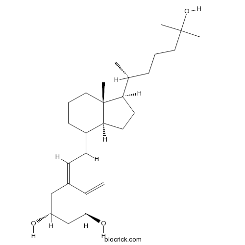
CalcitriolTNF-α and IL-1β production inhibitor CAS# 32222-06-3 |

- 25,26-Dihydroxyvitamin D3
Catalog No.:BCC4201
CAS No.:29261-12-9
- Vitamin D4
Catalog No.:BCC2042
CAS No.:511-28-4
- Impurity B of Calcitriol
Catalog No.:BCC1645
CAS No.:66791-71-7
- Calcifediol-D6
Catalog No.:BCC4075
CAS No.:78782-98-6
Quality Control & MSDS
3D structure
Package In Stock
Number of papers citing our products

| Cas No. | 32222-06-3 | SDF | Download SDF |
| PubChem ID | 5280453 | Appearance | Powder |
| Formula | C27H44O3 | M.Wt | 416.64 |
| Type of Compound | N/A | Storage | Desiccate at -20°C |
| Synonyms | 1,25-Dihydroxyvitamin D3 | ||
| Solubility | DMSO : ≥ 100 mg/mL (240.02 mM) *"≥" means soluble, but saturation unknown. | ||
| Chemical Name | (1R,3S,5Z)-5-[(2E)-2-[(1R,3aS,7aR)-1-[(2R)-6-hydroxy-6-methylheptan-2-yl]-7a-methyl-2,3,3a,5,6,7-hexahydro-1H-inden-4-ylidene]ethylidene]-4-methylidenecyclohexane-1,3-diol | ||
| SMILES | CC(CCCC(C)(C)O)C1CCC2C1(CCCC2=CC=C3CC(CC(C3=C)O)O)C | ||
| Standard InChIKey | GMRQFYUYWCNGIN-NKMMMXOESA-N | ||
| Standard InChI | InChI=1S/C27H44O3/c1-18(8-6-14-26(3,4)30)23-12-13-24-20(9-7-15-27(23,24)5)10-11-21-16-22(28)17-25(29)19(21)2/h10-11,18,22-25,28-30H,2,6-9,12-17H2,1,3-5H3/b20-10+,21-11-/t18-,22-,23-,24+,25+,27-/m1/s1 | ||
| General tips | For obtaining a higher solubility , please warm the tube at 37 ℃ and shake it in the ultrasonic bath for a while.Stock solution can be stored below -20℃ for several months. We recommend that you prepare and use the solution on the same day. However, if the test schedule requires, the stock solutions can be prepared in advance, and the stock solution must be sealed and stored below -20℃. In general, the stock solution can be kept for several months. Before use, we recommend that you leave the vial at room temperature for at least an hour before opening it. |
||
| About Packaging | 1. The packaging of the product may be reversed during transportation, cause the high purity compounds to adhere to the neck or cap of the vial.Take the vail out of its packaging and shake gently until the compounds fall to the bottom of the vial. 2. For liquid products, please centrifuge at 500xg to gather the liquid to the bottom of the vial. 3. Try to avoid loss or contamination during the experiment. |
||
| Shipping Condition | Packaging according to customer requirements(5mg, 10mg, 20mg and more). Ship via FedEx, DHL, UPS, EMS or other couriers with RT, or blue ice upon request. | ||
| Description | Active metabolite of vitamin D3 that activates the vitamin D receptor (VDR). Displays calcemic actions; stimulates intestinal and renal Ca2+ absorption and regulates bone Ca2+ turnover. Exhibits antitumor activity; inhibits in vivo and in vitro cell proliferation in a wide range of cells including breast, prostate, colon, skin and brain carcinomas and myeloid leukemia cells. |

Calcitriol Dilution Calculator

Calcitriol Molarity Calculator
| 1 mg | 5 mg | 10 mg | 20 mg | 25 mg | |
| 1 mM | 2.4002 mL | 12.0008 mL | 24.0015 mL | 48.0031 mL | 60.0038 mL |
| 5 mM | 0.48 mL | 2.4002 mL | 4.8003 mL | 9.6006 mL | 12.0008 mL |
| 10 mM | 0.24 mL | 1.2001 mL | 2.4002 mL | 4.8003 mL | 6.0004 mL |
| 50 mM | 0.048 mL | 0.24 mL | 0.48 mL | 0.9601 mL | 1.2001 mL |
| 100 mM | 0.024 mL | 0.12 mL | 0.24 mL | 0.48 mL | 0.6 mL |
| * Note: If you are in the process of experiment, it's necessary to make the dilution ratios of the samples. The dilution data above is only for reference. Normally, it's can get a better solubility within lower of Concentrations. | |||||

Calcutta University

University of Minnesota

University of Maryland School of Medicine

University of Illinois at Chicago

The Ohio State University

University of Zurich

Harvard University

Colorado State University

Auburn University

Yale University

Worcester Polytechnic Institute

Washington State University

Stanford University

University of Leipzig

Universidade da Beira Interior

The Institute of Cancer Research

Heidelberg University

University of Amsterdam

University of Auckland

TsingHua University

The University of Michigan

Miami University

DRURY University

Jilin University

Fudan University

Wuhan University

Sun Yat-sen University

Universite de Paris

Deemed University

Auckland University

The University of Tokyo

Korea University
Calcitriol is a form of Vitamin D, is converted to metabolites more potent and rapidly acting than other forms of Vitamin D [1].
Calcitriol has played an important role in mineral and skeletal homeostasis by regulating the differentiation, growth and the function of the cell immune system. In vitro studies, Calcitriol has been reported to dose-dependently inhibit the production of tumor necrosis factor-α(TNF-α) and interleukin-1β(IL-1β) in human peripheral blood cells (PBMC) stimulated by LPS. Furthermore, Calcitriol has been revealed to significantly reduced the basal intradialytic increases of TNF-αand IL-1βin vivo studies. Apart from these, Calcitriol has also been reported to modulate the cytokine production indirectly through the effect on calcium and PTH metabolism [2].
References:
[1] Holick MF, Schnoes HK, DeLuca HF. Identification of 1,25-dihydroxycholecalciferol, a form of vitamin D3 metabolically active in the intestine. Proc Natl Acad Sci U S A. 1971 Apr;68(4):803-4.
[2] Panichi V1, De Pietro S, Andreini B, Bianchi AM, Migliori M, Taccola D, Giovannini L, Tetta C, Palla R. Calcitriol modulates in vivo and in vitro cytokine production: a role for intracellular calcium. Kidney Int. 1998 Nov;54(5):1463-9.
- Lupeol palmitate
Catalog No.:BCN7133
CAS No.:32214-80-5
- H-Asp(OMe)-OMe.HCl
Catalog No.:BCC2890
CAS No.:32213-95-9
- Heraclenol 3'-O-beta-D-glucopyranoside
Catalog No.:BCN1459
CAS No.:32207-10-6
- Triflusal
Catalog No.:BCC4443
CAS No.:322-79-2
- p-3-Methylamino propyl phenol
Catalog No.:BCN1802
CAS No.:32180-92-0
- 1,10:4,5-Diepoxy-7(11)-germacren-8-one
Catalog No.:BCN1460
CAS No.:32179-18-3
- Pilloin
Catalog No.:BCN6817
CAS No.:32174-62-2
- L002
Catalog No.:BCC8000
CAS No.:321695-57-2
- Poloxin
Catalog No.:BCC1867
CAS No.:321688-88-4
- BIBR 1532
Catalog No.:BCC1147
CAS No.:321674-73-1
- Cytosporone B
Catalog No.:BCN6791
CAS No.:321661-62-5
- N-Acetyl-4-piperidone
Catalog No.:BCC9079
CAS No.:32161-06-1
- 2alpha,7beta,13alpha-Triacetoxy-5alpha-cinnamoyloxy-9beta-hydroxy-2(3->20)abeotaxa-4(20),11-dien-10-one
Catalog No.:BCN7208
CAS No.:322471-42-1
- SCH 221510
Catalog No.:BCC7612
CAS No.:322473-89-2
- Methyl 3-cyclopropyl-3-oxopropionate
Catalog No.:BCC9038
CAS No.:32249-35-7
- 4-Methoxy-1-methylquinolin-2-one
Catalog No.:BCN4824
CAS No.:32262-18-3
- H-Threoninol
Catalog No.:BCC2705
CAS No.:3228-51-1
- Triphosgene
Catalog No.:BCC2848
CAS No.:32315-10-9
- SCS
Catalog No.:BCC7266
CAS No.:3232-36-8
- p-Cresyl sulfate
Catalog No.:BCC4013
CAS No.:3233-58-7
- 4-Nitrobenzyl carbamate
Catalog No.:BCN3286
CAS No.:32339-07-4
- 9,9-Bis(4-hydroxyphenyl)fluorene
Catalog No.:BCC8795
CAS No.:3236-71-3
- Medicarpin
Catalog No.:BCN5241
CAS No.:32383-76-9
- Imetit dihydrobromide
Catalog No.:BCC6768
CAS No.:32385-58-3
High concentration calcitriol induces endoplasmic reticulum stress related gene profile in breast cancer cells.[Pubmed:28177777]
Biochem Cell Biol. 2017 Apr;95(2):289-294.
Calcitriol, the active form of vitamin D, is known for its anticancer properties including induction of apoptosis as well as the inhibition of angiogenesis and metastasis. Understanding the mechanisms of action for Calcitriol will help with the development of novel treatment strategies. Since vitamin D exerts its cellular actions via binding to its receptor and by altering expressions of a set of genes, we aimed to evaluate the effect of Calcitriol on transcriptomic profile of breast cancer cells. We previously demonstrated that Calcitriol alters endoplasmic reticulum (ER) stress markers, therefore in this study we have focused on ER-stress-related genes to reveal Calcitriols action on these genes in particular. We have treated breast cancer cell lines MCF-7 and MDA-MB-231 with previously determined IC50 concentrations of Calcitriol and evaluated the transcriptomic alterations via microarray. During analysis, only genes altered by at least 2-fold with a P value < 0.05 were taken into consideration. Our findings revealed an ER-stress-associated transcriptomic profile induced by Calcitriol. Induced genes include genes with a pro-survival function (NUPR1, DNAJB9, HMOX1, LCN2, and LAMP3) and with a pro-death function (CHOP (DDIT3), DDIT4, NDGR1, NOXA, and CLGN). These results suggest that Calcitriol induces an ER-stress-like response inducing both pro-survival and pro-death transcripts in the process.
Pharmacologic Calcitriol Inhibits Osteoclast Lineage Commitment via the BMP-Smad1 and IkappaB-NF-kappaB Pathways.[Pubmed:28370465]
J Bone Miner Res. 2017 Jul;32(7):1406-1420.
Vitamin D is involved in a range of physiological processes and its active form and analogs have been used to treat diseases such as osteoporosis. Yet how vitamin D executes its function remains unsolved. Here we show that the active form of vitamin D Calcitriol increases the peak bone mass in mice by inhibiting osteoclastogenesis and bone resorption. Although Calcitriol modestly promoted osteoclast maturation, it strongly inhibited osteoclast lineage commitment from its progenitor monocyte by increasing Smad1 transcription via the vitamin D receptor and enhancing BMP-Smad1 activation, which in turn led to increased IkappaBalpha expression and decreased NF-kappaB activation and NFATc1 expression, with IkappaBalpha being a Smad1 target gene. Inhibition of BMP type I receptor or ablation of Bmpr1a in monocytes alleviated the inhibitory effects of Calcitriol on osteoclast commitment, bone resorption, and bone mass augmentation. These findings uncover crosstalk between the BMP-Smad1 and RANKL-NF-kappaB pathways during osteoclastogenesis that underlies the action of active vitamin D on bone health. (c) 2017 American Society for Bone and Mineral Research.
Hypoparathyroidism: Less Severe Hypocalcemia With Treatment With Vitamin D2 Compared With Calcitriol.[Pubmed:28324108]
J Clin Endocrinol Metab. 2017 May 1;102(5):1505-1510.
Context: Options for chronic treatment of hypoparathyroidism include Calcitriol, recombinant human parathyroid hormone, and high-dose vitamin D (D2). D2 is used in a minority of patients because of fear of prolonged hypercalcemia and renal toxicity. There is a paucity of recent data about D2 use in hypoparathyroidism. Objective: Compare renal function, hypercalcemia, and hypocalcemia in patients with hypoparathyroidism treated chronically with either D2 (D2 group) or Calcitriol. Design, Setting, and Patients: A retrospective study of patients with hypoparathyroidism treated at the University of Maryland Hospital. Participants were identified by a billing record search with diagnosis confirmed by chart review. Thirty patients were identified; 16 were treated chronically with D2, 14 with Calcitriol. Data were extracted from medical records. Main Outcome Measures: Serum creatinine and calcium, hospitalizations, and emergency department (ED) visits for hypercalcemia and hypocalcemia. Results: D2 and Calcitriol groups were similar in age (58.9 +/- 16.7 vs 50.9 +/- 22.6 years, P = 0.28), sex, and treatment duration (17.8 +/- 14.2 vs 8.5 +/- 4.4 years, P = 0.076). Hospitalization or ED visits for hypocalcemia occurred in none of the D2 group vs four of 14 in the Calcitriol group (P = 0.03); three in the Calcitriol group had multiple ED visits. There were no differences between D2 and Calcitriol groups in hospitalizations or ED visits for hypercalcemia, serum creatinine or calcium, or kidney stones. Conclusion: We found less morbidity from hypocalcemia in hypoparathyroid patients treated chronically with D2 compared with Calcitriol and found no difference in renal function or morbidity from hypercalcemia. Treatment with D2 should be considered in patients with hypoparathyroidism, particularly in those who experience recurrent hypocalcemia.
Synergistic Action of Genistein and Calcitriol in Immature Osteosarcoma MG-63 Cells by SGPL1 Up-Regulation.[Pubmed:28125641]
PLoS One. 2017 Jan 26;12(1):e0169742.
BACKGROUND: Phytoestrogens such as genistein, the most prominent isoflavone from soy, show concentration-dependent anti-estrogenic or estrogenic effects. High genistein concentrations (>10 muM) also promote proliferation of bone cancer cells in vitro. On the other hand, the most active component of the vitamin D family, Calcitriol, has been shown to be tumor protective in vitro and in vivo. The purpose of this study was to examine a putative synergism of genistein and Calcitriol in two osteosarcoma cell lines MG-63 (early osteoblast), Saos-2 (mature osteoblast) and primary osteoblasts. METHODS: Thus, an initial screening based on cell cycle phase alterations, estrogen (ER) and vitamin D receptor (VDR) expression, live cell metabolic monitoring, and metabolomics were performed. RESULTS: Exposure to the combination of 100 muM genistein and 10 nM Calcitriol reduced the number of proliferative cells to control levels, increased ERss and VDR expression, and reduced extracellular acidification (40%) as well as respiratory activity (70%), primarily in MG-63 cells. In order to identify the underlying cellular mechanisms in the MG-63 cell line, metabolic profiling via GC/MS technology was conducted. Combined treatment significantly influenced lipids and amino acids preferably, whereas metabolites of the energy metabolism were not altered. The comparative analysis of the log2-ratios revealed that after combined treatment only the metabolite ethanolamine was highly up-regulated. This is the result: a strong overexpression (350%) of the enzyme sphingosine-1-phosphate lyase (SGPL1), which irreversibly degrades sphingosine-1-phosphate (S1P), thereby, generating ethanolamine. S1P production and secretion is associated with an increased capability of migration and invasion of cancer cells. CONCLUSION: From these results can be concluded that the tumor promoting effect of high concentrations of genistein in immature osteosarcoma cells is reduced by the co-administration of Calcitriol, primarily by the breakdown of S1P. It should be tested whether this anti-metastatic pathway can be stimulated by combined treatment also in metastatic xenograft mice models.
TNF receptor superfamily-induced cell death: redox-dependent execution.[Pubmed:16873882]
FASEB J. 2006 Aug;20(10):1589-98.
Tumor necrosis factor (TNF) superfamily is a group of cytokines with important functions in immunity, inflammation, differentiation, control of cell proliferation, and apoptosis. TNFalpha is the founding member of the 19 different proteins that have so far been identified within this family. TNF family members exert their biological effects through the TNF receptor (TNFR) superfamily of cell surface receptors that share a stretch of approximately 80 amino acids within their cytoplasmic region, the death domain (DD), critical for recruiting the death machinery. Work over the last decade has unraveled critical signaling networks involved in TNFR-induced cell death, specifically using the constitutively expressed TNFR1 as a prototype. Of particular interest is the intermediary role of intracellular reactive oxygen species (ROS) in signal transduction after ligation of the TNFR1. With the increasing understanding of the of death receptor signaling pathways, the exact role of ROS in TNFalpha-induced execution is now believed to be far more complicated than originally thought. Recently, some important discoveries have underscored the critical role of ROS in TNFalpha signaling, notably in TNFalpha-mediated activation of nuclear factor-kappaB (NF-kappaB) and c-Jun N-terminal kinase (c-Jun NH2-terminal kinase, JNK), as well as in cell death (apoptotic and necrotic) pathways. Here we attempt to review the existing knowledge on the involvement of ROS in death receptor signaling using TNFalpha-TNFR1 as the model system, specifically addressing the involvement of intracellular ROS in TNFalpha-induced cell death and in TNFalpha-induced activation of NF-kappaB and JNK and their crosstalk.
Calcitriol in cancer treatment: from the lab to the clinic.[Pubmed:15026558]
Mol Cancer Ther. 2004 Mar;3(3):373-81.
1,25-Dihydroxyvitamin D (Calcitriol), the most active metabolite of vitamin D, has significant antineoplastic activity in preclinical models. Several mechanisms of activity have been proposed. These include inhibition of proliferation associated with cell cycle arrest and, in some models, differentiation, reduction in invasiveness and angiogenesis, and induction of apoptosis. Proposed mechanisms differ between tumor models and experimental conditions, and no unifying hypothesis about the mechanism of antineoplastic activity has emerged. Synergistic and/or additive effects with cytotoxic chemotherapy, radiation, and other cancer drugs have been reported. Significantly supraphysiological concentrations of Calcitriol are required for antineoplastic effects. Such concentrations are not achievable in patients when Calcitriol is dosed daily due to predictable hypercalcemia and hypercalcuria; however, phase I trials have demonstrated that intermittent dosing allows substantial dose escalation and has produced potentially therapeutic peak Calcitriol concentrations. Recently, a phase II study reported encouraging levels of activity for the combination of high-dose Calcitriol and docetaxel administered on a weekly schedule in patients with androgen-independent prostate cancer. This regimen is now under study in a placebo-controlled randomized trial in androgen-independent prostate cancer and in phase II studies in several other tumor types. Further work is needed to elucidate the molecular mechanisms of antineoplastic activity and optimal clinical applications of Calcitriol in cancer.
The in vitro effect of calcitriol on parathyroid cell proliferation and apoptosis.[Pubmed:11004217]
J Am Soc Nephrol. 2000 Oct;11(10):1865-72.
Calcitriol treatment is used to reduce parathyroid hormone levels in azotemic patients with secondary hyperparathyroidism (HPT). Whether long-term Calcitriol administration reduces parathyroid gland size in patients with severe secondary hyperparathyroidism is not clear. The aim of the study was to evaluate in vitro the effect of Calcitriol on parathyroid cell proliferation and apoptosis in normal parathyroid glands and in adenomatous and hyperplastic human parathyroid glands. Freshly harvested parathyroid glands from normal dogs and hyperplastic and adenomatous glands from patients with secondary (2 degrees) and primary (1 degree) HPT undergoing parathyroidectomy were studied. Flow cytometry was used to quantify the cell cycle and apoptosis of parathyroid cells. Apoptosis was also evaluated by DNA electrophoresis and light and electron microscopy. In normal dog parathyroid glands, culture with Calcitriol (10(-10) to 10(-7) M) for 24 h produced a dose-dependent inhibitory effect on the progression of cells into the cell cycle and into apoptosis. When glands from patients with 2 degrees HPT were cultured for 24 h, only high Calcitriol concentrations (10(-7) M) inhibited the progression through the cell cycle and the induction of apoptosis. In parathyroid adenomas (1 degrees HPT), even a high concentration of Calcitriol (10(-7) M) had no significant effect on the cell cycle or apoptosis. The present study shows that in vitro, Calcitriol inhibits in a dose-dependent manner in normal parathyroid glands both parathyroid cell proliferation and apoptosis. However, in secondary hyperplasia, only high concentrations of Calcitriol inhibited cell proliferation and apoptosis. In 1 degree HPT, even high concentrations of Calcitriol had no effect. Because Calcitriol simultaneously inhibits both cell proliferation and apoptosis, a reduction in the parathyroid gland mass may not occur as a direct effect of Calcitriol treatment.


