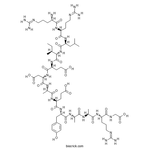
RR-srcTyrosine kinase substrate peptide CAS# 81156-93-6 |

- Perindopril Erbumine
Catalog No.:BCC3586
CAS No.:107133-36-8
- Losartan Potassium (DuP 753)
Catalog No.:BCC1080
CAS No.:124750-99-8
- Candesartan
Catalog No.:BCC2558
CAS No.:139481-59-7
- Telmisattan
Catalog No.:BCC3863
CAS No.:144701-48-4
- Imidapril HCl
Catalog No.:BCC3792
CAS No.:89396-94-1
Quality Control & MSDS
3D structure
Package In Stock
Number of papers citing our products

| Cas No. | 81156-93-6 | SDF | Download SDF |
| PubChem ID | 25078115 | Appearance | Powder |
| Formula | C64H106N22O21 | M.Wt | 1519.7 |
| Type of Compound | N/A | Storage | Desiccate at -20°C |
| Solubility | Soluble to 1 mg/ml in 20% acetonitrile / water | ||
| Sequence | RRLIEDAEYAARG | ||
| Chemical Name | (4S)-4-[[(2S)-2-[[(2S)-2-[[(2S)-2-[[(2S,3S)-2-[[(2S)-2-[[(2S)-2-[[(2S)-2-amino-5-(diaminomethylideneamino)pentanoyl]amino]-5-(diaminomethylideneamino)pentanoyl]amino]-4-methylpentanoyl]amino]-3-methylpentanoyl]amino]-4-carboxybutanoyl]amino]-3-carboxypropanoyl]amino]propanoyl]amino]-5-[[(2S)-1-[[(2S)-1-[[(2S)-1-[[(2S)-1-(carboxymethylamino)-5-(diaminomethylideneamino)-1-oxopentan-2-yl]amino]-1-oxopropan-2-yl]amino]-1-oxopropan-2-yl]amino]-3-(4-hydroxyphenyl)-1-oxopropan-2-yl]amino]-5-oxopentanoic acid | ||
| SMILES | CCC(C)C(C(=O)NC(CCC(=O)O)C(=O)NC(CC(=O)O)C(=O)NC(C)C(=O)NC(CCC(=O)O)C(=O)NC(CC1=CC=C(C=C1)O)C(=O)NC(C)C(=O)NC(C)C(=O)NC(CCCN=C(N)N)C(=O)NCC(=O)O)NC(=O)C(CC(C)C)NC(=O)C(CCCN=C(N)N)NC(=O)C(CCCN=C(N)N)N | ||
| Standard InChIKey | KEOPTZKJOKJEIM-VDNREOAASA-N | ||
| Standard InChI | InChI=1S/C64H106N22O21/c1-8-31(4)49(86-60(106)42(26-30(2)3)83-55(101)39(14-11-25-74-64(70)71)81-53(99)37(65)12-9-23-72-62(66)67)61(107)82-41(20-22-46(90)91)57(103)85-44(28-47(92)93)59(105)78-34(7)52(98)80-40(19-21-45(88)89)56(102)84-43(27-35-15-17-36(87)18-16-35)58(104)77-32(5)50(96)76-33(6)51(97)79-38(13-10-24-73-63(68)69)54(100)75-29-48(94)95/h15-18,30-34,37-44,49,87H,8-14,19-29,65H2,1-7H3,(H,75,100)(H,76,96)(H,77,104)(H,78,105)(H,79,97)(H,80,98)(H,81,99)(H,82,107)(H,83,101)(H,84,102)(H,85,103)(H,86,106)(H,88,89)(H,90,91)(H,92,93)(H,94,95)(H4,66,67,72)(H4,68,69,73)(H4,70,71,74)/t31-,32-,33-,34-,37-,38-,39-,40-,41-,42-,43-,44-,49-/m0/s1 | ||
| General tips | For obtaining a higher solubility , please warm the tube at 37 ℃ and shake it in the ultrasonic bath for a while.Stock solution can be stored below -20℃ for several months. We recommend that you prepare and use the solution on the same day. However, if the test schedule requires, the stock solutions can be prepared in advance, and the stock solution must be sealed and stored below -20℃. In general, the stock solution can be kept for several months. Before use, we recommend that you leave the vial at room temperature for at least an hour before opening it. |
||
| About Packaging | 1. The packaging of the product may be reversed during transportation, cause the high purity compounds to adhere to the neck or cap of the vial.Take the vail out of its packaging and shake gently until the compounds fall to the bottom of the vial. 2. For liquid products, please centrifuge at 500xg to gather the liquid to the bottom of the vial. 3. Try to avoid loss or contamination during the experiment. |
||
| Shipping Condition | Packaging according to customer requirements(5mg, 10mg, 20mg and more). Ship via FedEx, DHL, UPS, EMS or other couriers with RT, or blue ice upon request. | ||
| Description | Tyrosine kinase substrate peptide. |

RR-src Dilution Calculator

RR-src Molarity Calculator

Calcutta University

University of Minnesota

University of Maryland School of Medicine

University of Illinois at Chicago

The Ohio State University

University of Zurich

Harvard University

Colorado State University

Auburn University

Yale University

Worcester Polytechnic Institute

Washington State University

Stanford University

University of Leipzig

Universidade da Beira Interior

The Institute of Cancer Research

Heidelberg University

University of Amsterdam

University of Auckland

TsingHua University

The University of Michigan

Miami University

DRURY University

Jilin University

Fudan University

Wuhan University

Sun Yat-sen University

Universite de Paris

Deemed University

Auckland University

The University of Tokyo

Korea University
- Pravastatin sodium
Catalog No.:BCC2321
CAS No.:81131-70-6
- Cilastatin sodium
Catalog No.:BCC7457
CAS No.:81129-83-1
- (Z)-Lachnophyllum lactone
Catalog No.:BCN4746
CAS No.:81122-95-4
- N-Nonyldeoxynojirimycin
Catalog No.:BCC7752
CAS No.:81117-35-3
- Racecadotril
Catalog No.:BCC4614
CAS No.:81110-73-8
- Clarithromycin
Catalog No.:BCC9219
CAS No.:81103-11-9
- Cisapride
Catalog No.:BCC4207
CAS No.:81098-60-4
- Pravastatin
Catalog No.:BCC4141
CAS No.:81093-37-0
- Kauniolide
Catalog No.:BCC5313
CAS No.:81066-45-7
- EUK 134
Catalog No.:BCC4317
CAS No.:81065-76-1
- Methyl 4-prenyloxycinnamate
Catalog No.:BCN7520
CAS No.:81053-49-8
- 4-Hydroxy-4-(methoxycarbonylmethyl)cyclohexanone
Catalog No.:BCN1346
CAS No.:81053-14-7
- Imiloxan hydrochloride
Catalog No.:BCC6875
CAS No.:81167-22-8
- Apatinib
Catalog No.:BCC5099
CAS No.:811803-05-1
- Boc-D-Tyr(2-Br-Z)-OH
Catalog No.:BCC3464
CAS No.:81189-61-9
- Panaxynol
Catalog No.:BCN3833
CAS No.:81203-57-8
- L-741,626
Catalog No.:BCC6886
CAS No.:81226-60-0
- 15-Deoxoeucosterol
Catalog No.:BCN4348
CAS No.:81241-53-4
- ent-9-Hydroxy-15-oxo-16-kauren-19-oic acid beta-D-glucopyranosyl ester
Catalog No.:BCN1345
CAS No.:81263-96-9
- ent-6,11-Dihydroxy-15-oxo-16-kauren-19-oic acid beta-D-glucopyranosyl ester
Catalog No.:BCN1344
CAS No.:81263-97-0
- ent-6,9-Dihydroxy-15-oxo-16-kauren-19-oic acid beta-D-glucopyranosyl ester
Catalog No.:BCN1343
CAS No.:81263-98-1
- ent-6,9-Dihydroxy-15-oxo-16-kauren-19-oic acid
Catalog No.:BCN1342
CAS No.:81264-00-8
- D-AP7
Catalog No.:BCC6559
CAS No.:81338-23-0
- Amisulpride hydrochloride
Catalog No.:BCC4252
CAS No.:81342-13-4
Lipopolysaccharide-induced priming of the human neutrophil is not associated with a change in phosphotyrosine phosphatase activity.[Pubmed:10399319]
Int J Biochem Cell Biol. 1999 May;31(5):585-93.
The activation of the neutrophil respiratory burst is a two-step process involving an initial 'priming' phase followed by a 'triggering' event. The biochemical mechanisms which underlie these events are yet to be fully elucidated, but the evidence suggests a crucial role for stimulus-induced tyrosine phosphorylation. The enhanced tyrosine phosphorylation observed upon triggering primed cells may reflect an increase in tyrosine kinase activity or a reduction in the levels of the opposing phosphotyrosine phosphatases (PTPases). We have investigated the latter by examining the possibility that lipopolysaccharide (LPS)-induced priming of the neutrophil respiratory burst involves the suppression of cellular PTPase activity. Purified human neutrophils were incubated for 60 min with and without LPS. Priming of the respiratory burst was confirmed by fMet-Leu-Phe-induced cytochrome c reduction. The level of PTPase activity was assessed by dephosphorylation of [32P]RR-src peptide as substrate. Pretreatment of human neutrophils with 200 ng/ml LPS induced a 2.9 +/- 0.3 (mean +/- SEM, n = 3, P = 0.022) fold increase in the fMet-Leu-Phe-triggered respiratory burst. In the same cells, LPS did not induce a significant change in the total cellular PTPase activity (1.02 +/- 0.02-fold, mean +/- SEM, n = 3, P = 0.63). Similarly, stimulation of neutrophils with fMet-Leu-Phe or phorbol myristate acetate did not significantly affect the cellular PTPase activity (P = 0.94 and 0.68, respectively). Our results suggest that suppression of PTPase activity is not the mechanism underlying the priming and/or triggering of the neutrophil respiratory burst.
Interconversion of the kinetic identities of the tandem catalytic domains of receptor-like protein-tyrosine phosphatase PTPalpha by two point mutations is synergistic and substrate-dependent.[Pubmed:9786903]
J Biol Chem. 1998 Oct 30;273(44):28986-93.
The two tandem homologous catalytic domains of PTPalpha possess different kinetic properties, with the membrane proximal domain (D1) exhibiting much higher activity than the membrane distal (D2) domain. Sequence alignment of PTPalpha-D1 and -D2 with the D1 domains of other receptor-like PTPs, and modeling of the PTPalpha-D1 and -D2 structures, identified two non-conserved amino acids in PTPalpha-D2 that may account for its low activity. Mutation of each residue (Val-536 or Glu-671) to conform to its invariant counterpart in PTPalpha-D1 positively affected the catalytic efficiency of PTPalpha-D2 toward the in vitro substrates para-nitrophenylphosphate and the phosphotyrosyl-peptide RR-src. Together, they synergistically transformed PTPalpha-D2 into a phosphatase with catalytic efficiency for para-nitrophenylphosphate equal to PTPalpha-D1 but not approaching that of PTPalpha-D1 for the more complex substrate RR-src. In vivo, no gain in D2 activity toward p59(fyn) was effected by the double mutation. Alteration of the two corresponding invariant residues in PTPalpha-D1 to those in D2 conferred D2-like kinetics toward all substrates. Thus, these two amino acids are critical for interaction with phosphotyrosine but not sufficient to supply PTPalpha-D2 with a D1-like substrate specificity for elements of the phosphotyrosine microenvironment present in RR-src and p59(fyn). Whether the structural features of D2 can uniquely accommodate a specific phosphoprotein substrate or whether D2 has an alternate function in PTPalpha remains an open question.
Phosphotyrosine phosphatase activity in the macrophage is enhanced by lipopolysaccharide, tumor necrosis factor alpha, and granulocyte/macrophage-colony stimulating factor: correlation with priming of the respiratory burst.[Pubmed:9061005]
Biochim Biophys Acta. 1997 Mar 1;1355(3):343-52.
Tyrosine phosphorylation is now recognised as a key event in the activation of the macrophage respiratory burst. Since vanadate, a phosphotyrosine phosphatase (PTP) inhibitor is able to enhance the respiratory burst, we proposed that agents which prime the macrophage for enhance respiratory burst activity may do so by suppressing cellular PTP activity. The level of PTP activity in murine bone marrow-derived macrophages (BMM) was assessed by the ability of cell lysates to dephosphorylate 32P-labelled RR-src peptide. In contrast to our hypothesis, pretreatment of BMM with bacterial lipopolysaccharide (LPS), tumor necrosis factor alpha (TNF alpha) or granulocyte/macrophage-colony stimulating factor (GMCSF), agents which prime for enhanced respiratory burst activity, was found to dramatically increase the level of cellular PTP activity. The time-course for this increase correlated well with the time course of priming by these agents. In addition, colony stimulating factor-1, a cytokine which does not prime the macrophage respiratory burst, did not enhance PTP levels. The physiological relevance of the increased PTP activity was further supported by confirming it was active against endogenous tyrosine phosphorylated substrates. Interestingly, phorbol myristate acetate and zymosan, agents which trigger the macrophage respiratory burst, were found to inhibit the PTP activity of BMM. Our results demonstrate the regulation of cellular PTP activity by priming agents and further highlight the importance of tyrosine phosphorylation and dephosphorylation events in the regulation of macrophage function.
Isolation, sequence and expression of a novel mouse brain cDNA, mIA-2, and its relatedness to members of the protein tyrosine phosphatase family.[Pubmed:7980563]
Biochem Biophys Res Commun. 1994 Oct 28;204(2):930-6.
This study describes the isolation of a putative transmembrane protein tyrosine phosphatase (PTP), mIA-2, from a mouse brain cDNA library. The cDNA encodes 979 amino acids containing a unique extracellular domain and a single intracellular catalytic domain. Expression of mIA-2 was found primarily in the central nervous system and in neuroendocrine cells. The sequence shares a high degree of homology with its human counterpart (92% identity), especially in the intracellular domain, which shows 99.3% identity between the two species. In both human and mouse IA-2, several substitutions were found in the highly conserved regions including an Ala to Asp substitution in the core sequence. Bacterial expression of a glutathione S-transferase fusion protein showed that mIA-2 had no enzyme activity with conventional substrates such as Raytide, myelin basic protein, angiotensin, RR-src and pNpp. When tested with the total tyrosine-phosphorylated cellular proteins isolated on an anti-phosphotyrosine antibody column, it also showed little, if any, enzyme activity. These findings suggest that mIA-2 is a new member of the transmembrane PTP family that either has very narrow substrate specificity perhaps requiring post-translational modification for enzyme activity or has a still unknown biological function.
Modulation of the effector functions of cytolytic T-lymphocytes with synthetic peptide inhibitors of protein kinases.[Pubmed:1619567]
J Pharm Sci. 1992 Jan;81(1):37-44.
The hypothesis was tested that it is possible to influence cellular responses of intact cells using synthetic peptide substrates, pseudosubstrates, and inhibitors of protein kinases. Using cytotoxic T-cells (CTL), we demonstrate here that some basic amino acid-containing synthetic peptide substrates of protein kinases [e.g., of cGMP-dependent protein kinase (peptide PKG-S), synthetic peptide inhibitor of cGMP-dependent protein kinase (peptide PKG-I), and peptide corresponding to the tyrosine phosphorylation site in pp60src (peptide RR-src)] were strongly inhibitory in T-cell receptor (TCR) and T-cell growth factor, interleukin 2 (IL-2)-triggered proliferation of CTL. These peptides also inhibited other cellular responses of CTL. Peptides which contain basic amino acids, but do not have substrate specificity determinants for protein kinase, were not inhibitory. The inhibition with peptides is not due to their toxicity, since no cell death was observed by the trypan blue exclusion test and by lactate dehydrogenase release. Use of the granule exocytosis assay provided opportunities to clarify the mechanism of the peptide action. Tested peptides inhibited not only cell-surface ligand-induced CTL activation, but also affected cell-surface receptor-independent CTL activation (granule exocytosis and gamma-interferon secretion) induced by the synergistic action of the protein kinase C activator (PMA) and ionophore A23187. It was found that minor changes in amino acid composition or amino acid position in the synthetic peptides dramatically change their ability to affect lymphocytes.(ABSTRACT TRUNCATED AT 250 WORDS)
The receptor-like protein tyrosine phosphatase HPTP alpha has two active catalytic domains with distinct substrate specificities.[Pubmed:1915292]
EMBO J. 1991 Nov;10(11):3231-7.
Cloning and expression of the homologous domains of the receptor-like tyrosine phosphatase HPTP alpha shows that both domain 1 (D1) and domain 2 (D2) are enzymatically active. The two domains display different substrate specificities with D1 preferentially dephosphorylating MBP approximately RR-src greater than PNPP while D2 favours PNPP much much greater than RR-src and is inactive towards MBP. Each domain has lower activity than an expressed protein containing both domains. Analysis of chimaeric D1/2 proteins suggests that no particular region of D2 is responsible for the low activity of D2 on RR-src and that the specificity differences of D1 and D2 reflect overall sequence dissimilarities. Activities of D1 and D2 are inhibited by zinc, vanadate and EDTA and differentially susceptible to inhibition by heparin and poly(Glu4:Tyr1). Unusually, the activity of the protein containing both domains is stimulated by these polyanions. Regions amino-terminal to each domain are important for catalysis since deletion of these sequences abolishes phosphatase activity. Activity of the double domain polypeptide was also lost upon deletion of the sequence amino-terminal to D1, indicating that inactivation of D1 may suppress D2 activity. Differences in substrate specificity and responses to effectors and the interdependence between the two domains are likely important properties in the function of this PTPase in signal transduction.
Specificity of protein phosphotyrosine phosphatases. Comparison with mammalian alkaline phosphatase using polypeptide substrates.[Pubmed:2982803]
J Biol Chem. 1985 Feb 25;260(4):2042-5.
The specificity of cytosolic protein phosphotyrosine (PPT) phosphatases was investigated using different peptides and proteins that were phosphorylated on tyrosine residues by the EGF receptor kinase. The acidic phosphoproteins, serum albumin, casein, and myosin light chains, were dephosphorylated by the PPT phosphatases with apparent Km values of 1.2 to 12.5 microM and apparent velocities of 0.2 to 18 mumol/min/mg. In contrast, [Tyr(32P)]histone and the phosphotyrosine peptides [Val5]angiotensin and RR-src, a peptide with sequence Arg-Arg-Leu-Ile-Glu-Asp-Ala-Glu-Tyr-Ala-Ala-Arg-Gly, were unreactive with the PPT phosphatases. However, each of these unreactive phosphopolypeptides was dephosphorylated under the same conditions by calf-intestine alkaline phosphatase. The data reveal how PPT phosphatase activity has been ascribed to different cellular enzymes. When acidic phosphotyrosine proteins were used as substrates in assays for PPT phosphatase activity the cytosolic enzymes were isolated, whereas when phosphotyrosine histones were used as substrates only the membrane-bound alkaline phosphatase was detected. Apparently the protein tyrosine kinase and the protein tyrosine phosphatases do not have the same specificity, so substrates such as histone, angiotensin, or RR-src are phosphorylated but not hydrolyzed. Therefore, these polypeptides would be ideal for the characterization of protein tyrosine kinases in cellular extracts.


