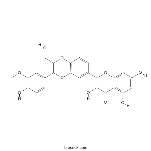
SilymarinCAS# 22888-70-6 |

Quality Control & MSDS
3D structure
Package In Stock
Number of papers citing our products

| Cas No. | 22888-70-6 | SDF | Download SDF |
| PubChem ID | 5213 | Appearance | White powder |
| Formula | C25H22O10 | M.Wt | 482.46 |
| Type of Compound | Flavonoids | Storage | Desiccate at -20°C |
| Synonyms | Silybin; Silibinin A; Silymarin I | ||
| Solubility | DMSO : ≥ 100 mg/mL (207.28 mM) *"≥" means soluble, but saturation unknown. | ||
| Chemical Name | 3,5,7-trihydroxy-2-[3-(4-hydroxy-3-methoxyphenyl)-2-(hydroxymethyl)-2,3-dihydro-1,4-benzodioxin-6-yl]-2,3-dihydrochromen-4-one | ||
| SMILES | COC1=C(C=CC(=C1)C2C(OC3=C(O2)C=C(C=C3)C4C(C(=O)C5=C(C=C(C=C5O4)O)O)O)CO)O | ||
| Standard InChIKey | SEBFKMXJBCUCAI-UHFFFAOYSA-N | ||
| Standard InChI | InChI=1S/C25H22O10/c1-32-17-6-11(2-4-14(17)28)24-20(10-26)33-16-5-3-12(7-18(16)34-24)25-23(31)22(30)21-15(29)8-13(27)9-19(21)35-25/h2-9,20,23-29,31H,10H2,1H3 | ||
| General tips | For obtaining a higher solubility , please warm the tube at 37 ℃ and shake it in the ultrasonic bath for a while.Stock solution can be stored below -20℃ for several months. We recommend that you prepare and use the solution on the same day. However, if the test schedule requires, the stock solutions can be prepared in advance, and the stock solution must be sealed and stored below -20℃. In general, the stock solution can be kept for several months. Before use, we recommend that you leave the vial at room temperature for at least an hour before opening it. |
||
| About Packaging | 1. The packaging of the product may be reversed during transportation, cause the high purity compounds to adhere to the neck or cap of the vial.Take the vail out of its packaging and shake gently until the compounds fall to the bottom of the vial. 2. For liquid products, please centrifuge at 500xg to gather the liquid to the bottom of the vial. 3. Try to avoid loss or contamination during the experiment. |
||
| Shipping Condition | Packaging according to customer requirements(5mg, 10mg, 20mg and more). Ship via FedEx, DHL, UPS, EMS or other couriers with RT, or blue ice upon request. | ||
| Description | Silymarin possesses hepatoprotective, antioxidant, anti-inflammatory and immunomodulatory properties. It is an effective anti-cancer and chemopreventive agent, has been shown to exert multiple effects on cancer cells, including inhibition of both cell proliferation and migration. Silymarin induces apoptosis primarily through a p53-dependent pathway involving Bcl-2/Bax, cytochrome c release, and caspase activation. It inhibits PGE2 -induced cell migration through inhibition of EP2 signaling pathways (G protein dependent PKA-CREB and G protein-independent Src-STAT3). |
| Targets | PGE | PKA | Src | STAT | ROS | Bcl-2/Bax | Caspase | p53 |
| In vitro | Silymarin suppresses the PGE2 -induced cell migration through inhibition of EP2 activation; G protein-dependent PKA-CREB and G protein-independent Src-STAT3 signal pathways.[Pubmed: 24127286]Mol Carcinog. 2015 Mar;54(3):216-28.Silymarin has been known as a chemopreventive agent, and possesses multiple anti-cancer activities including induction of apoptosis, inhibition of proliferation and growth, and blockade of migration and invasion. However, whether Silymarin could inhibit prostaglandin (PG) E2 -induced renal cell carcinoma (RCC) migration and what are the underlying mechanisms are not well elucidated.
|
| In vivo | Silymarin liposomes improves oral bioavailability of silybin besides targeting hepatocytes, and immune cells.[Pubmed: 25149982]Pharmacol Rep. 2014 Oct;66(5):788-98.Silymarin, a hepatoprotective agent, has poor oral bioavailability. However, the current dosage form of the drug does not target the liver and inflammatory cells selectively. The aim of the present study was to develop lecithin-based carrier system of Silymarin by incorporating phytosomal-liposomal approach to increase its oral bioavailability and to make it target-specific to the liver for enhanced hepatoprotection.
Silymarin and skin cancer prevention: anti-inflammatory, antioxidant and immunomodulatory effects (Review).[Pubmed: 15586237]Int J Oncol. 2005 Jan;26(1):169-76.Several environmental and genetic factors are involved in skin cancer induction, however exposure to chemical carcinogens and solar ultraviolet (UV) radiation are primarily responsible for several skin diseases including skin cancer. Chronic exposure of solar UV radiation to the skin leads to basal cell and squamous cell carcinoma, and melanoma. Chemoprevention of skin cancer by consumption of naturally occurring botanicals appears a practical approach and therefore world-wide interest is considerably increasing to use these botanicals. Sunscreens are useful but their protection is not ideal because of inadequate use, incomplete spectral protection and toxicity.
|
| Cell Research | Silymarin induces apoptosis primarily through a p53-dependent pathway involving Bcl-2/Bax, cytochrome c release, and caspase activation.[Pubmed: 15713892]Mol Cancer Ther. 2005 Feb;4(2):207-16.Silymarin, a plant flavonoid, has been shown to inhibit skin carcinogenesis in mice. However, the mechanism responsible for the anti-skin carcinogenic effects of Silymarin is not clearly understood.
|
| Animal Research | Cytoprotective effect of silymarin against diabetes-induced cardiomyocyte apoptosis in diabetic rats.[Pubmed: 25566861]Biomed Environ Sci. 2015 Jan;28(1):36-43.The beneficial effects of Silymarin have been extensively studied in the context of inflammation and cancer treatment, yet much less is known about its therapeutic effect on diabetes. The present study was aimed to investigate the cytoprotective activity of Silymarin against diabetes-induced cardiomyocyte apoptosis.
|

Silymarin Dilution Calculator

Silymarin Molarity Calculator
| 1 mg | 5 mg | 10 mg | 20 mg | 25 mg | |
| 1 mM | 2.0727 mL | 10.3636 mL | 20.7271 mL | 41.4542 mL | 51.8178 mL |
| 5 mM | 0.4145 mL | 2.0727 mL | 4.1454 mL | 8.2908 mL | 10.3636 mL |
| 10 mM | 0.2073 mL | 1.0364 mL | 2.0727 mL | 4.1454 mL | 5.1818 mL |
| 50 mM | 0.0415 mL | 0.2073 mL | 0.4145 mL | 0.8291 mL | 1.0364 mL |
| 100 mM | 0.0207 mL | 0.1036 mL | 0.2073 mL | 0.4145 mL | 0.5182 mL |
| * Note: If you are in the process of experiment, it's necessary to make the dilution ratios of the samples. The dilution data above is only for reference. Normally, it's can get a better solubility within lower of Concentrations. | |||||

Calcutta University

University of Minnesota

University of Maryland School of Medicine

University of Illinois at Chicago

The Ohio State University

University of Zurich

Harvard University

Colorado State University

Auburn University

Yale University

Worcester Polytechnic Institute

Washington State University

Stanford University

University of Leipzig

Universidade da Beira Interior

The Institute of Cancer Research

Heidelberg University

University of Amsterdam

University of Auckland

TsingHua University

The University of Michigan

Miami University

DRURY University

Jilin University

Fudan University

Wuhan University

Sun Yat-sen University

Universite de Paris

Deemed University

Auckland University

The University of Tokyo

Korea University
Silibinin, an effective anti-cancer and chemopreventive agent, has been shown to exert multiple effects on cancer cells, including inhibition of both cell proliferation and migration. IC50 value: Target: anticancer in vitro: silibinin significantly induced the expression of the non-steroidal anti-inflammatory drug-activated gene-1 (NAG-1) in both p53 wild-type and p53-null cancer cell lines, suggesting that silibinin-induced NAG-1 up-regulation is p53-independent manner.Silibinin up-regulates early growth response-1 (EGR-1) expression [1]. silibinin induced cell death in human breast cancer cell lines MCF7 and MDA-MB-231. Silibinininduced cell death was attenuated by antioxidants, N-acetylcysteine (NAC) and Trolox, suggesting that the effect of silibinin was dependent on generation of reactive oxygen species (ROS) [2]. SIL treatment resulted in a dose- and time-dependent inhibition of HCC cell viability, SIL exhibited strong antitumor activity, as evidenced not only by reductions in tumor cell adhesion, migration, intracellular glutathione (GSH) levels and total antioxidant capability (T-AOC) but also by increases in the apoptotic index, caspase3 activity, and reactive oxygen species (ROS). SIL treatment decreased the expression of the Notch1 intracellular domain (NICD), RBP-Jκ, and Hes1 proteins, upregulated the apoptosis pathway-related protein Bax, and downregulated Bcl2, survivin, and cyclin D1. Notch1 siRNA (in vitro) or DAPT (a known Notch1 inhibitor, in vivo) further enhanced the antitumor activity of SIL, and recombinant Jagged1 protein (a known Notch ligand in vitro) attenuated the antitumor activity of SIL [3]. in vivo: Topical application of silibinin at the dose of 9 mg/mouse effectively suppressed oxidative stress and deregulated activation of inflammatory mediators and tumorigenesis[4]. The kidney cortex of vehicle-treated control OVE26 mice displayed greater Nox4 expression and twice as much superoxide production than cortex of silybin-treated mice. The glomeruli of control OVE26 mice displayed 35% podocyte drop out that was not present in the silybin-treated mice [5].
References:
[1]. Woo SM, et al. Silibinin induces apoptosis of HT29 colon carcinoma cells through early growth response-1 (EGR-1)-mediated non-steroidal anti-inflammatory drug-activated gene-1 (NAG-1) up-regulation. Chem Biol Interact. 2014 Jan 16;211C:36-43.
[2]. Kim TH, et al. Silibinin induces cell death through ROS-dependent down-regulation of Notch-1/ERK/Akt signaling in human breast cancer cells. J Pharmacol Exp Ther. 2014 Jan 28.
[3]. Zhang S, et al. Silybin-mediated inhibition of Notch signaling exerts antitumor activity in human hepatocellular carcinoma cells. PLoS One. 2013 Dec 27;8(12):e83699.
[4]. Khan AQ, et al. Silibinin Inhibits Tumor Promotional Triggers and Tumorigenesis Against Chemically Induced Two-Stage Skin Carcinogenesis in Swiss Albino Mice: Possible Role of Oxidative Stress and Inflammation. Nutr Cancer. 2013 Dec 23.
[5]. Khazim K, et al. The antioxidant silybin prevents high glucose-induced oxidative stress and podocyte injury in vitro and in vivo. Am J Physiol Renal Physiol. 2013 Sep 1;305(5):F691-700.
- Famprofazone
Catalog No.:BCC3779
CAS No.:22881-35-2
- 6-Acetonyldihydrochelerythrine
Catalog No.:BCN5076
CAS No.:22864-92-2
- Anisomycin
Catalog No.:BCC7007
CAS No.:22862-76-6
- Ki8751
Catalog No.:BCC1116
CAS No.:228559-41-9
- VULM 1457
Catalog No.:BCC7533
CAS No.:228544-65-8
- 9-Epiblumenol B
Catalog No.:BCN5075
CAS No.:22841-42-5
- Pratensein
Catalog No.:BCN2918
CAS No.:2284-31-3
- Aspartame
Catalog No.:BCC8836
CAS No.:22839-47-0
- Boc-D-Val-OH
Catalog No.:BCC3466
CAS No.:22838-58-0
- Miconazole nitrate
Catalog No.:BCC9047
CAS No.:22832-87-7
- Hypoglaunine A
Catalog No.:BCN3086
CAS No.:228259-16-3
- Boc-Met-OH.DCHA
Catalog No.:BCC2602
CAS No.:22823-50-3
- 4-Amino-3,5-dichloropyridine
Catalog No.:BCC8679
CAS No.:22889-78-7
- TAK-779
Catalog No.:BCC4137
CAS No.:229005-80-5
- R 892
Catalog No.:BCC5992
CAS No.:229030-05-1
- Ginkgolic acid C15:1
Catalog No.:BCN2307
CAS No.:22910-60-7
- Neferine
Catalog No.:BCN6338
CAS No.:2292-16-2
- Desmethoxycentaureidin
Catalog No.:BCN5077
CAS No.:22934-99-2
- Abn-CBD
Catalog No.:BCC7011
CAS No.:22972-55-0
- GW9662
Catalog No.:BCC2260
CAS No.:22978-25-2
- AGN 194310
Catalog No.:BCC5416
CAS No.:229961-45-9
- Atazanavir sulfate (BMS-232632-05)
Catalog No.:BCC2114
CAS No.:229975-97-7
- MPTP hydrochloride
Catalog No.:BCC1778
CAS No.:23007-85-4
- Neoglycyrol
Catalog No.:BCN2907
CAS No.:23013-84-5
Silymarin suppresses the PGE2 -induced cell migration through inhibition of EP2 activation; G protein-dependent PKA-CREB and G protein-independent Src-STAT3 signal pathways.[Pubmed:24127286]
Mol Carcinog. 2015 Mar;54(3):216-28.
Silymarin has been known as a chemopreventive agent, and possesses multiple anti-cancer activities including induction of apoptosis, inhibition of proliferation and growth, and blockade of migration and invasion. However, whether Silymarin could inhibit prostaglandin (PG) E2 -induced renal cell carcinoma (RCC) migration and what are the underlying mechanisms are not well elucidated. Here, we found that Silymarin markedly inhibited PGE2 -stimulated migration. PGE2 induced G protein-dependent CREB phosphorylation via protein kinase A (PKA) signaling, and PKA inhibitor (H89) inhibited PGE2 -mediated migration. Silymarin reduced PGE2 -induced CREB phosphorylation and CRE-promoter activity. PGE2 also activated G protien-independent signaling pathways (Src and STAT3) and Silymarin reduced PGE2 -induced phosphorylation of Src and STAT3. Inhibitor of Src (Saracatinib) markedly reduced PGE2 -mediated migration. We found that EP2, a PGE2 receptor, is involved in PGE2 -mediated cell migration. Down regulation of EP2 by EP2 siRNA and EP2 antagonist (AH6809) reduced PGE2 -inudced migration. In contrast, EP2 agonist (Butaprost) increased cell migration and Silymarin effectively reduced butaprost-mediated cell migration. Moreover, PGE2 increased EP2 expression through activation of positive feedback mechanism, and PGE2 -induced EP2 expression, as well as basal EP2 levels, were reduced in Silymarin-treated cells. Taken together, our study demonstrates that Silymarin inhibited PGE2 -induced cell migration through inhibition of EP2 signaling pathways (G protein dependent PKA-CREB and G protein-independent Src-STAT3).
Cytoprotective effect of silymarin against diabetes-induced cardiomyocyte apoptosis in diabetic rats.[Pubmed:25566861]
Biomed Environ Sci. 2015 Jan;28(1):36-43.
OBJECTIVE: The beneficial effects of Silymarin have been extensively studied in the context of inflammation and cancer treatment, yet much less is known about its therapeutic effect on diabetes. The present study was aimed to investigate the cytoprotective activity of Silymarin against diabetes-induced cardiomyocyte apoptosis. METHODS: Rats were randomly divided into: control group, untreated diabetes group and diabetes group treated with Silymarin (120 mg/kg*d) for 10 d. Rats were sacrificed, and the cardiac muscle specimens and blood samples were collected. The immunoreactivity of caspase-3 and Bcl-2 in the cardiomyocytes was measured. Total proteins, glucose, insulin, creatinine, AST, ALT, cholesterol, and triglycerides levels were estimated. RESULTS: Unlike the treated diabetes group, cardiomyocyte apoptosis increased in the untreated rats, as evidenced by enhanced caspase-3 and declined Bcl-2 activities. The levels of glucose, creatinine, AST, ALT, cholesterol, and triglycerides declined in the treated rats. The declined levels of insulin were enhanced again after treatment of diabetic rats with Silymarin, reflecting a restoration of the pancreatic beta-cells activity. CONCLUSION: The findings of this study are of great importance, which confirmed for the first time that treatment of diabetic subjects with Silymarin may protect cardiomyocytes against apoptosis and promote survival-restoration of the pancreatic beta-cells.
Silymarin and skin cancer prevention: anti-inflammatory, antioxidant and immunomodulatory effects (Review).[Pubmed:15586237]
Int J Oncol. 2005 Jan;26(1):169-76.
Several environmental and genetic factors are involved in skin cancer induction, however exposure to chemical carcinogens and solar ultraviolet (UV) radiation are primarily responsible for several skin diseases including skin cancer. Chronic exposure of solar UV radiation to the skin leads to basal cell and squamous cell carcinoma, and melanoma. Chemoprevention of skin cancer by consumption of naturally occurring botanicals appears a practical approach and therefore world-wide interest is considerably increasing to use these botanicals. Sunscreens are useful but their protection is not ideal because of inadequate use, incomplete spectral protection and toxicity. Silymarin, a plant flavonoid isolated from the seeds of milk thistle (Silybum marianum), has been shown to have chemopreventive effects against chemical carcinogenesis as well as photocarcinogenesis in various animal tumor models. Topical treatment of Silymarin inhibited 7,12-dimethylbenz(a)anthracene-initiated and several tumor promoters, like 12-O-tetradecanoylphorbol-13-acetate, mezerein, benzoyal peroxide and okadaic acid, induced skin carcinogenesis in mouse models. Similarly, Silymarin also prevented UVB-induced skin carcinogenesis. Wide range of in vivo mechanistic studies indicated that Silymarin possesses antioxidant, anti-inflammatory and immunomodulatory properties which may lead to the prevention of skin cancer in in vivo animal models. The available experimental information suggests that Silymarin is a promising chemopreventive and pharmacologically safe agent which can be exploited or tested against skin cancer in human system. Moreover, Silymarin may favorably supplement sunscreen protection and provide additional anti-photocarcinogenic protection.
Silymarin liposomes improves oral bioavailability of silybin besides targeting hepatocytes, and immune cells.[Pubmed:25149982]
Pharmacol Rep. 2014 Oct;66(5):788-98.
BACKGROUND: Silymarin, a hepatoprotective agent, has poor oral bioavailability. However, the current dosage form of the drug does not target the liver and inflammatory cells selectively. The aim of the present study was to develop lecithin-based carrier system of Silymarin by incorporating phytosomal-liposomal approach to increase its oral bioavailability and to make it target-specific to the liver for enhanced hepatoprotection. METHODS: The formulation was prepared by film hydration method. Release of drug was assessed at pH 1.2 and 7.4. Formulation was assessed for in vitro hepatoprotection on Chang liver cells, lipopolysaccharide-induced reactive oxygen species (ROS) production by RAW 267.4 (murine macrophages), in vivo efficacy against paracetamol-induced hepatotoxicity and pharmacokinetic study by oral route in Wistar rat. RESULTS: The formulation showed maximum entrapment (55%) for a lecithin-cholesterol ratio of 6:1. Comparative release profile of formulation was better than Silymarin at pH 1.2 and pH 7.4. In vitro studies showed a better hepatoprotection efficacy for formulation (one and half times) and better prevention of ROS production (ten times) compared to Silymarin. In in vivo model, paracetamol showed significant hepatotoxicity in Wistar rats assessed through LFT, antioxidant markers and inflammatory markers. The formulation was found more efficacious than Silymarin suspension in protecting the liver against paracetamol toxicity and the associated inflammatory conditions. The liposomal formulation yielded a three and half fold higher bioavailability of Silymarin as compared with Silymarin suspension. CONCLUSIONS: Incorporating the phytosomal form of Silymarin in liposomal carrier system increased the oral bioavailability and showed better hepatoprotection and better anti-inflammatory effects compared with Silymarin suspension.
Silymarin induces apoptosis primarily through a p53-dependent pathway involving Bcl-2/Bax, cytochrome c release, and caspase activation.[Pubmed:15713892]
Mol Cancer Ther. 2005 Feb;4(2):207-16.
Silymarin, a plant flavonoid, has been shown to inhibit skin carcinogenesis in mice. However, the mechanism responsible for the anti-skin carcinogenic effects of Silymarin is not clearly understood. Here, we report that treatment of JB6 C141 cells (preneoplastic epidermal keratinocytes) and p53+/+ fibroblasts with Silymarin and silibinin (a major constituent of Silymarin) resulted in a dose-dependent inhibition of cell viability and induction of apoptosis in an identical manner. Silymarin-induced apoptosis was determined by fluorescence staining (8-64% apoptosis) and flow cytometry (12-76% apoptosis). The Silymarin-induced apoptosis was primarily p53 dependent because apoptosis occurred to a much greater extent in the cells expressing wild-type p53 (p53+/+, 9-61%) than in p53-deficient cells (p53-/-, 6-20%). The induction of apoptosis in JB6 C141 cells was associated with increased expression of the tumor suppressor protein, p53, and its phosphorylation at Ser15. The constitutive expression of antiapoptotic proteins Bcl-2 and Bcl-xl were decreased after Silymarin treatment, whereas the expression of the proapoptotic protein Bax was increased. There was a shift in Bax/Bcl-2 ratio in favor of apoptotic signal in Silymarin-treated cells, which resulted in increased levels of cytochrome c release, apoptotic protease-activating factor-1, and cleaved caspase-3 and poly(ADP-ribose) polymerase in JB6 C141 cells. The shift in Bax/Bcl-2 ratio was more prominent in p53+/+ fibroblasts than in p53-/- cells. Silymarin-induced apoptosis was blocked by the caspase inhibitor (Z-VAD-FMK) in JB6 C141 cells which suggested the role of caspase activation in the induction of apoptosis. These observations show that Silymarin-induced apoptosis is primarily p53 dependent and mediated through the activation of caspase-3.


