
Acanthoic acidCAS# 119290-87-8 |
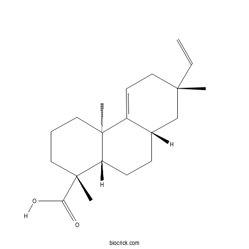
| Cas No. | 119290-87-8 | SDF | File under preparation. |
| PubChem ID | 9817887.0 | Appearance | Powder |
| Formula | C20H30O2 | M.Wt | 302.4 |
| Type of Compound | N/A | Storage | Desiccate at -20°C |
| Solubility | Soluble in Chloroform,Dichloromethane,Ethyl Acetate,DMSO,Acetone,etc. | ||
| Chemical Name | (1R,4aR,7S,8aS,10aS)-7-ethenyl-1,4a,7-trimethyl-3,4,6,8,8a,9,10,10a-octahydro-2H-phenanthrene-1-carboxylic acid | ||
| SMILES | CC12CCCC(C1CCC3C2=CCC(C3)(C)C=C)(C)C(=O)O | ||
| Standard InChIKey | TVHDZSRRHQKNEZ-MGFONVBGSA-N | ||
| Standard InChI | InChI=1S/C20H30O2/c1-5-18(2)12-9-15-14(13-18)7-8-16-19(15,3)10-6-11-20(16,4)17(21)22/h5,9,14,16H,1,6-8,10-13H2,2-4H3,(H,21,22)/t14-,16-,18-,19-,20+/m0/s1 | ||
| General tips | For obtaining a higher solubility , please warm the tube at 37 ℃ and shake it in the ultrasonic bath for a while.Stock solution can be stored below -20℃ for several months. We recommend that you prepare and use the solution on the same day. However, if the test schedule requires, the stock solutions can be prepared in advance, and the stock solution must be sealed and stored below -20℃. In general, the stock solution can be kept for several months. Before use, we recommend that you leave the vial at room temperature for at least an hour before opening it. |
||
| About Packaging | 1. The packaging of the product may be reversed during transportation, cause the high purity compounds to adhere to the neck or cap of the vial.Take the vail out of its packaging and shake gently until the compounds fall to the bottom of the vial. 2. For liquid products, please centrifuge at 500xg to gather the liquid to the bottom of the vial. 3. Try to avoid loss or contamination during the experiment. |
||
| Shipping Condition | Packaging according to customer requirements(5mg, 10mg, 20mg and more). Ship via FedEx, DHL, UPS, EMS or other couriers with RT, or blue ice upon request. | ||
Quality Control & MSDS
3D structure
Package In Stock
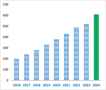
Number of papers citing our products

Acanthoic acid Dilution Calculator
Acanthoic acid Molarity Calculator
| 1 mg | 5 mg | 10 mg | 20 mg | 25 mg | |
| 1 mM | 3.3069 mL | 16.5344 mL | 33.0688 mL | 66.1376 mL | 82.672 mL |
| 5 mM | 0.6614 mL | 3.3069 mL | 6.6138 mL | 13.2275 mL | 16.5344 mL |
| 10 mM | 0.3307 mL | 1.6534 mL | 3.3069 mL | 6.6138 mL | 8.2672 mL |
| 50 mM | 0.0661 mL | 0.3307 mL | 0.6614 mL | 1.3228 mL | 1.6534 mL |
| 100 mM | 0.0331 mL | 0.1653 mL | 0.3307 mL | 0.6614 mL | 0.8267 mL |
| * Note: If you are in the process of experiment, it's necessary to make the dilution ratios of the samples. The dilution data above is only for reference. Normally, it's can get a better solubility within lower of Concentrations. | |||||
- 1,2-Dilinoleoyl-3-palmitin
Catalog No.:BCX2844
CAS No.:2190-15-0
- Amygdalinic acid
Catalog No.:BCX2843
CAS No.:66427-92-7
- Benzyl β-gentiobioside
Catalog No.:BCX2842
CAS No.:56775-64-5
- Xanthalin
Catalog No.:BCX2841
CAS No.:21800-48-6
- Apigenin-7-O-(6′′-E-p-coumaroyl-β-D-glucopyranoside)
Catalog No.:BCX2840
CAS No.:105815-90-5
- (E)-Ligustilide
Catalog No.:BCX2839
CAS No.:81944-08-3
- β-Curcumene
Catalog No.:BCX2838
CAS No.:451-56-9
- Cistanoside C
Catalog No.:BCX2837
CAS No.:94492-22-5
- (7S)-Hellicoside
Catalog No.:BCX2836
CAS No.:132278-04-7
- 3-Butylidene-7-hydroxyphthalide
Catalog No.:BCX2835
CAS No.:93236-67-0
- Molephantinin
Catalog No.:BCX2834
CAS No.:56221-98-8
- Molephantin
Catalog No.:BCX2833
CAS No.:50656-66-1
- Penicillide
Catalog No.:BCX2846
CAS No.:55303-92-9
- Gingerglycolipid B
Catalog No.:BCX2847
CAS No.:88168-90-5
- Atractylodinol
Catalog No.:BCX2848
CAS No.:61642-89-5
- Dendronobiloside A
Catalog No.:BCX2849
CAS No.:370557-49-6
- N-Methylhernovine
Catalog No.:BCX2850
CAS No.:5550-65-2
- Hosenkoside L
Catalog No.:BCX2851
CAS No.:161016-50-8
- 1,3-Dioleoyl-2-linoleoylglycerol
Catalog No.:BCX2852
CAS No.:2190-19-4
- (-)-3-O -Acetylcatechin
Catalog No.:BCX2853
CAS No.:116935-88-7
- 4-Hydroxy-3-butylphthalide
Catalog No.:BCX2854
CAS No.:74459-23-7
- (Z)-α-Santalol
Catalog No.:BCX2855
CAS No.:115-71-9
- Tenuifoliose Q
Catalog No.:BCX2856
CAS No.:662166-93-0
- (-)-Epigallocatechin 3'-O-methylether gallate
Catalog No.:BCX2857
CAS No.:298700-56-8
Biological activities of acanthoic acid and its pharmacological potential: a literature review.[Pubmed:40255093]
Nat Prod Res. 2026 Sep;40(18):5808-5824.
Plant-derived compounds can influence human physiology and have been harnessed through infusions and teas, which addressed human health since prehistory. Thus, Acanthoic acid is a natural pimaradiene diterpene found in species of the Annonaceae family. This botanical family includes fruit-bearing trees such as cherimoyas, custard apples, soursops, and rollinia, popularly using these species for antiparasitic, anti-leishmanial, antimicrobial, cytotoxic, and antioxidant purposes. This review aims to consolidate key findings in the literature regarding Acanthoic acid and its biological activities. Original articles published in English between the years 2013 and 2023 were included. The results indicated that only 15 articles were identified during the investigated period, all focused on preclinical or in silico studies. Among the main effects observed, antimicrobial, anti-inflammatory, hepatoprotective, and antitumor activities were highlighted. However, additional studies examining other facets are imperative, including toxicity, pharmacokinetics, and systemic effects, such as those on the central nervous and renal systems.
Gouregine, an alpha-Gem-Dimethyltetradehydrocularine Alkaloid, and Other Aporphinoid Alkaloids from the Bark of Guatteria olivacea (Annonaceae) and Their In Vitro Cytotoxic Activities.[Pubmed:39202913]
Molecules. 2024 Aug 13;29(16):3834.
Guatteria olivacea R.E. Fries is an Amazonian species known as 'envira-bobo' and 'envira-fofa' and is common in the states of Amazonas, Acre, and Para. Recently, the essential oil from the leaves of this species has shown promising antitumor activity both in vitro and in vivo. The presence of isoquinoline-derived alkaloids, including aporphinoids and tetrahydroprotoberberine alkaloids, has also been previously reported. In our ongoing search for bioactive compounds from Annonaceae Amazonian plants, the bark of G. olivacea was investigated via classical chromatography techniques, which revealed nine compounds, eight isoquinoline-derived alkaloids, a rare alkaloid with a alpha-gem-dimethyltetradehydrocularine structure known as gouregine, seven known aporphinoid alkaloids: isopiline, O-methylisopiline, melosmine, 9-hydroxyiguattescine, dihydromelosmine, lysicamine, and guattouregidine, and one known pimaradiene diterpene: Acanthoic acid. All the isolated compounds were described for the first time in the bark of G. olivacea, and their structures were elucidated by extensive analyses of their 1D and 2D NMR spectra in combination with MS data. The NMR data of the alkaloids isopiline, O-methylisopiline, melosmine, dihydromelosmine, and guattouregidine were revised due to incomplete data in the literature and some ambiguities. The in vitro cytotoxic activities of the isolated compounds were evaluated against human cancer (HepG2, KG-1a, and HCT116) and noncancerous (MRC-5) cell lines via the Alamar blue assay after 72 h of incubation. Among the compounds evaluated against human cancer cell lines, the most active was the oxoaporphine alkaloid lysicamine, which has strong activity against HCT116 cells, with an IC(50) value of 6.64 microg/mL (22.79 micromol/L). Melosmine had a moderate effect on HCT116 cells, with an IC(50) value of 16.77 microg/mL (49.70 micromol/L), whereas Acanthoic acid had moderate effects on HepG2 and HCT116 cells, with IC(50) values of 14.63 microg/mL (48.37 micromol/L) and 21.25 microg/mL (70.25 micromol/L), respectively.
Assessing the Efficacy of Acanthoic Acid Isolated from Acanthopanax koreanum Nakai in Male Infertility: An In Vivo and In Silico Approach.[Pubmed:39057081]
Curr Issues Mol Biol. 2024 Jul 13;46(7):7411-7429.
Acanthoic acid, a diterpene isolated from the root bark of Acanthopanax koreanum Nakai, possesses diverse pharmacological activities, including anti-inflammatory, anti-diabetic, gastrointestinal protection, and cardiovascular protection. This study is the first to investigate the egg-hatching rates of Drosophila melanogaster affected by Acanthoic acid. Notably, male flies supplemented with 10 muM Acanthoic acid exhibited a strong increase in hatching rates compared with controls under adverse temperature conditions, suggesting a potential protective effect against environmental stressors. Molecular docking simulations revealed the binding affinities and specific interactions between Acanthoic acid and proteins related to male infertility, including SHBG, ADAM17, and DNase I, with binding affinity values of -10.2, -6.8, and -5.8 kcal/mol, respectively. Following the docking studies, molecular dynamic simulations were conducted for a duration of 100 ns to examine the stability of these interactions. Additionally, a total binding energy analysis and decomposition analysis offered insights into the underlying energetic components and identified key contributing residues.
Antimicrobial Activity and Molecular Docking Studies of the Biotransformation of Diterpene Acanthoic Acid Using the Fungus Xylaria sp.[Pubmed:37627751]
Antibiotics (Basel). 2023 Aug 18;12(8):1331.
Biotransformations are reactions mediated by microorganisms, such as fungi. These bioreactions have high chemo- and stereoselectivity on organic substrates and can be applied in the search for new bioactive compounds. In this study, Acanthoic acid (AA) was biotransformed using the fungus Xylaria sp., giving the novel compound 3beta,7beta-dihydroxyAcanthoic acid (S1). Both the AA and the product S1 were tested against Gram-positive and Gram-negative bacteria. To identify and validate possible biological targets as enzymes or proteins involved in the activity observed in vitro, we used the molecular docking method. Hydroxylation at the C-3 and C-7 positions of the biotransformation product enhanced its activity against Escherichia coli as well as its binding affinity and interactions with superoxide dismutase 1 (SOD1; PDB ID 4A7G). Based on our results, the SOD1 enzyme was suggested to be a possible target for the antioxidant activity of product S1.
Identification of the antibacterial action mechanism of diterpenoids through transcriptome profiling.[Pubmed:35958135]
Front Microbiol. 2022 Jul 26;13:945023.
Effective antibacterial substances of Aralia continentalis have anti-biofilm and bactericidal activity to the oral pathogen Streptococcus mutans. In this study, three compounds extracted from A. continentalis were identified as Acanthoic acid, continentalic acid, and kaurenoic acid by NMR and were further investigated how these diterpenoids affect the physiology of the S. mutans. When S. mutans was exposed to individual or mixed fraction of diterpenoids, severe growth defects and unique morphology were observed. The proportion of unsaturated fatty acids in the cell membrane was increased compared to that of saturated fatty acids in the presence of diterpenoids. Genome-wide gene expression profiles with RNA-seq were compared to reveal the mode of action of diterpenoids. Streptococcus mutans commonly enhanced the expression of 176 genes in the presence of the individual diterpenoids, whereas the expression of 232 genes was considerably reduced. The diterpenoid treatment modulated the expression of genes or operon(s) involved in cell membrane synthesis, cell division, and carbohydrate metabolism of S. mutans. Collectively, these findings provide novel insights into the antibacterial effect of diterpenoids to control S. mutans infection, which causes human dental caries.
Acanthoic acid, unique potential pimaradiene diterpene isolated from Acanthopanax koreanum Nakai (Araliaceae): A review on its pharmacology, molecular mechanism, and structural modification.[Pubmed:35597316]
Phytochemistry. 2022 Aug;200:113247.
Acanthoic acid (AA) is a pimaradiene diterpene isolated from the root bark of Acanthopanax koreanum Nakai (Araliaceae) with a wide range of pharmacological activities, including anti-cancer, anti-inflammatory, anti-diabetes, liver protection, gastrointestinal protection, and cardiovascular protection. In addition, AA promotes its pharmacological effects by targeting liver X receptors (LXRs), nuclear factor-kappa B (NF-kappaB), Toll-Like Receptor 4 (TLR4) and IL-1 receptor-associated kinase (IRAK) signaling pathways, or AMP-activated protein kinase (AMPK) signaling pathway, etc. Also, some studies focus on the structural modification of AA to improve its pharmacological activities. The review summarizes the pharmacological activities, molecular mechanism, and the structural modification of AA, which might supply information for the development of AA in the future.
Antitumor effect of acanthoic acid against primary effusion lymphoma via inhibition of c-FLIP.[Pubmed:34779075]
Phytother Res. 2021 Dec;35(12):7018-7026.
Acanthoic acid (AA) is an active substance that is extracted from Croton oblongifolius Roxb., a traditional plant in Thailand. The antiinflammatory effect of AA on NF-kappaB pathway has been exclusively reported, however, its anticancer effect is still lacking. PEL is a B cell lymphoma that is mostly found in HIV patients. The prognosis and progression of PEL patients are terribly poor with a median survival time less than 6 months, so the new effective treatment is urgently needed. In this study, we found that AA effectively inhibited PEL cell proliferation with IC50s at 120-130 muM in well-representative cells, while the IC50s of AA in PBMC were higher (>200 muM). AA increased percentages of Annexin V/PI positive cells, whereas adding of caspase inhibitor (Q-VD-OPh) prevented AA-induced cell death. The antiapoptotic protein, c-FLIP, was downregulated by AA which leading to the activation of caspase-8 and -3. Combination of AA and TRAIL dramatically enhanced apoptotic cell death. In PEL xenograft model, AA at the dose of 250 mg/kg effectively inhibited PEL tumor growth without detectable toxicities assessed by mice weight and appearance.
A novel synthetic acanthoic acid analogues and their cytotoxic activity in cholangiocarcinoma cells.[Pubmed:33290909]
Bioorg Med Chem. 2021 Jan 1;29:115886.
A novel series of Acanthoic acid analogues containing triazole moiety were synthesized through esterification and CuAAC reaction. Evaluation of their biological activities against four cell lines of cholangiocarcinoma cells showed that 3d exhibited the strongest activity with an IC(50) value of 18 microM against KKU-213 cell line, which was 8 fold more potent than Acanthoic acid. Interestingly, the triazole ring and nitro group on benzyl ring play very significant role in cytotoxic activity. The computational studies revealed that 3d occupies the binding energy of -12.7 and -10.8 kcal/mol with CDK-2 and EGFR protein kinases, respectively. This result might provide a beginning for the development of Acanthoic acid analogues as an anticancer agent.
Acanthoic acid modulates lipogenesis in nonalcoholic fatty liver disease via FXR/LXRs-dependent manner.[Pubmed:31421115]
Chem Biol Interact. 2019 Sep 25;311:108794.
Acanthoic acid (AA) is a pimaradiene diterpene isolated from Acanthopanax koreanum Nakai (Araliaceae), with anti-inflammatory and hepatic-protective effects. The present study intended to reveal the effect and mechanism of AA on nonalcoholic fatty liver disease (NAFLD) associated with lipid accumulation by activating Farnesoid X receptor (FXR) and liver X receptors (LXRs) signaling. C57BL/6 mice were received a modified Lieber-DeCarli diet with 71% high-fat (L-D) and treated with AA (20 and 40 mg/kg) or equal volume of saline for 12 weeks. The regulation of AA on lipid accumulation was also detected in pro-steatotic stimulated AML12 cells with palmitic acid (PA). When L-D diet-fed mice were treated with AA, loss in body weight, liver index, and liver lipid droplet were observed along with reduced triglyceride (TG) and serum transaminase. Furthermore, AA decreased sterol regulatory element binding protein 1 (SREBP-1) and target genes expression, regulated PPARalpha and PPARgamma expressions, ameliorated hepatic fibrosis markers, enhanced hepatic FXR and LXR, and regulated AMPK-LKB1 and SIRT1 signaling pathway. Moreover, AA attenuated lipid accumulation via FXR and LXR activation in steatotic AML-12 cells, which was confirmed by guggulsterones (FXR antagonist) or GW3965 (LXR agonist). Activation of FXR and LXR signaling caused by AA might increase AMPK-SIRT1 signaling and then contribute to modulating lipid accumulation and fatty acid synthesis, which suggested that activated FXR-LXR axis by AA represented an effective strategy for relieving NAFLD.
Acanthoic acid suppresses lipin1/2 via TLR4 and IRAK4 signalling pathways in EtOH- and lipopolysaccharide-induced hepatic lipogenesis.[Pubmed:29341132]
J Pharm Pharmacol. 2018 Mar;70(3):393-403.
OBJECTIVES: In alcoholic liver disease, alcohol and lipopolysaccharide (LPS) are major stimulation factors of hepatic lipogenesis. Our objective was to determine the protective mechanism of Acanthoic acid (AA) in EtOH- and LPS-induced hepatic lipogenesis. METHODS: HSC-T6 cells were treated with ethanol (200 mm) plus LPS (1 mug/ml) for 1 h, followed by AA (10 or 20 mum) for another 6 h. C57BL/6 mice were pretreated with of AA (20 and 40 mg/kg) or equal volume of saline and then exposed to three doses of ethanol (5 g/kg body weight) within 24 h. The mice were sacrificed at 6 h after the last ethanol dosing. KEY FINDINGS: Acanthoic acid significantly decreased the expressions of alpha-SMA, collagen-I, SREBP-1, and lipin1/2 induced, also decreased fat droplets caused by EtOH/LPS. AA treatment decreased the protein expressions of TLR4, CD14, IRAK4, TRAF3, p-TAK1 and NF-kappaB increased by EtOH/LPS on HSC cells. Results in vivo were consistent with results in vitro. CONCLUSIONS: Our data demonstrated that AA might modulate hepatic fibrosis and lipid deposition in HSC-T6 cell stimulated with ethanol combined with LPS by decreasing lipin1/2 via TLR4 and IRAK4 signalling pathways, and AA might be considered as a potential therapeutic candidate for alcoholic liver disease.
Acanthoic acid protectsagainst ethanol-induced liver injury: Possible role of AMPK activation and IRAK4 inhibition.[Pubmed:28964808]
Toxicol Lett. 2017 Nov 5;281:127-138.
The aim of this study was to investigate the effects of Acanthoic acid (AA) on the regulation of inflammatory response, lipid accumulation, and fibrosis via AMPK- IRAK4 signaling against chronic alcohol consumption in mice. Ethanol-induced liver injury was induced in male mice by Lieber-DeCarli diet for 28d. And mice in AA groups were gavaged with AA (20 or 40mg/kg) for 28d. AA treatment significantly decreased serum AST and TG, hepatic TG levels, serum ethanol and LPS levels compared with chronic ethanol administration. AA ameliorated histological changes, lipid droplets, hepatic fibrosis, and inflammation induced by ethanol. AA significantly increased the expressions of p-LKB1, p-AMPK, and SIRT1 caused by chronic ethanol administration, and attenuated the increasing protein expressions of IRAK1 and IRAK4.siRNA against AMPKalpha1 blocked AMPKalpha1 and increased IRAK4 protein expressions, compared with control-siRNA-transfected group, while AA treatment significantly decreased IRAK4 expressions compared with AMPKalpha1-siRNA-transfected group. AMPK-siRNA also blocked the decreased effect of AA on inflammatory factors. AA decreased over-expression of IRAK4 and inflammation under ethanol plus LPS challenge. AA recruited LKB1-AMPK phosphorylation and activated SIRT1 to regulate alcoholic liver injury, especially, inhibited IRAK1/4 signaling pathway to regulate lipid metabolism, hepatic fibrosis and inflammation caused by alcohol consumption.
Acanthoic Acid Can Partially Prevent Alcohol Exposure-Induced Liver Lipid Deposition and Inflammation.[Pubmed:28360860]
Front Pharmacol. 2017 Mar 16;8:134.
Aims: The present study aims to detect the effect of Acanthoic acid (AA) on alcohol exposure-induced liver lipid deposition and inflammation, and to explore the mechanisms. Methods: C57BL/6 mice were pretreated with single dose of AA (20 and 40 mg/kg) by oral gavage or equal volume of saline, and then exposed to three doses of ethanol (5 g/kg body weight, 25%, w/v) by gavage within 24 h. The mice were sacrificed at 6 h after the last ethanol dosing. Serum and hepatic indexes were detected by western blot, RT-PCR, and histopathological assay. AML-12 cells were pretreated with AA (5, 10, 20 muM), or AICAR (500 muM), GW3965 (1 muM), SRT1720 (6 muM), Nicotinamide (20 mM) for 2 h, respectively, and then following treated with EtOH (200 mM) and lipopolysaccharide (LPS) (10 ng/ml) for additional 48 h. Cell protein and mRNA were collected for western blot and RT-PCR. Cytokines interleukin-1beta (IL-1beta) and tumor necrosis factor-alpha (TNF-alpha) release were detected by ELISA assay. Results: It was found that AA significantly decreased acute ethanol-induced increasing of the serum ALT/AST, LDH, ALP levels, and hepatic and serum triglyceride levels, and reduced fat droplets accumulation in mice liver. AA significantly suppressed the levels of sterol regulatory element binding protein 1 (SREBP-1), cytochrome P4502E1 (CYP2E1), IL-1beta, and caspase-1 induced by ethanol. Furthermore, a significant decline of sirtuin 1 (Sirt1) and liver X receptors (LXRs) levels was observed in EtOH group, compared with normal group mice. And AA pretreatment increased the Sirt1 and LXRs levels, and also ameliorated phosphorylation of liver kinase B-1 (LKB-1), adenosine monophosphate-activated protein kinase (AMPK), acetyl CoA carboxylase (ACC) proteins, compared with EtOH group. However, the levels of peroxisome proliferator activated receptor -alpha or -gamma (PPAR-alpha or PPAR-gamma) induced by acute ethanol were reversed by AA. In EtOH/LPS cultivated AML-12 cells, AA decreased IL-1beta and TNF-alpha levels, lipid droplets, and SREBP-1 and CYP2E1 expressions, compared with EtOH/LPS treatment. AA also significantly increased protein expressions of Sirt1, p-LKB1, p-ACC, PPARalpha, and decreased protein expression of PPARgamma, compared with EtOH/LPS treatment. Conclusion: Acanthoic acid can partially prevent alcohol exposure-induced liver lipid deposition and inflammation via regulation of LKB1/Sirt1/AMPK/ACC and LXRs pathways.
Acanthoic acid inhibits LPS-induced inflammatory response by activating LXRalpha in human umbilical vein endothelial cells.[Pubmed:26803523]
Int Immunopharmacol. 2016 Mar;32:111-115.
Acanthoic acid, a pimaradiene diterpene isolated from Acanthopanax koreanum, has been reported to have anti-inflammatory activities. However, the effect of Acanthoic acid on vascular inflammation has not been investigated. The aim of this study was to investigate the anti-inflammatory effects of Acanthoic acid on lipopolysaccharide (LPS)-induced inflammatory response in human umbilical vein endothelial cells (HUVECs). The production of cytokines TNF-alpha and IL-8 was detected by ELISA. The expression of VCAM-1, ICAM-1, E-selectin, NF-kappaB and LXRalpha were detected by Western blotting. Adhesion of monocytes to HUVECs was detected by monocytic cell adhesion assay. The results showed that Acanthoic acid dose-dependently inhibited LPS-induced TNF-alpha and IL-8 production. Acanthoic acid also inhibited TNF-alpha-induced IL-8 and IL-6 production. LPS-induced endothelial cell adhesion molecules, VCAM-1 and ICAM-1 were also inhibited by Acanthoic acid. Acanthoic acid inhibited LPS-induced NF-kappaB activation. Furthermore, Acanthoic acid dose-dependently up-regulated the expression of LXRalpha. In addition, our results showed that the anti-inflammatory effect of Acanthoic acid was attenuated by transfection with LXRalpha siRNA. In conclusion, the anti-inflammatory effect of Acanthoic acid is due to its ability to activate LXRalpha. Acanthoic acid may be a therapeutic agent for inflammatory cardiovascular disease.
Acanthoic acid ameliorates lipopolysaccharide-induced acute lung injury.[Pubmed:25620130]
Eur J Pharmacol. 2015 Mar 5;750:32-8.
Acanthoic acid, a pimaradiene diterpene isolated from Acanthopanax koreanum, has been reported to have anti-inflammatory activities. However, the effects of Acanthoic acid on LPS-induced acute lung injury have not been reported. The purpose of this study was to investigate the protective effect of Acanthoic acid on LPS-induced ALI and to clarify the possible anti-inflammatory mechanisms. In vivo, an LPS-induced ALI model in mice was used to assess the protective effects of Acanthoic acid on ALI. Meanwhile, mouse alveolar macrophages MH-S were stimulated with LPS in the presence or absence of Acanthoic acid. The expressions of TNF-alpha, IL-6 and IL-1beta were measured by ELISA. LXRalpha and NF-kappaB expression were detected by Western blot analysis. The results showed that Acanthoic acid downregulated LPS-induced TNF-alpha, IL-6 and IL-1beta production in BALF. MPO activity and lung wet-to-dry ratio were also inhibited by Acanthoic acid. In addition, Acanthoic acid attenuated lung histopathologic changes. In vitro, Acanthoic acid inhibited inflammatory cytokines TNF-alpha, IL-6 and IL-1beta production and NF-kappaB activation in LPS-stimulated alveolar macrophages. Acanthoic acid was found to up-regulated the expression of LXRalpha. The inhibition of Acanthoic acid on LPS-induced cytokines and NF-kappaB activation can be abolished by LXRalpha siRNA. In conclusion, our results suggested that the protective effect of Acanthoic acid on LPS-induced ALI was due to its ability to activate LXRalpha, thereby inhibiting LPS-induced inflammatory response.

Calcutta University

University of Minnesota

University of Maryland School of Medicine

University of Illinois at Chicago

The Ohio State University

University of Zurich

Harvard University

Colorado State University

Auburn University

Yale University

Worcester Polytechnic Institute

Washington State University

Stanford University

University of Leipzig

Universidade da Beira Interior

The Institute of Cancer Research

Heidelberg University

University of Amsterdam

University of Auckland

TsingHua University

The University of Michigan

Miami University

DRURY University

Jilin University

Fudan University

Wuhan University

Sun Yat-sen University

Universite de Paris

Deemed University

Auckland University

The University of Tokyo

Korea University