HeptasaccharideCAS# 121591-98-8 |

Quality Control & MSDS
Package In Stock

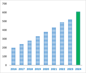
Number of papers citing our products
| Cas No. | 121591-98-8 | SDF | File under preparation. |
| PubChem ID | N/A | Appearance | Powder |
| Formula | C39H66O33 | M.Wt | 1062.92 |
| Type of Compound | N/A | Storage | Desiccate at -20°C |
| Solubility | Soluble in Chloroform,Dichloromethane,Ethyl Acetate,DMSO,Acetone,etc. | ||
| General tips | For obtaining a higher solubility , please warm the tube at 37 ℃ and shake it in the ultrasonic bath for a while.Stock solution can be stored below -20℃ for several months. We recommend that you prepare and use the solution on the same day. However, if the test schedule requires, the stock solutions can be prepared in advance, and the stock solution must be sealed and stored below -20℃. In general, the stock solution can be kept for several months. Before use, we recommend that you leave the vial at room temperature for at least an hour before opening it. |
||
| About Packaging | 1. The packaging of the product may be reversed during transportation, cause the high purity compounds to adhere to the neck or cap of the vial.Take the vail out of its packaging and shake gently until the compounds fall to the bottom of the vial. 2. For liquid products, please centrifuge at 500xg to gather the liquid to the bottom of the vial. 3. Try to avoid loss or contamination during the experiment. |
||
| Shipping Condition | Packaging according to customer requirements(5mg, 10mg, 20mg and more). Ship via FedEx, DHL, UPS, EMS or other couriers with RT, or blue ice upon request. | ||

Heptasaccharide Dilution Calculator
Heptasaccharide Molarity Calculator
| 1 mg | 5 mg | 10 mg | 20 mg | 25 mg | |
| 1 mM | 0.9408 mL | 4.704 mL | 9.408 mL | 18.8161 mL | 23.5201 mL |
| 5 mM | 0.1882 mL | 0.9408 mL | 1.8816 mL | 3.7632 mL | 4.704 mL |
| 10 mM | 0.0941 mL | 0.4704 mL | 0.9408 mL | 1.8816 mL | 2.352 mL |
| 50 mM | 0.0188 mL | 0.0941 mL | 0.1882 mL | 0.3763 mL | 0.4704 mL |
| 100 mM | 0.0094 mL | 0.047 mL | 0.0941 mL | 0.1882 mL | 0.2352 mL |
| * Note: If you are in the process of experiment, it's necessary to make the dilution ratios of the samples. The dilution data above is only for reference. Normally, it's can get a better solubility within lower of Concentrations. | |||||

Calcutta University

University of Minnesota

University of Maryland School of Medicine

University of Illinois at Chicago

The Ohio State University

University of Zurich

Harvard University

Colorado State University

Auburn University

Yale University

Worcester Polytechnic Institute

Washington State University

Stanford University

University of Leipzig

Universidade da Beira Interior

The Institute of Cancer Research

Heidelberg University

University of Amsterdam

University of Auckland

TsingHua University

The University of Michigan

Miami University

DRURY University

Jilin University

Fudan University

Wuhan University

Sun Yat-sen University

Universite de Paris

Deemed University

Auckland University

The University of Tokyo

Korea University
- Licorice glycoside C2
Catalog No.:BCX0824
CAS No.:202657-55-4
- Picfeltarraegenin I
Catalog No.:BCX0823
CAS No.:82145-63-9
- 6''- methyl glycyrrhizinate
Catalog No.:BCX0822
CAS No.:1186016-30-7
- Tribuloside
Catalog No.:BCX0821
CAS No.:22153-44-2
- (25R)-26-O-β-D-Glucopyranosyl-22-hydroxy-5β-furost-3β,26-diol-3-O-β-D-glucopyranosyl-(1→2)-β-D-galactopyranoside
Catalog No.:BCX0820
CAS No.:897386-27-5
- Coreoside B
Catalog No.:BCX0819
CAS No.:1580464-83-0
- Xylohexaose
Catalog No.:BCX0818
CAS No.:49694-21-5
- Hirudonucleodisulfide A
Catalog No.:BCX0817
CAS No.:1072789-37-7
- Xylopentaose
Catalog No.:BCX0816
CAS No.:49694-20-4
- Kuwanon U
Catalog No.:BCX0815
CAS No.:123702-95-4
- Nepetalacton
Catalog No.:BCX0814
CAS No.:21651-62-7
- Xylotetraose
Catalog No.:BCX0813
CAS No.:22416-58-6
- 6',6''- dimethyl glycyrrhizinate
Catalog No.:BCX0826
CAS No.:114006-81-4
- Platycoside F
Catalog No.:BCX0827
CAS No.:314756-03-1
- Quercetin 3-O-β-D-Glucuronide 6''-Methyl Ester
Catalog No.:BCX0828
CAS No.:79543-28-5
- 4′′,5′′-dehydroisopsoralidin
Catalog No.:BCX0829
CAS No.:65639-51-2
- Paeonoside
Catalog No.:BCX0830
CAS No.:20309-70-0
- Artemisitene
Catalog No.:BCX0831
CAS No.:101020-89-7
- 6''-O-Acetyldaidzin
Catalog No.:BCX0832
CAS No.:71385-83-6
- Aquilarone B
Catalog No.:BCX0833
CAS No.:1404479-45-3
- (S)-Goitrin
Catalog No.:BCX0834
CAS No.:500-12-9
- 6''-O-acetyl-saikosaponin B2
Catalog No.:BCX0835
CAS No.:104121-82-6
- 4'-Methoxyisoagarotetrol
Catalog No.:BCX0836
CAS No.:104901-10-2
- 8-epi-helenalin
Catalog No.:BCX0837
CAS No.:97643-91-9
Evaluation of a FlpA Glycoconjugate Vaccine with Ten N-Heptasaccharide Glycan Moieties to reduce Campylobacter jejuni Colonisation in Chickens.[Pubmed:38675777]
Vaccines (Basel). 2024 Apr 9;12(4):395.
Campylobacter is a major cause of acute gastroenteritis in humans, and infections can be followed by inflammatory neuropathies and other sequelae. Handling or consumption of poultry meat is the primary risk factor for human campylobacteriosis, and C. jejuni remains highly prevalent in retail chicken in many countries. Control of Campylobacter in the avian reservoir is expected to limit the incidence of human disease. Toward this aim, we evaluated a glycoconjugate vaccine comprising the fibronectin-binding adhesin FlpA conjugated to up to ten moieties of the conserved N-linked Heptasaccharide glycan of C. jejuni or with FlpA alone. The glycan dose significantly exceeded previous trials using FlpA with two N-glycan moieties. Vaccinated birds were challenged with C. jejuni orally or by exposure to seeder-birds colonised by C. jejuni to mimic natural transmission. No protection against caecal colonisation was observed with FlpA or the FlpA glycoconjugate vaccine. FlpA-specific antibody responses were significantly induced in vaccinated birds at the point of challenge relative to mock-vaccinated birds. A slight but significant antibody response to the N-glycan was detected after vaccination with FlpA-10xGT and challenge. As other laboratories have reported protection against Campylobacter with FlpA and glycoconjugate vaccines in chickens, our data indicate that vaccine-mediated immunity may be sensitive to host- or study-specific variables.
Molecular characterization of Rft1, a membrane protein associated with congenital disorder of glycosylation type 1N.[Pubmed:38617304]
bioRxiv [Preprint]. 2024 Apr 3:2024.04.03.587922.
The oligosaccharide needed for protein N-glycosylation is assembled on a lipid carrier via a multi-step pathway. Synthesis is initiated on the cytoplasmic face of the endoplasmic reticulum (ER) and completed on the luminal side after transbilayer translocation of a Heptasaccharide lipid intermediate. More than 30 Congenital Disorders of Glycosylation (CDGs) are associated with this pathway, including CDG 1N which results from defects in the membrane protein Rft1. Rft1 is essential for the viability of yeast and mammalian cells and was proposed as the transporter needed to flip the Heptasaccharide lipid intermediate across the ER membrane. However, other studies indicated that Rft1 is not required for Heptasaccharide lipid flipping in microsomes or unilamellar vesicles reconstituted with ER membrane proteins, nor is it required for the viability of at least one eukaryote. It is therefore not known what role Rft1 plays in N-glycosylation. Here, we present a molecular characterization of human Rft1, using yeast cells as a reporter system. We show that it is a multi-spanning membrane protein located in the ER, with its N and C-termini facing the cytoplasm. It is not N-glycosylated. The majority of CDG 1N mutations map to highly conserved regions of the protein. We identify key residues that are important for Rft1's ability to support glycosylation and cell viability. Our results provide a necessary platform for future work on this enigmatic protein.
Chemical Synthesis of a Branched Nonasaccharide Fragment from Helicobacter pylori Lipopolysaccharide.[Pubmed:38443201]
Org Lett. 2024 Mar 15;26(10):2103-2107.
A chemical synthesis of a unique nanosaccharide fragment from Helicobacter pylori lipopolysaccharide was achieved via a convergent glycosylation method. Challenges involved in the synthesis include the highly stereoselective construction of beta-3-deoxy-d-manno-oct-2-ulosonic acid (Kdo) and two 1,2-cis-glycosidic linkages, as well as the formation of a branched 2,7-disubstituted heptose subunit. Hydrogen-bond mediated aglycone delivery strategy and benzoyl-directing remote participation effect were employed, respectively, for the efficient generation of the desired beta-Kdo glycoside and 1,2-cis-alpha-l-fucoside/d-glucoside. Moreover, the key branched framework was successfully established through a [(7 + 1) + 1] assembly approach involving the stepwise glycosylation of the Heptasaccharide alcohol with two monosaccharide donors. The synthesized 1 containing a propylamine linker at the reducing end can be covalently bound to a carrier protein for further immunological studies.
Enzymatic construction of a library of even- and odd-numbered heparosan oligosaccharides and their N-sulfonated derivatives.[Pubmed:38442831]
Int J Biol Macromol. 2024 Apr;264(Pt 1):130501.
Low-molecular-weight heparins (LMWHs), especially the specific-sized heparin oligosaccharides, are attractive for the therapeutic applications, while their synthesis remains challenging. In the present study, unsaturated even-numbered heparosan oligosaccharides were firstly prepared by cleaving high-molecular-weight heparosan using recombinant heparinase III (HepIII). The conversion rates of the unsaturated disaccharides, tetrasaccharides, hexasaccharides, octasaccharides, and decasaccharides were 33.9 %, 47.9 %, 78.7 %, 71.8 %, and 53.4 %, respectively. After processing the aforementioned heparosan oligosaccharides with the Delta4,5 unsaturated glycuronidase, saturated odd-numbered heparosan trisaccharides, pentasaccharides, Heptasaccharides, and nonasaccharides were produced. It was observed that among them, the pentasaccharides were the smallest units of saturated odd-numbered oligosaccharides recognized by HepIII. These oligosaccharides were further catalyzed with bifunctional heparan sulfate N-deacetylase/N-sulfotransferase (NDST) under optimized reaction conditions. It was found that the tetrasaccharide was defined as the smallest recognition unit for NDST, obtaining the N-sulfonated heparosan tetrasaccharides, pentasaccharides, and hexasaccharides with a single sulfonate group, as well as N-sulfonated heparosan Heptasaccharides, octasaccharides, and nonasaccharides with multiple sulfonate groups. These results provide an easy pathway for constructing a library of specific-sized N-sulfonated heparosan oligosaccharides that can be used as the substrates for the enzymatic synthesis of LMWHs and heparin oligosaccharides, shedding new light on the substrate preference of NDST.
The Acinetobacter baumannii K239 capsular polysaccharide includes heptasaccharide units that are structurally related to K86 but joined by different linkages formed by different Wzy polymerases.[Pubmed:38336317]
Int J Biol Macromol. 2024 Mar;262(Pt 1):130045.
The K239 type capsular polysaccharide (CPS) isolated from Acinetobacter baumannii isolate MAR19-4435 was studied by sugar analysis, one- and two-dimensional (1)H and (13)C NMR spectroscopy. K239 consists of branched Heptasaccharide repeats (K-units) comprised of five residues of l-rhamnose (l-Rhap), and one residue each of d-glucuronic acid (d-GlcpA) and N-acetyl-d-glucosamine (d-GlcpNAc). The structure of K239 is closely related to that of the A. baumannii K86 CPS type, though the two differ in the 2,3-substitution patterns on the l-Rhap residue that is involved in the linkage between K-units in the CPS polymer. This structural difference was attributed to the presence of a gtr221 glycosyltransferase gene and a wzy(KL239) polymerase gene in KL239 that replaces the gtr80 and wzy(KL86) genes in the KL86 CPS biosynthesis gene cluster. Comparison of the two structures established the role of a novel Wzy(KL239) polymerase encoded by KL239 that forms the beta-d-GlcpNAc-(1-->2)-l-Rhap linkage between K239 units. A. baumannii MAR19-4435 was found to be non-susceptible to infection by the APK86 bacteriophage, which encodes a depolymerase that specifically cleaves the linkage between K-units in the K86 CPS, indicating that the difference in 2,3-substitution of l-Rhap influences the susceptibility of this isolate to bacteriophage activity.
Characterization of Dichloroisoeverninic Acid Biosynthesis and Chemoenzymatic Synthesis of New Orthosomycins.[Pubmed:38289021]
ACS Chem Biol. 2024 Feb 16;19(2):526-535.
The orthosomycins are highly modified oligosaccharide natural products with a broad spectrum and potent antimicrobial activities. These include everninomicins and avilamycins, which inhibit protein translation by binding a unique site on the bacterial ribosome. Notably, ribosomal bound structures reveal a network of interactions between the 50S subunit and dichloroisoeverninic acid (DCIE), the aromatic A(1)-ring conserved across orthosomycins, but the relationship of these interactions to their antimicrobial activity remains undetermined. Genetic functional analysis of three genes putatively associated with DCIE biosynthesis in the everninomicin producer Micromonospora carbonacea delineates the native biosynthetic pathway and provides previously unreported advanced biosynthetic intermediates. Subsequent in vitro biochemical analyses demonstrate the complete DCIE biosynthetic pathway and provide access to novel everninomicin analogs. In addition to the orsellinate synthase EvdD3 and a flavin-dependent halogenase EvdD2, our results identified a key acyltransferase, EvdD1, responsible for transferring orsellinate from the acyl carrier protein domain of EvdD3 to a Heptasaccharide orthosomycin biosynthetic intermediate. We have also shown that EvdD1 is able to transfer unnatural aryl groups via their N-acyl cysteamine thioesters to the everninomicin scaffold and used this as a biocatalyst to generate a panel of unnatural aryl analogs. The impact of diverse aryl functional group substitution on both ribosome inhibition and antibacterial activities demonstrates the importance of the DCIE moiety in the pharmacology of orthosomycins, notably revealing an uncoupling between ribosomal engagement and antibiotic activity. Control of A(1)-ring functionality in this class of molecules provides a potential handle to explore and address pharmacological roles of the DCIE ring in this potent and unique class of antibiotics.
Moraxella ovis and Moraxella bovoculi lipooligosaccharide biosynthesis genes, and structural characterisation of oligosaccharide from M. ovis 354T.[Pubmed:38281396]
Carbohydr Res. 2024 Feb;536:109043.
Moraxella ovis is a Gram-negative bacterium isolated from sheep conjunctivitis cases and is a rare isolate of infectious bovine keratoconjunctivitis (IBK). This species is closely related to M. bovoculi, another species which can also be isolated from IBK, or cattle upper respiratory tract (URT). Prior to molecular identification techniques, M. bovoculi was frequently misclassified as M. ovis. We previously described the structure of two oligosaccharides (lipooligosaccharide-derived, minor and major glycoforms) from M. bovoculi 237T (type strain, also ATCC BAA-1259T). Here, we have identified the genetic loci for lipooligosaccharide synthesis in M. ovis 354T (NCTC11227) and compared it with M. bovoculi 237T. We identified genes encoding the known glycosyltransferases Lgt6 and Lgt3 in M.ovis. These genes are conserved in Moraxella spp., including M bovoculi. We identified three further putative OS biosynthesis genes that are restricted to M. ovis and M. bovoculi. These encode enzymes predicted to function as GDP-mannose synthases, namely a mannosyltransferase and a glycosyltransferase. Adding insight into the genetic relatedness of M.ovis and M. bovoculi, the M. ovis genes have higher similarity to those in M. bovoculi genotype 2 (nasopharyngeal isolates from asymptomatic cattle), than to M. bovoculi genotype 1 (isolates from eyes of IBK-affected cattle). Sequence analysis confirmed that the predicted mannosyltransferase in M. bovoculi 237T is interrupted by a C>T polymorphism. This mutation is not present in other M. bovoculi strains sequenced to date. We isolated and characterised LOS-derived oligosaccharide from M. ovis 354T. GLC-MS and NMR spectroscopy data revealed a Heptasaccharide structure with three beta-D-Glcp residues attached as branches to the central 3,4,6-alpha-D-Glcp, with subsequent attachment to Kdo. This inner core arrangement is consistent with the action of Lgt6 and Lgt3 glycosyltransferases. Two alpha-D-Manp residues are linearly attached to the 4-linked beta-D-Glcp, consistent with the presence of the two identified glycosyltransferases. This oligosaccharide structure is consistent with the previously reported minor glycoform isolated from M. bovoculi 237T.
Isolation, phytochemistry, characterization, biological activity, and application of Morinda officinalis How oligosaccharide: a review.[Pubmed:37991722]
J Pharm Pharmacol. 2023 Nov 22:rgad096.
Morinda officinalis How oligosaccharide (MOO) stands as one of the principal active constituents of M. officinalis How, widely employed in traditional Chinese medicine. The methods for MOO extraction predominantly encompass hot water extraction, ethanol extraction, ultrasonic-assisted extraction, and microwave-assisted extraction. Distinct extraction techniques yield varying MOO quantities. MOO encompasses a diversity of oligosaccharides, including bajijiasu, sucrose, 1-kestose, nystose, mannose, 1F-fructofuranosylnystose, 1,1,1,1-kestohexose, fructoHeptasaccharide, inulin-type hexasaccharide, inulin-type Heptasaccharide, inulotriose, inulotetraose, inulopentaose, and mannose. MOO exhibits a wide spectrum of biological activities, exerting specific effects on the nervous system, cardiovascular system, motor system, reproductive system, and immune system. It demonstrates antidepressant properties, offers potential in mitigating Alzheimer's disease, stimulates angiogenesis, and possesses anti-osteoporotic and other pharmacological effects. Clinically, when combined with various antidepressants, MOO exhibits specific therapeutic efficacy across multiple forms of depression. As a naturally occurring plant oligosaccharide, MOO holds diverse pharmaceutical applications. This article conducts a review of the latest extraction and purification methodologies, structural characterization analysis, biological activity assessment, and clinical applications of MOO. Such a comprehensive analysis yields innovative insights for advancing the research and application of MOO in the future.
Branch distribution pattern and anticoagulant activity of a fucosylated chondroitin sulfate from Phyllophorella kohkutiensis.[Pubmed:37739534]
Carbohydr Polym. 2023 Dec 1;321:121304.
Fucosylated chondroitin sulfate (FCS) extracted from Phyllophorella kohkutiensis (PkFCS) is composed of d-GalNAc, d-GlcA, l-Fuc and -SO(4)(2-). According to the defined structures revealed by NMR spectra of the branches released by mild acid hydrolysis and oligosaccharides generated by beta-eliminative depolymerization, the backbone of PkFCS is CS-E, and the branch types attached to C-3 of d-GlcA include l-Fuc(2S4S), l-Fuc(3S4S), l-Fuc(4S,) and the disaccharide alpha-d-GalNAc-1,2-alpha-l-Fuc(3S4S) with the ratio of 43:13:22:22. Notably, novel Heptasaccharide and hendecasaccharide were identified that are branched with continuous distribution of the disaccharide. The structural sequences of the oligosaccharides indicate that three unique structural motifs are present in the entire PkFCS polymer, including a motif branched with randomly distributed different sulfated l-Fuc units, a motif containing regular l-Fuc(2S4S) branches and a motif enriched in alpha-d-GalNAc-1,2-alpha-l-Fuc(3S4S). This is the first report about the distribution pattern of diverse branches in natural FCS. Natural PkFCS exhibited potent anticoagulant activity on APTT prolonging and anti-iXase activity. Regarding the structurally defined oligosaccharides with sulfated fucosyl side chains, octasaccharide (Pk4b) is the minimum fragment responsible for its anticoagulant activity correlated with anti-iXase. However, further glycosyl modification with a non-sulfated d-GalNAc at the C-2 position of l-Fuc(3S4S) could significantly decrease the anticoagulant and anti-iXase activity.
A concise chemoenzymatic total synthesis of neutral Globo-series glycosphingolipids Globo A and Globo B, and Forssman and para-Forssman antigens.[Pubmed:37606864]
Glycoconj J. 2023 Oct;40(5):551-563.
Globo A is a neutral Globo-series glycosphingolipid (GSL) that shows natural properties of a cytotoxicity receptor NKp44 binding ligand. The highly complex Heptasaccharide glycan structure of Globo A combined with its biological profile provides a unique target for the development of a synthetic method to facilitate its bioactivity studies. Here, a concise chemoenzymatic route to the synthesis of Globo A and its alpha1,3-galactose-linked congener Globo B is reported. The key to success was the use of a synthetic azido beta-Globo H sphingosine (Globo H-betaSph) as an acceptor substrate and two glycosyl transferases, an alpha1,3-N-acetylgalactosaminyltransferase from Helicobacter mustelae (BgtA) and a human blood group B alpha1,3-galactosyltransferase (h1,3GTB), for stereoselective construction of the terminal alpha1,3-GalNAc and alpha1,3-Gal linkages, respectively. The azido-Sph lipid sidechain is further elaborated by reduction and a chemoselective N-acylation to complete the total synthesis of the neutral Globo-series GSLs. In addition, the synthesis of Forssman and para-Forssman antigens were prepared. The strategy may be suitable for accessing other complex GSLs and related lipid-modified GSL derivatives.
A repertoire of alginate lyases in the alginate polysaccharide utilization loci of marine bacterium Wenyingzhuangia fucanilytica: biochemical properties and action pattern.[Pubmed:37540808]
J Sci Food Agric. 2024 Jan 15;104(1):134-140.
BACKGROUND: Alginate lyases are important tools for alginate biodegradation and oligosaccharide production, which have great potential in food and biofuel fields. The alginate polysaccharide utilization loci (PUL) typically encode a series of alginate lyases with a synergistic action pattern. Exploring valuable alginate lyases and revealing the synergistic effect of enzymes in the PUL is of great significance. RESULTS: An alginate PUL was discovered from the marine bacterium Wenyingzhuangia fucanilytica CZ1127(T) , and a repertoire of alginate lyases within it was cloned, expressed and characterized. The four alginate lyases in PUL demonstrated similar optimal reaction conditions: maximum enzyme activity at 35-50 degrees C and pH 8.0-9.0. The results of action pattern indicated that they were two PL7 endolytic bifunctional enzymes (Aly7A and Aly7B), a PL6 exolytic bifunctional enzyme (Aly6A) and a PL17 exolytic M-specific enzyme (Aly17A). Ultra-performance liquid chromatography-mass spectrometry was employed to reveal the synergistic effect of the four enzymes. The end products of Aly7A were further degraded by Aly7B and eventually generated oligosaccharides, from disaccharide to Heptasaccharide. The oligosaccharide products were completely degraded to monosaccharides by Aly6A, but it was unable to directly degrade alginate. Aly17A could also produce monosaccharides by cleaving the M-blocks of oligosaccharide products, as well as the M-blocks of polysaccharides. The combination of these enzymes resulted in the complete degradation of alginate to monosaccharides. CONCLUSION: A new alginate PUL was mined and four novel alginate lyases in the PUL were expressed and characterized. The four cooperative alginate lyases provide novel tools for alginate degradation and biological fermentation. (c) 2023 Society of Chemical Industry.
Chemoenzymatic Preparation of a Campylobacter jejuni Lipid-Linked Heptasaccharide on an Azide-Linked Polyisoprenoid.[Pubmed:37151508]
ACS Omega. 2023 Apr 22;8(17):15790-15798.
Complex poly- and oligosaccharides on the surface of bacteria provide a unique fingerprint to different strains of pathogenic and symbiotic microbes that could be exploited for therapeutics or sensors selective for specific glycans. To discover reagents that can selectively interact with specific bacterial glycans, a system for both the chemoenzymatic preparation and immobilization of these materials would be ideal. Bacterial glycans are typically synthesized in nature on the C55 polyisoprenoid bactoprenyl (or undecaprenyl) phosphate. However, this long-chain isoprenoid can be difficult to work with in vitro. Here, we describe the addition of a chemically functional benzylazide tag to polyisoprenoids. We have found that both the organic-soluble and water-soluble benzylazide isoprenoid can serve as a substrate for the well-characterized system responsible for Campylobacter jejuni N-linked Heptasaccharide assembly. Using the organic-soluble analogue, we demonstrate the use of an N-acetyl-glucosamine epimerase that can be used to lower the cost of glycan assembly, and using the water-soluble analogue, we demonstrate the immobilization of the C. jejuni Heptasaccharide on magnetic beads. These conjugated beads are then shown to interact with soybean agglutinin, a lectin known to interact with N-acetyl-galactosamine in the C. jejuni Heptasaccharide. The methods provided could be used for a wide variety of applications including the discovery of new glycan-interacting partners.
A combinatorial DNA assembly approach to biosynthesis of N-linked glycans in E. coli.[Pubmed:36637423]
Glycobiology. 2023 Mar 6;33(2):138-149.
Glycoengineering of recombinant glycans and glycoconjugates is a rapidly evolving field. However, the production and exploitation of glycans has lagged behind that of proteins and nucleic acids. Biosynthetic glycoconjugate production requires the coordinated cooperation of three key components within a bacterial cell: a substrate protein, a coupling oligosaccharyltransferase, and a glycan biosynthesis locus. While the acceptor protein and oligosaccharyltransferase are the products of single genes, the glycan is a product of a multigene metabolic pathway. Typically, the glycan biosynthesis locus is cloned and transferred en bloc from the native organism to a suitable Escherichia coli strain. However, gene expression within these pathways has been optimized by natural selection in the native host and is unlikely to be optimal for heterologous production in an unrelated organism. In recent years, synthetic biology has addressed the challenges in heterologous expression of multigene systems by deconstructing these pathways and rebuilding them from the bottom up. The use of DNA assembly methods allows the convenient assembly of such pathways by combining defined parts with the requisite coding sequences in a single step. In this study, we apply combinatorial assembly to the heterologous biosynthesis of the Campylobacter jejuni N-glycosylation (pgl) pathway in E. coli. We engineered reconstructed biosynthesis clusters that faithfully reproduced the C. jejuni Heptasaccharide glycan. Furthermore, following a single round of combinatorial assembly and screening, we identified pathway clones that outperform glycan and glycoconjugate production of the native unmodified pgl cluster. This platform offers a flexible method for optimal engineering of glycan structures in E. coli.
Characterisation of N-linked protein glycosylation in the bacterial pathogen Campylobacter hepaticus.[Pubmed:36604449]
Sci Rep. 2023 Jan 5;13(1):227.
Campylobacter hepaticus is an important pathogen which causes Spotty Liver Disease (SLD) in layer chickens. SLD results in an increase in mortality and a significant decrease in egg production and therefore is an important economic concern of the global poultry industry. The human pathogen Campylobacter jejuni encodes an N-linked glycosylation system that plays fundamental roles in host colonization and pathogenicity. While N-linked glycosylation has been extensively studied in C. jejuni and is now known to occur in a range of Campylobacter species, little is known about C. hepaticus glycosylation. In this study glycoproteomic analysis was used to confirm the functionality of the C. hepaticus N-glycosylation system. It was shown that C. hepaticus HV10(T) modifies > 35 proteins with an N-linked Heptasaccharide glycan. C. hepaticus shares highly conserved glycoproteins with C. jejuni that are involved in host colonisation and also possesses unique glycoproteins which may contribute to its ability to survive in challenging host environments. C. hepaticus N-glycosylation may function as an important virulence factor, providing an opportunity to investigate and develop a better understanding the system's role in poultry infection.
Synthesis of a B-Antigen Hexasaccharide, a B-Lewis b Heptasaccharide and Glycoconjugates Thereof to Investigate Binding Properties of Helicobacter pylori.[Pubmed:36562295]
Chemistry. 2023 Mar 16;29(16):e202203672.
Infecting the stomach of almost 50 % of people, Helicobacter pylori is a causative agent of gastritis, peptic ulcers and stomach cancers. Interactions between bacterial membrane-bound lectin, Blood group Antigen Binding Adhesin (BabA), and human blood group antigens are key in the initiation of infection. Herein, the synthesis of a B-antigen hexasaccharide (B6) and a B-Lewis b Heptasaccharide (BLeb7) and Bovine Serum Albumin glycoconjugates thereof is reported to assess the binding properties and preferences of BabA from different strains. From a previously reported trisaccharide acceptor a versatile key Lacto-N-tetraose tetrasaccharide intermediate was synthesized, which allowed us to explore various routes to the final targets, either via initial introduction of fucosyl residues followed by introduction of the B-determinant or vice versa. The first approach proved unsuccessful, whereas the second afforded the target structures in good yields. Protein conjugation using isothiocyanate methodology allowed us to reach high glycan loadings (up to 23 per protein) to mimic multivalent displays encountered in Nature. Protein glycoconjugate inhibition binding studies were performed with H. pylori strains displaying high or low affinity for Lewis b hexasaccharide structures showing that the binding to the high affinity strain was reduced due to the presence of the B-determinant in the Bleb7-conjugates and further reduced by the absence of the Lewis fucose residue in the B6-conjugate.