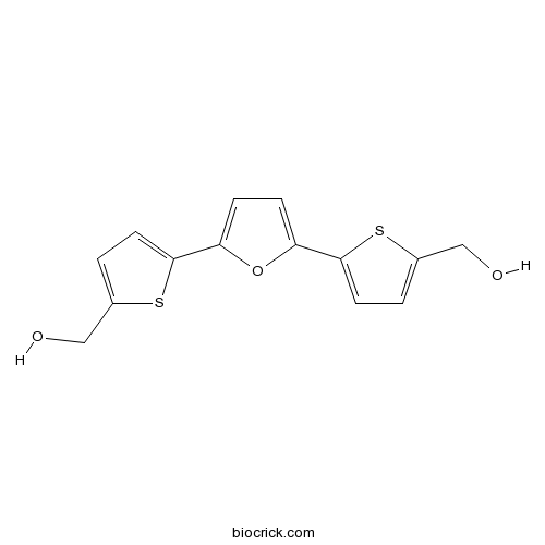
RITA (NSC 652287)Mdm2-p53 interaction and p53 ubiquitination blocking CAS# 213261-59-7 |

- AMG232
Catalog No.:BCC3992
CAS No.:1352066-68-2
- NSC 146109 hydrochloride
Catalog No.:BCC2410
CAS No.:59474-01-0
- Nutlin-3a chiral
Catalog No.:BCC1812
CAS No.:675576-98-4
- Nutlin-3
Catalog No.:BCC2254
CAS No.:890090-75-2
- p53 and MDM2 proteins-interaction-inhibitor chiral
Catalog No.:BCC1830
CAS No.:939981-37-0
- p53 and MDM2 proteins-interaction-inhibitor racemic
Catalog No.:BCC1831
CAS No.:939983-14-9
Quality Control & MSDS
3D structure
Package In Stock
Number of papers citing our products

| Cas No. | 213261-59-7 | SDF | Download SDF |
| PubChem ID | 374536 | Appearance | Powder |
| Formula | C14H12O3S2 | M.Wt | 292.4 |
| Type of Compound | N/A | Storage | Desiccate at -20°C |
| Synonyms | NSC 652287 | ||
| Solubility | DMSO : ≥ 100 mg/mL (342.03 mM) *"≥" means soluble, but saturation unknown. | ||
| Chemical Name | [5-[5-[5-(hydroxymethyl)thiophen-2-yl]furan-2-yl]thiophen-2-yl]methanol | ||
| SMILES | C1=C(SC(=C1)C2=CC=C(O2)C3=CC=C(S3)CO)CO | ||
| Standard InChIKey | KZENBFUSKMWCJF-UHFFFAOYSA-N | ||
| Standard InChI | InChI=1S/C14H12O3S2/c15-7-9-1-5-13(18-9)11-3-4-12(17-11)14-6-2-10(8-16)19-14/h1-6,15-16H,7-8H2 | ||
| General tips | For obtaining a higher solubility , please warm the tube at 37 ℃ and shake it in the ultrasonic bath for a while.Stock solution can be stored below -20℃ for several months. We recommend that you prepare and use the solution on the same day. However, if the test schedule requires, the stock solutions can be prepared in advance, and the stock solution must be sealed and stored below -20℃. In general, the stock solution can be kept for several months. Before use, we recommend that you leave the vial at room temperature for at least an hour before opening it. |
||
| About Packaging | 1. The packaging of the product may be reversed during transportation, cause the high purity compounds to adhere to the neck or cap of the vial.Take the vail out of its packaging and shake gently until the compounds fall to the bottom of the vial. 2. For liquid products, please centrifuge at 500xg to gather the liquid to the bottom of the vial. 3. Try to avoid loss or contamination during the experiment. |
||
| Shipping Condition | Packaging according to customer requirements(5mg, 10mg, 20mg and more). Ship via FedEx, DHL, UPS, EMS or other couriers with RT, or blue ice upon request. | ||
| Description | Anti-tumor agent that binds wild-type p53 (Kd = 1.5 nM) preventing p53-MDM2 (HDM2) interaction. Induces p53 accumulation and stimulates apoptosis in tumor cell lines expressing wild-type p53 in vitro and in vivo. Inhibits HPV-E6-mediated proteasomal degradation. Suppresses expression of cervical carcinoma HeLa xenografts in vivo. |

RITA (NSC 652287) Dilution Calculator

RITA (NSC 652287) Molarity Calculator
| 1 mg | 5 mg | 10 mg | 20 mg | 25 mg | |
| 1 mM | 3.42 mL | 17.0999 mL | 34.1997 mL | 68.3995 mL | 85.4993 mL |
| 5 mM | 0.684 mL | 3.42 mL | 6.8399 mL | 13.6799 mL | 17.0999 mL |
| 10 mM | 0.342 mL | 1.71 mL | 3.42 mL | 6.8399 mL | 8.5499 mL |
| 50 mM | 0.0684 mL | 0.342 mL | 0.684 mL | 1.368 mL | 1.71 mL |
| 100 mM | 0.0342 mL | 0.171 mL | 0.342 mL | 0.684 mL | 0.855 mL |
| * Note: If you are in the process of experiment, it's necessary to make the dilution ratios of the samples. The dilution data above is only for reference. Normally, it's can get a better solubility within lower of Concentrations. | |||||

Calcutta University

University of Minnesota

University of Maryland School of Medicine

University of Illinois at Chicago

The Ohio State University

University of Zurich

Harvard University

Colorado State University

Auburn University

Yale University

Worcester Polytechnic Institute

Washington State University

Stanford University

University of Leipzig

Universidade da Beira Interior

The Institute of Cancer Research

Heidelberg University

University of Amsterdam

University of Auckland

TsingHua University

The University of Michigan

Miami University

DRURY University

Jilin University

Fudan University

Wuhan University

Sun Yat-sen University

Universite de Paris

Deemed University

Auckland University

The University of Tokyo

Korea University
IC50: 2 nM and 20 nM for A-498 and TK-10, respectively
A series of naturally occurring and synthetic compounds containing one or more thiophene moieties have been tested in the NCI Anticancer Drug Screen and have demonstrated differential antiproliferative activity. Thiophene derivatives as a class exhibit very similar patterns of differential sensitivity, the molecular basis for which is not clear. The compound 2,5-bis(5-hydroxymethyl-2-thienyl) furan (NSC 652287), is the most potent thiophene derivative and has been selected as the lead compound for mechanistic studies.
In vitro: A number of cell lines showed a striking differential sensitivity to NSC 652287 when compared with the other cell lines in the panel, with GI50 values of 10–60 nM. The compound was found to decrease the initial number of cells by 50% (LC50) at a concentration of 100 nM in the A-498 cell line, compared with ~100 mM for the majority of the tumor cell lines. The A-498 and TK-10 cell lines were particularly sensitive to NSC 652287-induced cytotoxicity compared with ACHN and UO-31 cell lines [1].
In vivo: NSC 652287 was evaluated against A-498 tumor cell xenografts grown subcutaneously in nude mice. When NSC 652287 was administered twice a day, all three doses resulted in complete tumor regression in 100% of the treated mice by the end of the third treatment period. The tumors did not regrow during the remaining 40 days of the study, and no gross evidence of toxicity was observed. Studies with xenografts derived from other sensitive cell lines including the renal CAKI-1, melanoma UACC-257, ovarian OVCAR-5, and colon HCC-2998, showed moderate or minimal in vivo activity [2].
Clinical trials: Currenlty no clinical data are available.
Reference:
[1] Rivera MI, Stinson SF, Vistica DT, Jorden JL, Kenney S, Sausville EA. Selective toxicity of the tricyclic thiophene NSC 652287 in renal carcinoma cell lines: differential accumulation and metabolism. Biochem Pharmacol. 1999;57(11):1283-95.
- [Phe1Ψ(CH2-NH)Gly2]Nociceptin(1-13)NH2
Catalog No.:BCC5701
CAS No.:213130-17-7
- Ceanothic acid
Catalog No.:BCN4918
CAS No.:21302-79-4
- Org 37684
Catalog No.:BCC6291
CAS No.:213007-95-5
- Boc-Tyr(Bzl)-OH
Catalog No.:BCC3461
CAS No.:2130-96-3
- Tetrahymanol acetate
Catalog No.:BCN6933
CAS No.:2130-22-5
- Tetrahymanol
Catalog No.:BCN6934
CAS No.:2130-17-8
- (S)-(+)-Abscisic acid
Catalog No.:BCN2210
CAS No.:21293-29-8
- Purvalanol B
Catalog No.:BCC3887
CAS No.:212844-54-7
- Purvalanol A
Catalog No.:BCC5654
CAS No.:212844-53-6
- Cowaxanthone B
Catalog No.:BCN3892
CAS No.:212842-64-3
- Kumokirine
Catalog No.:BCN2011
CAS No.:21284-20-8
- Kuramerine
Catalog No.:BCN1806
CAS No.:21284-19-5
- H-Pro-OMe.HCl
Catalog No.:BCC3022
CAS No.:2133-40-6
- 15,18-Dihydroxyabieta-8,11,13-trien-7-one
Catalog No.:BCN1495
CAS No.:213329-45-4
- 18-Nor-4,15-dihydroxyabieta-8,11,13-trien-7-one
Catalog No.:BCN1494
CAS No.:213329-46-5
- 1-Methyl-L-tryptophan
Catalog No.:BCN8341
CAS No.:21339-55-9
- Ercalcidiol
Catalog No.:BCC1555
CAS No.:21343-40-8
- Flumethasone
Catalog No.:BCC8986
CAS No.:2135-17-3
- Drim-7-ene-11,12-diol acetonide
Catalog No.:BCN4919
CAS No.:213552-47-7
- 5-Iodo-A-85380, 5-trimethylstannyl N-BOC derivative
Catalog No.:BCC7102
CAS No.:213766-21-3
- 6,8-Cyclo-1,4-eudesmanediol
Catalog No.:BCN4920
CAS No.:213769-80-3
- Zedoarofuran
Catalog No.:BCN3527
CAS No.:213833-34-2
- Nifenazone
Catalog No.:BCC3822
CAS No.:2139-47-1
- 1-Acetoxy-9,17-octadecadiene-12,14-diyne-11,16-diol
Catalog No.:BCN1493
CAS No.:213905-35-2
The conserved Trp114 residue of thioredoxin reductase 1 has a redox sensor-like function triggering oligomerization and crosslinking upon oxidative stress related to cell death.[Pubmed:25611390]
Cell Death Dis. 2015 Jan 22;6:e1616.
The selenoprotein thioredoxin reductase 1 (TrxR1) has several key roles in cellular redox systems and reductive pathways. Here we discovered that an evolutionarily conserved and surface-exposed tryptophan residue of the enzyme (Trp114) is excessively reactive to oxidation and exerts regulatory functions. The results indicate that it serves as an electron relay communicating with the FAD moiety of the enzyme, and, when oxidized, it facilitates oligomerization of TrxR1 into tetramers and higher multimers of dimers. A covalent link can also be formed between two oxidized Trp114 residues of two subunits from two separate TrxR1 dimers, as found both in cell extracts and in a crystal structure of tetrameric TrxR1. Formation of covalently linked TrxR1 subunits became exaggerated in cells on treatment with the pro-oxidant p53-reactivating anticancer compound RITA, in direct correlation with triggering of a cell death that could be prevented by antioxidant treatment. These results collectively suggest that Trp114 of TrxR1 serves a function reminiscent of an irreversible sensor for excessive oxidation, thereby presenting a previously unrecognized level of regulation of TrxR1 function in relation to cellular redox state and cell death induction.
CRISPR-Cas9-based target validation for p53-reactivating model compounds.[Pubmed:26595461]
Nat Chem Biol. 2016 Jan;12(1):22-8.
Inactivation of the p53 tumor suppressor by Mdm2 is one of the most frequent events in cancer, so compounds targeting the p53-Mdm2 interaction are promising for cancer therapy. Mechanisms conferring resistance to p53-reactivating compounds are largely unknown. Here we show using CRISPR-Cas9-based target validation in lung and colorectal cancer that the activity of nutlin, which blocks the p53-binding pocket of Mdm2, strictly depends on functional p53. In contrast, sensitivity to the drug RITA, which binds the Mdm2-interacting N terminus of p53, correlates with induction of DNA damage. Cells with primary or acquired RITA resistance display cross-resistance to DNA crosslinking compounds such as cisplatin and show increased DNA cross-link repair. Inhibition of FancD2 by RNA interference or pharmacological mTOR inhibitors restores RITA sensitivity. The therapeutic response to p53-reactivating compounds is therefore limited by compound-specific resistance mechanisms that can be resolved by CRISPR-Cas9-based target validation and should be considered when allocating patients to p53-reactivating treatments.
The p53-reactivating small molecule RITA induces senescence in head and neck cancer cells.[Pubmed:25119136]
PLoS One. 2014 Aug 13;9(8):e104821.
TP53 is the most commonly mutated gene in head and neck cancer (HNSCC), with mutations being associated with resistance to conventional therapy. Restoring normal p53 function has previously been investigated via the use of RITA (reactivation of p53 and induction of tumor cell apoptosis), a small molecule that induces a conformational change in p53, leading to activation of its downstream targets. In the current study we found that RITA indeed exerts significant effects in HNSCC cells. However, in this model, we found that a significant outcome of RITA treatment was accelerated senescence. RITA-induced senescence in a variety of p53 backgrounds, including p53 null cells. Also, inhibition of p53 expression did not appear to significantly inhibit RITA-induced senescence. Thus, this phenomenon appears to be partially p53-independent. Additionally, RITA-induced senescence appears to be partially mediated by activation of the DNA damage response and SIRT1 (Silent information regulator T1) inhibition, with a synergistic effect seen by combining either ionizing radiation or SIRT1 inhibition with RITA treatment. These data point toward a novel mechanism of RITA function as well as hint to its possible therapeutic benefit in HNSCC.
Cholesterol Retards Senescence in Bone Marrow Mesenchymal Stem Cells by Modulating Autophagy and ROS/p53/p21(Cip1/Waf1) Pathway.[Pubmed:27703600]
Oxid Med Cell Longev. 2016;2016:7524308.
In the present study, we demonstrated that bone marrow mesenchymal stem cells (BMSCs) of the 3rd passage displayed the senescence-associated phenotypes characterized with increased activity of SA-beta-gal, altered autophagy, and increased G1 cell cycle arrest, ROS production, and expression of p53 and p21(Cip1/Waf1) compared with BMSCs of the 1st passage. Cholesterol (CH) reduced the number of SA-beta-gal positive cells in a dose-dependent manner in aging BMSCs induced by H2O2 and the 3rd passage BMSCs. Moreover, CH inhibited the production of ROS and expression of p53 and p21(Cip1/Waf1) in both cellular senescence models and decreased the percentage of BMSCs in G1 cell cycle in the 3rd passage BMSCs. CH prevented the increase in SA-beta-gal positive cells induced by RITA (reactivation of p53 and induction of tumor cell apoptosis, a p53 activator) or 3-MA (3-methyladenine, an autophagy inhibitor). Our results indicate that CH not only is a structural component of cell membrane but also functionally contributes to regulating cellular senescence by modulating cell cycle, autophagy, and the ROS/p53/p21(Cip1/Waf1) signaling pathway.
Targeting NF-kappaB RelA/p65 phosphorylation overcomes RITA resistance.[Pubmed:27721021]
Cancer Lett. 2016 Dec 28;383(2):261-271.
Inactivation of p53 occurs frequently in various cancers. RITA is a promising anticancer small molecule that dissociates p53-MDM2 interaction, reactivates p53 and induces exclusive apoptosis in cancer cells, but acquired RITA resistance remains a major drawback. This study found that the site-differential phosphorylation of nuclear factor-kappaB (NF-kappaB) RelA/p65 creates a barcode for RITA chemosensitivity in cancer cells. In naive MCF7 and HCT116 cells where RITA triggered vast apoptosis, phosphorylation of RelA/p65 increased at Ser536, but decreased at Ser276 and Ser468; oppositely, in RITA-resistant cells, RelA/p65 phosphorylation decreased at Ser536, but increased at Ser276 and Ser468. A phosphomimetic mutation at Ser536 (p65/S536D) or silencing of endogenous RelA/p65 resensitized the RITA-resistant cells to RITA while the phosphomimetic mutant at Ser276 (p65/S276D) led to RITA resistance of naive cells. In mouse xenografts, intratumoral delivery of the phosphomimetic p65/S536D mutant increased the antitumor activity of RITA. Furthermore, in the RITA-resistant cells ATP-binding cassette transporter ABCC6 was upregulated, and silencing of ABCC6 expression in these cells restored RITA sensitivity. In the naive cells, ABCC6 delivery led to RITA resistance and blockage of p65/S536D mutant-induced RITA sensitivity. Taken together, these data suggest that the site-differential phosphorylation of RelA/p65 modulates RITA sensitivity in cancer cells, which may provide an avenue to manipulate RITA resistance.
Rescue of p53 function by small-molecule RITA in cervical carcinoma by blocking E6-mediated degradation.[Pubmed:20395210]
Cancer Res. 2010 Apr 15;70(8):3372-81.
Proteasomal degradation of p53 by human papilloma virus (HPV) E6 oncoprotein plays a pivotal role in the survival of cervical carcinoma cells. Abrogation of HPV-E6-dependent p53 destruction can therefore be a good strategy to combat cervical carcinomas. Here, we show that a small-molecule reactivation of p53 and induction of tumor cell apoptosis (RITA) is able to induce the accumulation of p53 and rescue its tumor suppressor function in cells containing high-risk HPV16 and HPV18 by inhibiting HPV-E6-mediated proteasomal degradation. RITA blocks p53 ubiquitination by preventing p53 interaction with E6-associated protein, required for HPV-E6-mediated degradation. RITA activates the transcription of proapoptotic p53 targets Noxa, PUMA, and BAX, and repressed the expression of pro-proliferative factors CyclinB1, CDC2, and CDC25C, resulting in p53-dependent apoptosis and cell cycle arrest. Importantly, RITA showed substantial suppression of cervical carcinoma xenografts in vivo. These results provide a proof of principle for the treatment of cervical cancer in a p53-dependent manner by using small molecules that target p53.
Selective toxicity of the tricyclic thiophene NSC 652287 in renal carcinoma cell lines: differential accumulation and metabolism.[Pubmed:10230772]
Biochem Pharmacol. 1999 Jun 1;57(11):1283-95.
The tricyclic compound 2,5-bis(5-hydroxymethyl-2-thienyl)furan (NSC 652287) has shown a highly selective pattern of differential cytotoxic activity in the tumor cell lines comprising the National Cancer Institute (NCI) Anticancer Drug Screen. The mechanism underlying the selective cytotoxicity is unknown. We hypothesized that differential sensitivity to the compound observed in several renal tumor cell lines could be the result of selective accumulation or differential metabolism of this agent. We demonstrated here that the capacity of certain renal cell lines to accumulate and retain the compound, determined by accumulation of [14C]NSC 652287-derived radioactivity and by flow cytometric determination of unlabeled compound, paralleled the sensitivity of the renal cell lines to growth inhibition by NSC 652287: A-498 > TK-10 >> ACHN approximately/= to UO-31. The ability of the cell lines to metabolize [14C]NSC 652287 to a reactive species capable of binding covalently to cellular macromolecules also directly correlated with sensitivity to the compound. Different patterns of metabolites were generated by relatively more drug-sensitive cell lines in comparison with drug-resistant cell lines. The metabolizing capacity for NSC 652287 was localized primarily to the cytosolic (S100) fraction. The rate of metabolism in the cytosolic fraction from the most sensitive renal cell line, A-498, was faster than that observed in the cytosolic fractions from the other, less sensitive cell lines. The data support the hypothesis that both selective cellular accumulation and the capacity to metabolize NSC 652287 to a reactive species by certain renal carcinoma cell types are the basis for the differential cytotoxicity of this compound class.


