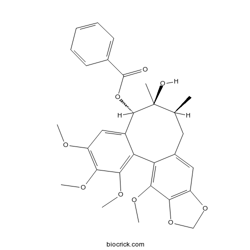
Schisantherin ACAS# 58546-56-8 |

- Schisanwilsonin H
Catalog No.:BCN3315
CAS No.:1181216-83-0
- Benzoylgomisin P
Catalog No.:BCN8803
CAS No.:129445-43-8
Quality Control & MSDS
3D structure
Package In Stock
Number of papers citing our products

| Cas No. | 58546-56-8 | SDF | Download SDF |
| PubChem ID | 151529 | Appearance | White powder |
| Formula | C30H32O9 | M.Wt | 536.56 |
| Type of Compound | Lignans | Storage | Desiccate at -20°C |
| Synonyms | Gomisin-C; Schizantherin-A; Wuweizi ester-A | ||
| Solubility | Soluble in methanol; insoluble in water | ||
| SMILES | CC1CC2=CC3=C(C(=C2C4=C(C(=C(C=C4C(C1(C)O)OC(=O)C5=CC=CC=C5)OC)OC)OC)OC)OCO3 | ||
| Standard InChIKey | UFCGDBKFOKKVAC-DSASHONVSA-N | ||
| Standard InChI | InChI=1S/C30H32O9/c1-16-12-18-13-21-25(38-15-37-21)26(35-5)22(18)23-19(14-20(33-3)24(34-4)27(23)36-6)28(30(16,2)32)39-29(31)17-10-8-7-9-11-17/h7-11,13-14,16,28,32H,12,15H2,1-6H3/t16-,28-,30-/m0/s1 | ||
| General tips | For obtaining a higher solubility , please warm the tube at 37 ℃ and shake it in the ultrasonic bath for a while.Stock solution can be stored below -20℃ for several months. We recommend that you prepare and use the solution on the same day. However, if the test schedule requires, the stock solutions can be prepared in advance, and the stock solution must be sealed and stored below -20℃. In general, the stock solution can be kept for several months. Before use, we recommend that you leave the vial at room temperature for at least an hour before opening it. |
||
| About Packaging | 1. The packaging of the product may be reversed during transportation, cause the high purity compounds to adhere to the neck or cap of the vial.Take the vail out of its packaging and shake gently until the compounds fall to the bottom of the vial. 2. For liquid products, please centrifuge at 500xg to gather the liquid to the bottom of the vial. 3. Try to avoid loss or contamination during the experiment. |
||
| Shipping Condition | Packaging according to customer requirements(5mg, 10mg, 20mg and more). Ship via FedEx, DHL, UPS, EMS or other couriers with RT, or blue ice upon request. | ||
| Description | Schisantherin A exhibits anti-tussive, sedative, anti-inflammatory, antioxidant, anti-osteoporotic, neuroprotective, cognition enhancing, and cardioprotective activities. Schisantherin A can significantly attenuate Aβ1-42-induced learning and memory impairment and noticeably improve the histopathological changes in the hippocampus. Schisantherin A exhibits neuroprotection against 1-methyl-4-phenylpyridinium ion (MPP(+)) through the regulation of two distinct pathways including increasing CREB-mediated Bcl-2 expression and activating PI3K/Akt survival signaling. |
| Targets | ROS | NO | NOS | MAPK | PI3K | GSK-3 | IL Receptor | PGE | TNF-α | COX | ERK | p38MAPK | JNK | NF-kB | Beta Amyloid | Akt |
| In vivo | Protective role of deoxyschizandrin and schisantherin A against myocardial ischemia-reperfusion injury in rats.[Pubmed: 23620773]PLoS One. 2013 Apr 19;8(4):e61590.
Schisantherin A suppresses osteoclast formation and wear particle-induced osteolysis via modulating RANKL signaling pathways.[Pubmed: 2484538]Biochem Biophys Res Commun. 2014 Jul 4;449(3):344-50.
Receptor activator of NF-κB ligand (RANKL) plays critical role in osteoclastogenesis. Targeting RANKL signaling pathways has been a promising strategy for treating osteoclast related bone diseases such as osteoporosis and aseptic prosthetic loosening. Schisantherin A (SA), a dibenzocyclooctadiene lignan isolated from the fruit of Schisandra sphenanthera, has been used as an antitussive, tonic, and sedative agent, but its effect on osteoclasts has been hitherto unknown. |
| Kinase Assay | Strong inhibition of deoxyschizandrin and schisantherin A toward UDP-glucuronosyltransferase (UGT) 1A3 indicating UGT inhibition-based herb–drug interaction.[Pubmed: 23339253]Schisantherin A exhibits anti-inflammatory properties by down-regulating NF-kappaB and MAPK signaling pathways in lipopolysaccharide-treated RAW 264.7 cells.[Pubmed: 20238486 ]Inflammation. 2010 Apr;33(2):126-36.Schisantherin A, a dibenzocyclooctadiene lignan isolated from the fruit of Schisandra sphenanthera, has been used as an antitussive, tonic, and sedative agent under the name of Wuweizi in Chinese traditional medicine. Fitoterapia. 2012 Dec;83(8):1415-9.Deoxyschizandrin and Schisantherin A are major bioactive lignans isolated from Fructusschisandrae which has been widely used as a tonic in traditional Chinese medicine for manyyears. Inhibition of UDP-glucuronosyltransferases (UGTs) by herbal components might be animportant reason for clinical herb–drug interaction. The aim of the present study is toinvestigate the inhibitory effect of deoxyschizandrin and Schisantherin A on major UGTisoforms. |
| Cell Research | Schisantherin A protects against 6-OHDA-induced dopaminergic neuron damage in zebrafish and cytotoxicity in SH-SY5Y cells through the ROS/NO and AKT/GSK3β pathways.[Pubmed: 25934514]J Ethnopharmacol. 2015 Apr 29. pii: S0378-8741(15)00306-2.The fruit of Schisandra chinensis (Turcz.) Baill, has been traditionally used in management of liver diseases and ageing associated neurodegeneration. The bioactive compound from this medicinal plant would be valuable for its potential use in prevention and treatment of Parkinson׳s disease. The overall objective of the present study was to understand the neuroprotective effect of Schisantherin A, a dibenzocyclooctadiene lignan from the fruit of S. chinensis (Turcz.) Baill, and to elucidate its underlying mechanism of action. |
| Animal Research | Schisantherin A recovers Aβ-induced neurodegeneration with cognitive decline in mice.[Pubmed: 24813830]Physiol Behav. 2014 Jun 10;132:10-6.Schisantherin A (STA) is a main bioactive lignan isolated from Schisandra chinensis (Turcz.) Baill., which has been widely used as a tonic in traditional Chinese medicine for many years. Lots of studies have reported that STA exhibited anti-inflammatory and antioxidant effects. |

Schisantherin A Dilution Calculator

Schisantherin A Molarity Calculator
| 1 mg | 5 mg | 10 mg | 20 mg | 25 mg | |
| 1 mM | 1.8637 mL | 9.3186 mL | 18.6372 mL | 37.2745 mL | 46.5931 mL |
| 5 mM | 0.3727 mL | 1.8637 mL | 3.7274 mL | 7.4549 mL | 9.3186 mL |
| 10 mM | 0.1864 mL | 0.9319 mL | 1.8637 mL | 3.7274 mL | 4.6593 mL |
| 50 mM | 0.0373 mL | 0.1864 mL | 0.3727 mL | 0.7455 mL | 0.9319 mL |
| 100 mM | 0.0186 mL | 0.0932 mL | 0.1864 mL | 0.3727 mL | 0.4659 mL |
| * Note: If you are in the process of experiment, it's necessary to make the dilution ratios of the samples. The dilution data above is only for reference. Normally, it's can get a better solubility within lower of Concentrations. | |||||

Calcutta University

University of Minnesota

University of Maryland School of Medicine

University of Illinois at Chicago

The Ohio State University

University of Zurich

Harvard University

Colorado State University

Auburn University

Yale University

Worcester Polytechnic Institute

Washington State University

Stanford University

University of Leipzig

Universidade da Beira Interior

The Institute of Cancer Research

Heidelberg University

University of Amsterdam

University of Auckland

TsingHua University

The University of Michigan

Miami University

DRURY University

Jilin University

Fudan University

Wuhan University

Sun Yat-sen University

Universite de Paris

Deemed University

Auckland University

The University of Tokyo

Korea University
Schisantherin A is a dibenzocyclooctadiene lignan isolated from the fruit of Schisandra sphenanthera. Schisantherin A inhibits p65-NF-κB translocation into the nucleus by IκBα degradation.
In Vitro:The concentrations of TNF-α and IL-6 in the supernatant of cells pretreated with 2.5 or 25 mg/L of Schisantherin A are significantly decreased compared to the LPS control group (p<0.05, p<0.01). The potential cytotoxicity of Schisantherin A is evaluated by the MTT assay after incubating cells for 24 h in the absence or presence of LPS, result shows cell viabilities are not affected by the cytokines at concentrations used (0.5, 2.5, 25 mg/L). RAW 264.7 murine macrophage cells are pre-incubated with Schisantherin A for 1 h and then stimulated with 1 mg/L LPS for 12 h. Both LPS and samples are untreated in control group. After the cell culture media are collected, nitrite and PGE2 levels are determined, and Schisantherin A is found to reduce NO and PGE2 production in a dose-dependent manner[1].
In Vivo:Schisantherin A, a dibenzocyclooctadiene lignan isolated from the fruit of Schisandra sphenanthera, has been reported to possess varied beneficial pharmacological effects. Schisantherin A protects lipopolysaccharide-induced acute respiratory distress syndrome in mice through inhibiting NF-κB and MAPKs signaling pathways. Pretreatment with Schisantherin A markedly ameliorates LPS-induced histopathologic changes and decreases the levels of TNF-α, IL-6 and IL-1β in the BALF. In addition, the phosphorylation of NF-κB p65, IκB-α, JNK, ERK and p38 induced by LPS are suppressed by Schisantherin A. The lung wet/dry weight ratio is evaluated at 7 h after the intranasal instillation of LPS. The results show that there are no differences between control group and Schisantherin A (40 mg/kg) group (p>0.05). LPS causes a significant increase in lung wet/dry weight ratio (p<0.01) compared with the control group. Schisantherin A dose-dependently decreases the lung wet/dry weight ratio (p<0.05) compared to those in the LPS group[1].
References:
[1]. Ci X, et al. Schisantherin A exhibits anti-inflammatory properties by down-regulating NF-kappaB and MAPK signaling pathways in lipopolysaccharide-treated RAW 264.7 cells. Inflammation. 2010 Apr;33(2):126-36.
[2]. Zhou E, et al. Schisantherin A protects lipopolysaccharide-induced acute respiratory distress syndrome in mice through inhibiting NF-κB and MAPKs signaling pathways. Int Immunopharmacol. 2014 Sep;22(1):133-40.
- Schisantherin B
Catalog No.:BCN1023
CAS No.:58546-55-7
- Gomisin A
Catalog No.:BCN5794
CAS No.:58546-54-6
- Cucurbitacin IIA
Catalog No.:BCN5019
CAS No.:58546-34-2
- Rebaudioside B
Catalog No.:BCN2612
CAS No.:58543-17-2
- Rebaudioside A
Catalog No.:BCN5900
CAS No.:58543-16-1
- (-)-Cephaeline dihydrochloride
Catalog No.:BCN8323
CAS No.:5853-29-2
- Pseurotin A
Catalog No.:BCN7246
CAS No.:58523-30-1
- IOX 1
Catalog No.:BCC6192
CAS No.:5852-78-8
- ent-16beta,17-Isopropylidenedioxykaurane
Catalog No.:BCN1408
CAS No.:58493-71-3
- Olvanil
Catalog No.:BCC6855
CAS No.:58493-49-5
- Lemannine
Catalog No.:BCN3742
CAS No.:58480-54-9
- Platycodin D
Catalog No.:BCN4982
CAS No.:58479-68-8
- Confluentin
Catalog No.:BCN5795
CAS No.:585534-03-8
- Losmapimod
Catalog No.:BCC5368
CAS No.:585543-15-3
- Saikosaponin B1
Catalog No.:BCN5917
CAS No.:58558-08-0
- Saikosaponin B4
Catalog No.:BCN8516
CAS No.:58558-09-1
- Anagrelide HCl
Catalog No.:BCC2306
CAS No.:58579-51-4
- m-Anisic acid
Catalog No.:BCC9015
CAS No.:586-38-9
- H- ß-HoGlu-OH.HCl
Catalog No.:BCC3232
CAS No.:58610-41-6
- Boc-ON
Catalog No.:BCC2797
CAS No.:58632-95-4
- PH-797804
Catalog No.:BCC3672
CAS No.:586379-66-0
- Boc-Cys(Acm)-ONp
Catalog No.:BCC3375
CAS No.:58651-76-6
- 2-C-Methyl-D-erythritol
Catalog No.:BCC8570
CAS No.:58698-37-6
- 3,4-Dichloro-Phe-OH
Catalog No.:BCC2636
CAS No.:587-56-4
Lead levels in ancient and contemporary Japanese bones.[Pubmed:2484538]
Biol Trace Elem Res. 1988 Jun;16(1):77-85.
During the past few centuries, lead production, consumption and emissions, to our total environment have increased remarkably. We have determined the concentrations of lead in 41 well-preserved ancient and 11 contemporary rib bones of a mature age (40-60 y), with a view of historically evaluating lead exposure in humans. The oldest Japanese bones (1000-300 B.C.) were found to contain a mean of 0.58 microgram Pb/g dry wt and a mean molar ratio of lead to calcium of 0.6 x 10(-6), compared with 4.7-5.2 x 10(-6) in the bones of the Edo era (1600-1867 A.D.) and contemporary residents in Japan. The mean molar ratios of female bones were always higher than those of male bones for each era. From this fact we may assume that facial cosmetics were one of the main routes of lead exposure among the ancient Japanese, especially those who lived during the Edo era.
Schisantherin A recovers Abeta-induced neurodegeneration with cognitive decline in mice.[Pubmed:24813830]
Physiol Behav. 2014 Jun 10;132:10-6.
Schisantherin A (STA) is a main bioactive lignan isolated from Schisandra chinensis (Turcz.) Baill., which has been widely used as a tonic in traditional Chinese medicine for many years. Lots of studies have reported that STA exhibited anti-inflammatory and antioxidant effects. This paper was designed to investigate the effects of STA on cognitive function and neurodegeneration in the mouse control of Alzheimer's disease (AD) induced by Abeta1-42. It was found that successive intracerebroventricular (ICV) administration of STA (0.01 and 0.1mg/kg) for 5days significantly attenuated Abeta1-42-induced learning and memory impairment as measured by the Y-maze test, shuttle-box test and Morris water maze test. Furthermore, STA at a dose of 0.1mg/kg restored the activities of superoxide dismutase (SOD) and glutathione peroxidase (GSH-Px) as well as the levels of Abeta1-42, malondialdehyde (MDA) and glutathione (GSH) to some extent in the hippocampus and cerebral cortex. It also noticeably improved the histopathological changes in the hippocampus. The results suggested that STA might protect against cognitive deficits, oxidative stress and neurodegeneration induced by Abeta1-42, and serve as a potential agent in treatment of AD.
Schisantherin A protects against 6-OHDA-induced dopaminergic neuron damage in zebrafish and cytotoxicity in SH-SY5Y cells through the ROS/NO and AKT/GSK3beta pathways.[Pubmed:25934514]
J Ethnopharmacol. 2015 Jul 21;170:8-15.
ETHNOPHARMACOLOGICAL RELEVANCE: The fruit of Schisandra chinensis (Turcz.) Baill, has been traditionally used in management of liver diseases and ageing associated neurodegeneration. The bioactive compound from this medicinal plant would be valuable for its potential use in prevention and treatment of Parkinsons disease. AIM OF THE STUDY: The overall objective of the present study was to understand the neuroprotective effect of Schisantherin A, a dibenzocyclooctadiene lignan from the fruit of S. chinensis (Turcz.) Baill, and to elucidate its underlying mechanism of action. MATERIAL AND METHODS: This study investigated the protective effect of Schisantherin A against selective dopaminergic neurotoxin 6-hydroxydopamine (6-OHDA)-induced neural damage in human neuroblastoma SH-SY5Y cells and zebrafish models. Oxidative stress and related signaling pathways underlying the neuroprotective effect were determined by multiple biochemical assays and Western blot. RESULTS: Pretreatment with Schisantherin A offered neuroprotection against 6-OHDA-induced SH-SY5Y cytotoxicity. Moreover, Schisantherin A could prevent 6-OHDA-stimulated dopaminergic neuron loss in zebrafish. Our mechanistic study showed that Schisantherin A can regulate intracellular ROS accumulation, and inhibit NO overproduction by down-regulating the over-expression of iNOS in 6-OHDA treated SH-SY5Y cells. Schisantherin A also protects against 6-OHDA-mediated activation of MAPKs, PI3K/Akt and GSK3beta. CONCLUSION: These findings demonstrate that Schisantherin A may have potential therapeutic value for neurodegenerative diseases associated with abnormal oxidative stress such as Parkinsons disease.
Protective role of deoxyschizandrin and schisantherin A against myocardial ischemia-reperfusion injury in rats.[Pubmed:23620773]
PLoS One. 2013 Apr 19;8(4):e61590.
BACKGROUND: Our previous studies suggested that deoxyschizandrin (DSD) and Schisantherin A (STA) may have cardioprotective effects, but information in this regard is lacking. Therefore, we explored the protective role of DSD and STA in myocardial ischemia-reperfusion (I/R) injury. METHODOLOGY/PRINCIPAL FINDINGS: Anesthetized male rats were treated once with DSD and STA (each 40 micromol/kg) through the tail vein after 45 min of ischemia, followed by 2-h reperfusion. Cardiac function, infarct size, biochemical markers, histopathology and apoptosis were measured and mRNA expression of gp91 (phox) in myocardial tissue assessed by RT-PCR. Neonatal rat cardiomyocytes were pretreated with DSD and STA and then damaged by H2O2. Cell apoptosis was tested by a flow cytometric assay. Compared with the I/R group: (i) DSD and STA could significantly reduce the abnormalities of LVSP, LVEDP, +/-dp/dtmax and arrhythmias, thereby showing their protective roles in cardiac function; (ii) DSD and STA could significantly attenuate the infarct size and MDA release while increasing SOD activity, suggesting a role in reducing myocardial injury; (iii) tissue morphology and myocardial textual analysis revealed that DSD and STA mitigated changes in myocardial histopathology; (iv) DSD and STA decreased apoptosis (33.56+/-2.58% to 10.28+/-2.80% and 10.98+/-1.99%, respectively) and caspase-3 activity in the myocardium (0.62+/-0.02 OD/mg to 0.38+/-0.02 OD/mg and 0.32+/-0.02 OD/mg, respectively), showing their protective effects upon cardiomyocytes; and (v) DSD and STA had similar protective effects on I/R injury as those seen with the positive control metoprolol. In vitro, DSD and STA could significantly decrease the apoptosis of neonatal cardiomyocytes. CONCLUSIONS/SIGNIFICANCE: These data suggest that DSD and STA can protect against myocardial I/R injury. The underlining mechanism may be related to their role in inhibiting cardiomyocyte apoptosis.
Strong inhibition of deoxyschizandrin and schisantherin A toward UDP-glucuronosyltransferase (UGT) 1A3 indicating UGT inhibition-based herb-drug interaction.[Pubmed:23339253]
Fitoterapia. 2012 Dec;83(8):1415-9.
Deoxyschizandrin and Schisantherin A are major bioactive lignans isolated from Fructusschisandrae which has been widely used as a tonic in traditional Chinese medicine for manyyears. Inhibition of UDP-glucuronosyltransferases (UGTs) by herbal components might be animportant reason for clinical herb-drug interaction. The aim of the present study is toinvestigate the inhibitory effect of deoxyschizandrin and Schisantherin A on major UGTisoforms. Recombinant UGT isoforms were used as enzyme source, and a nonspecific substrate4-methylumbelliferone (4-MU) was utilized as substrate. The results showed that 100 muM ofdeoxyschizandrin and Schisantherin A exhibited strong inhibition on UGT1A3, and negligibleinhibition on other tested UGT isoforms. Furthermore, deoxyschizandrin and Schisantherin Awere demonstrated to inhibit UGT1A3 in a concentration-dependent manner, with IC50 valueof 10.8+/-0.4 muM and 12.5+/-0.5 muM, respectively. Dixon and Lineweaver-Burk plots showedthat inhibition of UGT1A3 by deoxyschizandrin was best fit to competitive inhibition type, andinhibition kinetic parameter (Ki) was calculated to be 0.48 muM. Inhibition of UGT1A3 bySchisantherin A gave the best fit for types of noncompetitive inhibition, and the results showedKi to be 11.3 muM. All these experimental data suggested that herb-drug interaction might occurwhen deoxyschizandrin or Schisantherin A containing herbs were co-administered with drugswhich mainly undergo UGT1A3-mediated metabolism. However, given that many in vivofactors could influence the in vitro-in vivo extrapolation (IVIVE), these in vitro inhibitoryparameters should be considered with caution.
Schisantherin A exhibits anti-inflammatory properties by down-regulating NF-kappaB and MAPK signaling pathways in lipopolysaccharide-treated RAW 264.7 cells.[Pubmed:20238486]
Inflammation. 2010 Apr;33(2):126-36.
Schisantherin A, a dibenzocyclooctadiene lignan isolated from the fruit of Schisandra sphenanthera, has been used as an antitussive, tonic, and sedative agent under the name of Wuweizi in Chinese traditional medicine. In the present study, we carry out a screening program to identify the anti-inflammatory potentials of Schisantherin A. We found that Schisantherin A reduced lipopolysaccharide (LPS (1 mg/L))-induced levels of TNF-alpha, IL-6, NO, and PGE2 (p<0.01 or p<0.05), and also reduced levels of iNOS and COX-2 in RAW 264.7 macrophages in a concentration-dependent manner. We further investigated signal transduction mechanisms to determine how Schisantherin A affects. RAW264.7 cells were pretreated with 0.5, 2.5, or 25 mg/L of Schisantherin A 1 h prior to treatment with 1 mg/L of LPS. Thirty minutes later, cells were harvested and mitogen activated protein kinases (MAPKs) activation and I kappaB alpha was measured by Western blot. Alternatively, cells were fixed and nuclear factor-kappaB (NF-kappaB) activation was measured using immunocytochemical analysis. Signal transduction studies showed that Schisantherin A significantly inhibited extracellular signal-regulated kinase (ERK), p38, and c-jun NH2-terminal kinase (JNK) phosphorylation protein expression. Schisantherin A also inhibited p65-NF-kappaB translocation into the nucleus by I kappaB alpha degradation. By using specific inhibitors of ERK, JNK and p38, we found that Schisantherin A may inhibit TNF-alpha mostly through ERK pathway. Therefore, Schisantherin A may inhibit LPS-induced production of inflammatory cytokines by blocking NF-kappaB and MAPKs signaling in RAW264.7 cells.


