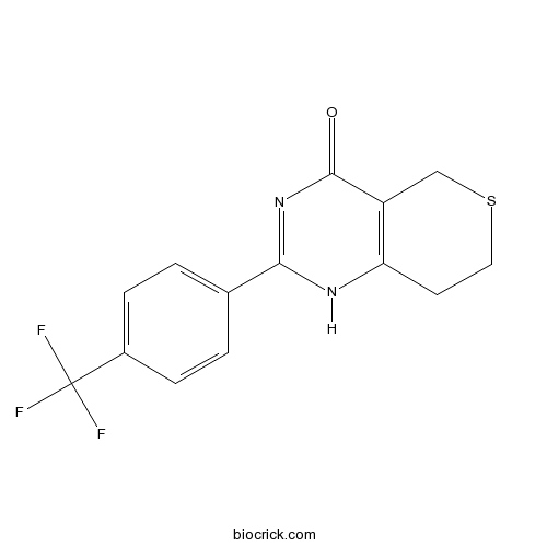
XAV-939Tankyrase 1/2 inhibitor CAS# 284028-89-3 |

Quality Control & MSDS
3D structure
Package In Stock
Number of papers citing our products

| Cas No. | 284028-89-3 | SDF | Download SDF |
| PubChem ID | 2726824 | Appearance | Powder |
| Formula | C14H11F3N2OS | M.Wt | 312.31 |
| Type of Compound | N/A | Storage | Desiccate at -20°C |
| Solubility | DMSO : 21.5 mg/mL (68.84 mM; Need ultrasonic and warming) H2O : < 0.1 mg/mL (insoluble) | ||
| Chemical Name | 2-[4-(trifluoromethyl)phenyl]-1,5,7,8-tetrahydrothiopyrano[4,3-d]pyrimidin-4-one | ||
| SMILES | C1CSCC2=C1NC(=NC2=O)C3=CC=C(C=C3)C(F)(F)F | ||
| Standard InChIKey | KLGQSVMIPOVQAX-UHFFFAOYSA-N | ||
| Standard InChI | InChI=1S/C14H11F3N2OS/c15-14(16,17)9-3-1-8(2-4-9)12-18-11-5-6-21-7-10(11)13(20)19-12/h1-4H,5-7H2,(H,18,19,20) | ||
| General tips | For obtaining a higher solubility , please warm the tube at 37 ℃ and shake it in the ultrasonic bath for a while.Stock solution can be stored below -20℃ for several months. We recommend that you prepare and use the solution on the same day. However, if the test schedule requires, the stock solutions can be prepared in advance, and the stock solution must be sealed and stored below -20℃. In general, the stock solution can be kept for several months. Before use, we recommend that you leave the vial at room temperature for at least an hour before opening it. |
||
| About Packaging | 1. The packaging of the product may be reversed during transportation, cause the high purity compounds to adhere to the neck or cap of the vial.Take the vail out of its packaging and shake gently until the compounds fall to the bottom of the vial. 2. For liquid products, please centrifuge at 500xg to gather the liquid to the bottom of the vial. 3. Try to avoid loss or contamination during the experiment. |
||
| Shipping Condition | Packaging according to customer requirements(5mg, 10mg, 20mg and more). Ship via FedEx, DHL, UPS, EMS or other couriers with RT, or blue ice upon request. | ||
| Description | Potent tankyrase (TNKS) inhibitor (IC50 values are 4 and 11 nM for TNKS2 and TNKS1 respectively). Antagonizes Wnt signaling via stimulation of β-catenin degradation and stabilization of axin. Inhibits proliferation of the β-catenin-dependent colon carcinoma cell line DLD-1. Promotes cardiomyogenic development in mesoderm progenitor cells. |

XAV-939 Dilution Calculator

XAV-939 Molarity Calculator
| 1 mg | 5 mg | 10 mg | 20 mg | 25 mg | |
| 1 mM | 3.2019 mL | 16.0097 mL | 32.0195 mL | 64.0389 mL | 80.0487 mL |
| 5 mM | 0.6404 mL | 3.2019 mL | 6.4039 mL | 12.8078 mL | 16.0097 mL |
| 10 mM | 0.3202 mL | 1.601 mL | 3.2019 mL | 6.4039 mL | 8.0049 mL |
| 50 mM | 0.064 mL | 0.3202 mL | 0.6404 mL | 1.2808 mL | 1.601 mL |
| 100 mM | 0.032 mL | 0.1601 mL | 0.3202 mL | 0.6404 mL | 0.8005 mL |
| * Note: If you are in the process of experiment, it's necessary to make the dilution ratios of the samples. The dilution data above is only for reference. Normally, it's can get a better solubility within lower of Concentrations. | |||||

Calcutta University

University of Minnesota

University of Maryland School of Medicine

University of Illinois at Chicago

The Ohio State University

University of Zurich

Harvard University

Colorado State University

Auburn University

Yale University

Worcester Polytechnic Institute

Washington State University

Stanford University

University of Leipzig

Universidade da Beira Interior

The Institute of Cancer Research

Heidelberg University

University of Amsterdam

University of Auckland

TsingHua University

The University of Michigan

Miami University

DRURY University

Jilin University

Fudan University

Wuhan University

Sun Yat-sen University

Universite de Paris

Deemed University

Auckland University

The University of Tokyo

Korea University
XAV-939 is a small-molecule inhibitor of tankyrase 1/2 with IC50 values of 4 and 11 nM, respectively [1].
XAV-939 was screened out as an inhibitor of Wnt/β-catenin pathway. In HEK293 cells, XAV-939 significantly inhibited the Wnt3a-stimulated STF activity and β-catenin accumulation. In SW480 cells, XAV-939 also inhibited STF activity and increased β-catenin phosphorylation. XAV-939 was demonstrated to stabilize the axin levels through inhibiting tankyrases and subsequently inhibit the Wnt signaling. It tightly bound to the catalytic domains of tankyrase 1 and tankyrase 2 with Kd values of 99 and 93 nM, respectively. In addition, XAV-939 significantly inhibited colony formation of β-catenin-dependent DLD-1 cells but not β-catenin-independent RKO cells [1].
References:
[1] Huang S M A, Mishina Y M, Liu S, et al. Tankyrase inhibition stabilizes axin and antagonizes Wnt signalling. Nature, 2009, 461(7264): 614-620.
- FR 236924
Catalog No.:BCC7564
CAS No.:28399-31-7
- 1(10)-Aristolen-2-one
Catalog No.:BCN7634
CAS No.:28398-06-3
- Bumetanide
Catalog No.:BCC1119
CAS No.:28395-03-1
- 7-Neohesperidosides
Catalog No.:BCN8200
CAS No.:28383-41-7
- Aloin B
Catalog No.:BCN2576
CAS No.:28371-16-6
- Chrysin 6-C-glucoside
Catalog No.:BCN3324
CAS No.:28368-57-2
- Mahanine
Catalog No.:BCN3176
CAS No.:28360-49-8
- Canolol
Catalog No.:BCC8371
CAS No.:28343-22-8
- Oxychelerythrine
Catalog No.:BCN4864
CAS No.:28342-33-8
- 6-Ethoxydihydrosanguinarine
Catalog No.:BCN7589
CAS No.:28342-31-6
- IVHD-valtrate
Catalog No.:BCN7125
CAS No.:28325-56-6
- sn-Glycero-3-phosphocholine
Catalog No.:BCC4168
CAS No.:28319-77-9
- DR 2313
Catalog No.:BCC2451
CAS No.:284028-90-6
- NPS-2143
Catalog No.:BCC4409
CAS No.:284035-33-2
- Ac9-25
Catalog No.:BCC5997
CAS No.:284040-76-2
- 6-Hydroxy-2,6-dimethyl-2,7-octadienoic acid
Catalog No.:BCN1463
CAS No.:28420-25-9
- Sorafenib
Catalog No.:BCN2174
CAS No.:284461-73-0
- 4-(4-Aminophenoxy)-N-methyl-2-pyridinecarboxamide
Catalog No.:BCC8649
CAS No.:284462-37-9
- Bavachalcone
Catalog No.:BCN3193
CAS No.:28448-85-3
- Tomentin
Catalog No.:BCN5180
CAS No.:28449-62-9
- 20-Deacetyltaxuspine X
Catalog No.:BCN7374
CAS No.:284672-76-0
- 9-Deacetyltaxinine E
Catalog No.:BCN7227
CAS No.:284672-78-2
- Delphinidin-3-O-galactoside chloride
Catalog No.:BCN3019
CAS No.:28500-00-7
- Petunidin-3-O-galactoside chloride
Catalog No.:BCN3024
CAS No.:28500-02-9
Chemoradiotherapy Resistance in Colorectal Cancer Cells is Mediated by Wnt/beta-catenin Signaling.[Pubmed:28811361]
Mol Cancer Res. 2017 Nov;15(11):1481-1490.
Activation of Wnt/beta-catenin signaling plays a central role in the development and progression of colorectal cancer. The Wnt-transcription factor, TCF7L2, is overexpressed in primary rectal cancers that are resistant to chemoradiotherapy and TCF7L2 mediates resistance to chemoradiotherapy. However, it is unclear whether the resistance is mediated by a TCF7L2 inherent mechanism or Wnt/beta-catenin signaling in general. Here, inhibition of beta-catenin by siRNAs or a small-molecule inhibitor (XAV-939) resulted in sensitization of colorectal cancer cells to chemoradiotherapy. To investigate the potential role of Wnt/beta-catenin signaling in controlling therapeutic responsiveness, nontumorigenic RPE-1 cells were stimulated with Wnt-3a, a physiologic ligand of Frizzled receptors, which increased resistance to chemoradiotherapy. This effect could be recapitulated by overexpression of a degradation-resistant mutant of beta-catenin (S33Y), also boosting resistance of RPE-1 cells to chemoradiotherapy, which was, conversely, abrogated by siRNA-mediated silencing of beta-catenin. Consistent with these findings, higher expression levels of active beta-catenin were observed as well as increased TCF/LEF reporter activity in SW1463 cells that evolved radiation resistance due to repeated radiation treatment. Global gene expression profiling identified several altered pathways, including PPAR signaling and other metabolic pathways, associated with cellular response to radiation. In summary, aberrant activation of Wnt/beta-catenin signaling not only regulates the development and progression of colorectal cancer, but also mediates resistance of rectal cancers to chemoradiotherapy.Implications: Targeting Wnt/beta-catenin signaling or one of the downstream pathways represents a promising strategy to increase response to chemoradiotherapy. Mol Cancer Res; 15(11); 1481-90. (c)2017 AACR.
[miR-218 Promoted the Apoptosis of Human Ovarian Carcinoma Cells via Suppression of the WNT/beta-Catenin Signaling Pathway].[Pubmed:28900081]
Mol Biol (Mosk). 2017 Jul-Aug;51(4):629-636.
MicroRNA-218 (miR-218) is a short, noncoding RNA, with multiple biological functions. In this study, we aimed to investigate the potential effects of miR-218 on the apoptosis of human ovarian carcinoma cells and the underlying mechanisms by which miR-218 exerted its actions. After over-expressing miR-218 in human ovarian carcinoma (OVCAR3) cells, cell viability was determined by MTT method, cell apoptosis was observed by flow cytometry (FCM), mRNA expression of miR-218, Bcl2, Bax was measured by RT-PCR and protein expression levels of Wnt, tankyrase and beta-catenin were quantified by Western blots. Over-expression of miR-218 potently suppressed cell viability and promoted the apoptosis of human ovarian carcinoma cells in a time-dependent manner. In addition, the down-regulation of tankyrase expression level was detected in miR-218-over-expressed cells. Following the block of the Wnt/beta-catenin signaling pathway using the inhibitor XAV-939, the effects of miR-218 on the proliferation and apoptosis of human ovarian carcinoma cells were significantly suppressed. Augmenting expression of miR-218 and/or miRNA-218 mimicking therapeutics may provide viable avenue for the treatment of ovarian cancer.
Extracellular matrix derived from periodontal ligament cells maintains their stemness and enhances redifferentiation via the wnt pathway.[Pubmed:28884507]
J Biomed Mater Res A. 2018 Jan;106(1):272-284.
Large numbers of viable cells cannot be obtained from periodontal ligament tissues of patients with periodontitis. Therefore, it is imperative to establish an ex vivo environment that can support cell proliferation and delay senescence. Here, we have successfully reconstructed a native extracellular matrix (ECM), derived from early-passage human periodontal ligament cells (PDLCs) using the NH4 OH/Triton X-100 protocol. The ECM was investigated by scanning electron microscopy and immunostaining for specific ECM proteins (collagen I and fibronectin). Late-passage ECM-expanded PDLCs exhibited a much higher proliferation index and lower levels of reactive oxygen species (ROS), confirmed by the increased expression of pluripotent markers and enhanced osteogenic capacity. Interestingly, the Wnt pathway was suppressed during the ECM expansion-mediated increase in pluripotency, but was activated in an osteogenic differentiation environment, as confirmed by treatment with the XAV-939 beta-catenin inhibitor or the SP600125 c-Jun N-terminal kinase (JNK) inhibitor. Cell sheets formed by ECM-expanded PDLCs exhibited an enhanced periodontal tissue regeneration capacity compared to those formed on tissue culture polystyrene (TCP) surfaces in vivo. Taken together, the cell-free ECM provides a tissue-specific cell niche for the ex vivo expansion of PDLCs while retaining stemness and osteogenic potential, partially via the Wnt pathway. This represents a promising matrix for future applications in periodontal tissue regeneration therapy. (c) 2017 Wiley Periodicals, Inc. J Biomed Mater Res Part A: 106A: 272-284, 2018.
Axin1 up-regulated 1 accelerates stress-induced cardiomyocytes apoptosis through activating Wnt/beta-catenin signaling.[Pubmed:28830684]
Exp Cell Res. 2017 Oct 15;359(2):441-448.
Stress-induced cardiomyocyte apoptosis contributes to the pathogenesis of a variety of cardiovascular diseases, but how stress induces cardiomyocyte apoptosis remains largely unclear. The present study aims to investigate the effects of Axin1 up-regulated 1 (Axud1), a novel pro-apoptotic protein, on the cardiomyocyte survival and the underlying mechanisms. To this end, a rat model under restraint stress (RS) was established and in vitro stress-induced cardiomyocytes culture was achieved. Our data showed that Axud1 was upregulated in the rat myocardia after exposure to RS. Anti-apoptotic Bcl-2 was decreased, whereas pro-apoptotic Bax and Cleaved caspase-3 (Cc3) were increased in a time-dependent manner. The Wnt/beta-catenin signaling was observed to be interestingly activated in heart undergoing RS. In addition, the treatment of norepinephrine (NE) to in vitro cardiomyocytes increased Axud1 level and induced cell apoptosis. Wnt/beta-catenin signaling was consistently activated. Knockdown of Axud1 using specific siRNA blunted NE-induced cardiomyocytes apoptosis and also inactivated the Wnt/beta-catenin signaling. XAV-939, an inhibitor of Wnt/beta-catenin signaling, partially reversed the pro-apoptotic effect of NE. In conclusion, Axud1 accelerated stress-induced cardiomyocytes apoptosis through activation of Wnt/beta-catenin signaling pathway. Our data provided novel evidence that therapeutic strategies against Axud1 or Wnt/beta-catenin signaling might be promising in relation to RS-induced myocardial injury.
Tankyrase inhibitors suppress hepatocellular carcinoma cell growth via modulating the Hippo cascade.[Pubmed:28877210]
PLoS One. 2017 Sep 6;12(9):e0184068.
Previous data indicate that Tankyrase inhibitors exert anti-growth functions in many cancer cell lines due to their ability to inactivate the YAP protooncogene. In the present manuscript, we investigated the effect of Tankyrase inhibitors on the growth of hepatocellular carcinoma (HCC) cell lines and the molecular mechanisms involved. For this purpose, we performed cell proliferation assay by colony-forming ability in seven human HCC cells subjected to XAV-939 and G007-LK Tankyrase inhibitors. Noticeably, the two Tankyrase inhibitors suppressed the HCC cell growth in a dose-dependent manner. Furthermore, we found that Tankyrase inhibitors synergized with MEK and AKT inhibitors to suppress HCC cell proliferation. At the molecular level, Tankyrase inhibitors significantly decreased YAP protein levels, reduced the expression of YAP target genes, and inhibited YAP/TEAD luciferase reporter activity. In addition, Tankyrase inhibitors administration was accompanied by upregulation of Angiomotin-like 1 (AMOTL1) and Angiomotin-like 2 (AMOTL2) proteins, two major negative regulators of YAP. Altogether, the present data indicate that XAV-939 and G007-LK Tankyrase inhibitors could suppress proliferation of hepatocellular carcinoma cells and downregulate YAP/TAZ by stabilizing AMOTL1 and AMOTL2 proteins, thus representing new potential anticancer drugs against hepatocellular carcinoma.
Cardiac induction of embryonic stem cells by a small molecule inhibitor of Wnt/beta-catenin signaling.[Pubmed:21077691]
ACS Chem Biol. 2011 Feb 18;6(2):192-7.
In vitro differentiation of embryonic stem cells is tightly regulated by the same key signaling pathways that control pattern formation during embryogenesis. Small molecules that selectively target these developmental pathways, including Wnt, and BMP signaling may be valuable for directing differentiation of pluripotent stem cells toward many desired tissue types, but to date only few such compounds have been shown to promote cardiac differentiation. Here, we show that XAV939, a recently discovered small molecule inhibitor of Wnt/beta-catenin signaling, can robustly induce cardiomyogenesis in mouse ES cells. Our results suggest that a timely administration of XAV939 immediately following the formation of mesoderm progenitor cells promotes cardiomyogenic development at the expense of other mesoderm derived lineages, including the endothelial, smooth muscle, and hematopoietic lineages. Given the critical role that Wnt/beta-catenin signaling plays in many aspects of embryogenesis and tissue regeneration, XAV939 is a valuable chemical probe to dissect in vitro differentiation of stem cells and to explore their regenerative potential in a variety of contexts.
Tankyrase inhibition stabilizes axin and antagonizes Wnt signalling.[Pubmed:19759537]
Nature. 2009 Oct 1;461(7264):614-20.
The stability of the Wnt pathway transcription factor beta-catenin is tightly regulated by the multi-subunit destruction complex. Deregulated Wnt pathway activity has been implicated in many cancers, making this pathway an attractive target for anticancer therapies. However, the development of targeted Wnt pathway inhibitors has been hampered by the limited number of pathway components that are amenable to small molecule inhibition. Here, we used a chemical genetic screen to identify a small molecule, XAV939, which selectively inhibits beta-catenin-mediated transcription. XAV939 stimulates beta-catenin degradation by stabilizing axin, the concentration-limiting component of the destruction complex. Using a quantitative chemical proteomic approach, we discovered that XAV939 stabilizes axin by inhibiting the poly-ADP-ribosylating enzymes tankyrase 1 and tankyrase 2. Both tankyrase isoforms interact with a highly conserved domain of axin and stimulate its degradation through the ubiquitin-proteasome pathway. Thus, our study provides new mechanistic insights into the regulation of axin protein homeostasis and presents new avenues for targeted Wnt pathway therapies.


