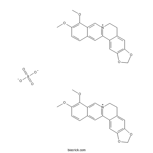
Berberine SulphateCAS# 316-41-6 |

Quality Control & MSDS
3D structure
Package In Stock
Number of papers citing our products

| Cas No. | 316-41-6 | SDF | Download SDF |
| PubChem ID | 9424 | Appearance | Powder |
| Formula | C40H36N2O12S | M.Wt | 768.8 |
| Type of Compound | N/A | Storage | Desiccate at -20°C |
| Solubility | Soluble in Chloroform,Dichloromethane,Ethyl Acetate,DMSO,Acetone,etc. | ||
| SMILES | COC1=C(C2=C[N+]3=C(C=C2C=C1)C4=CC5=C(C=C4CC3)OCO5)OC.COC1=C(C2=C[N+]3=C(C=C2C=C1)C4=CC5=C(C=C4CC3)OCO5)OC.[O-]S(=O)(=O)[O-] | ||
| Standard InChIKey | OJVABJMSSDUECT-UHFFFAOYSA-L | ||
| Standard InChI | InChI=1S/2C20H18NO4.H2O4S/c2*1-22-17-4-3-12-7-16-14-9-19-18(24-11-25-19)8-13(14)5-6-21(16)10-15(12)20(17)23-2;1-5(2,3)4/h2*3-4,7-10H,5-6,11H2,1-2H3;(H2,1,2,3,4)/q2*+1;/p-2 | ||
| General tips | For obtaining a higher solubility , please warm the tube at 37 ℃ and shake it in the ultrasonic bath for a while.Stock solution can be stored below -20℃ for several months. We recommend that you prepare and use the solution on the same day. However, if the test schedule requires, the stock solutions can be prepared in advance, and the stock solution must be sealed and stored below -20℃. In general, the stock solution can be kept for several months. Before use, we recommend that you leave the vial at room temperature for at least an hour before opening it. |
||
| About Packaging | 1. The packaging of the product may be reversed during transportation, cause the high purity compounds to adhere to the neck or cap of the vial.Take the vail out of its packaging and shake gently until the compounds fall to the bottom of the vial. 2. For liquid products, please centrifuge at 500xg to gather the liquid to the bottom of the vial. 3. Try to avoid loss or contamination during the experiment. |
||
| Shipping Condition | Packaging according to customer requirements(5mg, 10mg, 20mg and more). Ship via FedEx, DHL, UPS, EMS or other couriers with RT, or blue ice upon request. | ||

Berberine Sulphate Dilution Calculator

Berberine Sulphate Molarity Calculator
| 1 mg | 5 mg | 10 mg | 20 mg | 25 mg | |
| 1 mM | 1.3007 mL | 6.5036 mL | 13.0073 mL | 26.0146 mL | 32.5182 mL |
| 5 mM | 0.2601 mL | 1.3007 mL | 2.6015 mL | 5.2029 mL | 6.5036 mL |
| 10 mM | 0.1301 mL | 0.6504 mL | 1.3007 mL | 2.6015 mL | 3.2518 mL |
| 50 mM | 0.026 mL | 0.1301 mL | 0.2601 mL | 0.5203 mL | 0.6504 mL |
| 100 mM | 0.013 mL | 0.065 mL | 0.1301 mL | 0.2601 mL | 0.3252 mL |
| * Note: If you are in the process of experiment, it's necessary to make the dilution ratios of the samples. The dilution data above is only for reference. Normally, it's can get a better solubility within lower of Concentrations. | |||||

Calcutta University

University of Minnesota

University of Maryland School of Medicine

University of Illinois at Chicago

The Ohio State University

University of Zurich

Harvard University

Colorado State University

Auburn University

Yale University

Worcester Polytechnic Institute

Washington State University

Stanford University

University of Leipzig

Universidade da Beira Interior

The Institute of Cancer Research

Heidelberg University

University of Amsterdam

University of Auckland

TsingHua University

The University of Michigan

Miami University

DRURY University

Jilin University

Fudan University

Wuhan University

Sun Yat-sen University

Universite de Paris

Deemed University

Auckland University

The University of Tokyo

Korea University
- Heraclenol
Catalog No.:BCN5234
CAS No.:31575-93-6
- 4EGI-1
Catalog No.:BCC5337
CAS No.:315706-13-9
- PTC-209
Catalog No.:BCC5111
CAS No.:315704-66-6
- JK 184
Catalog No.:BCC3936
CAS No.:315703-52-7
- STF-62247
Catalog No.:BCC4960
CAS No.:315702-99-9
- TC-DAPK 6
Catalog No.:BCC1989
CAS No.:315694-89-4
- Kavain
Catalog No.:BCN8295
CAS No.:3155-48-4
- 1,18-Octadecanediol
Catalog No.:BCN5233
CAS No.:3155-43-9
- Matsukaze-lactone
Catalog No.:BCN7580
CAS No.:3153-73-9
- O-Nornuciferine
Catalog No.:BCN7074
CAS No.:3153-55-7
- 5-Hydroxyseselin
Catalog No.:BCN3428
CAS No.:31525-75-4
- Isobavachin
Catalog No.:BCN5232
CAS No.:31524-62-6
- Emetine dihydrochloride
Catalog No.:BCN8307
CAS No.:316-42-7
- Z-Asp(OMe)-OH
Catalog No.:BCC2790
CAS No.:3160-47-2
- Z-Ile-OH
Catalog No.:BCC2593
CAS No.:3160-59-6
- Moroxydine HCl
Catalog No.:BCC4802
CAS No.:3160-91-6
- SCH 442416
Catalog No.:BCC7372
CAS No.:316173-57-6
- Artocarpesin
Catalog No.:BCN8071
CAS No.:3162-09-2
- 5,7-Dihydroxy-6,8-dimethoxyflavone
Catalog No.:BCN5235
CAS No.:3162-45-6
- 9-Anthracenylmethyl acrylate
Catalog No.:BCC8798
CAS No.:31645-34-8
- Palifosfamide
Catalog No.:BCC1833
CAS No.:31645-39-3
- 6-Aminonicotinic acid
Catalog No.:BCC8764
CAS No.:3167-49-5
- Gatifloxacin mesylate
Catalog No.:BCC4225
CAS No.:316819-28-0
- Pinusolide
Catalog No.:BCN5236
CAS No.:31685-80-0
Distribution, density and histochemical profiles of the lung mast cells during the post-hatching period of Japanese quails (Coturnix coturnix japonica).[Pubmed:20537669]
Res Vet Sci. 2011 Feb;90(1):1-8.
The distribution, density and histochemical characteristics of mast cells in the lungs of the Japanese quail were investigated during the post-hatching period. In the period starting from the first to the 60th day post-hatching, based on proteoglycan content, three types of mast cells, which were alcian blue-positive, safranin O-positive and alcian blue/safranin O-positive, were found to exist in the lungs. The application of staining with Berberine Sulphate demonstrated that, similar to the distribution of safranin O-positive cells, the heparin-containing cells were located in the periphery of large blood vessels. The percentages of mast cells in different localization sites of the lungs were found to vary with age in the post-hatching period with toluidine blue staining. The lack of any statistically significant increase/decrease in the number of mast cells per unit area of the right and left lung lobes is partially in favour of the proposal that the mast cell number increases with the growth of the lung volume in the post-hatching period.
Characterization of aqueous pores in plant cuticles and permeation of ionic solutes.[Pubmed:16825315]
J Exp Bot. 2006;57(11):2471-91.
Plant cuticles are lipid membranes with separate diffusion paths for lipophilic non-electrolytes and hydrated ionic compounds. Ions are lipid insoluble and require an aqueous pathway across cuticles. Based on experimental data, the aqueous pathway in cuticles has been characterized. Aqueous pores arise by hydration of permanent dipoles and ionic functional groups. They can be localized using ionic fluorescent dyes, silver nitrate, and mercuric chloride. Aqueous pores preferentially occur in cuticular ledges, at the base of trichomes, and in cuticles over anticlinal walls. Average pore radii ranged from 0.45 to 1.18 nm. Penetration of ions was a first order process as the fraction of the salt remaining on the cuticle surface decreased exponentially with time. Permeability of cuticles to ions depended on humidity and was highest at 100% humidity. Wetting agents increased rate constants by factors of up to 12, which indicates that the pore openings are surrounded by waxes. The pores in cuticular ledges of Helxine soleirolii allowed passage of Berberine Sulphate, which has a molecular weight of 769 g mol(-1). Increasing the molecular weight of solutes from 100 to 500 g mol(-1) decreased the rate constants of penetration by factors of 7 (Vicia faba) and 13 (Populus canescens), respectively. Half-times of penetration of inorganic salts and organic ions across Populus cuticles and Vicia leaf surfaces varied between 1 and 12 h. This shows that penetration of ionic compounds can be fairly rapid, and ions with molecular weights of up to 800 g mol(-1) can penetrate cuticles that possess aqueous pores.
Distribution, histochemical and enzyme histochemical characterization of mast cells in dogs.[Pubmed:15328916]
J Mol Histol. 2004 Feb;35(2):123-32.
This study describes the distribution, proteoglycan properties and protease activity of mast cells from 15 different dog organs. In beagles and mixed breed dogs, staining with Alcian Blue-Safranin O revealed mast cells in all the organs examined. However, their numbers varied and they demonstrated unique localization patterns within some of these organs. Berberine Sulphate fluorescence-positive mast cells were observed in the submucosa, muscularis and serosa of the intestines, as well as the tongue and liver (within the connective tissue). Mast cells within the intestinal mucosa were negative for, or demonstrated weak, Berberine Sulphate staining. Heterogeneity of mast cells in terms of the proteoglycans contained within their granules was further confirmed by determination of critical electrolyte concentrations (CECs). The CECs of mast cells within the connective tissue of several organs, including the intestines (submucosal and muscularis-serosal layers) were all greater than 1.0 M. The results from CEC experiments together with berberine staining indicate that heparin was contained within their granules. Relative to the CECs of mast cells in other organs, mast cells in the intestinal mucosa exhibited lower CECs, suggesting that the proteoglycans within their granules were of lower charge density and/or molecular weight. Although mast cells were classified into two groups by proteoglycans within the granules, enzyme histochemical analysis in beagles revealed three subtypes of mast cells: chymase (MC(C)), tryptase (MC(T)) and dual positive (MC(TC)) cells. There was no correlation between the proteoglycan content and enzyme properties of the mast cell granules.


