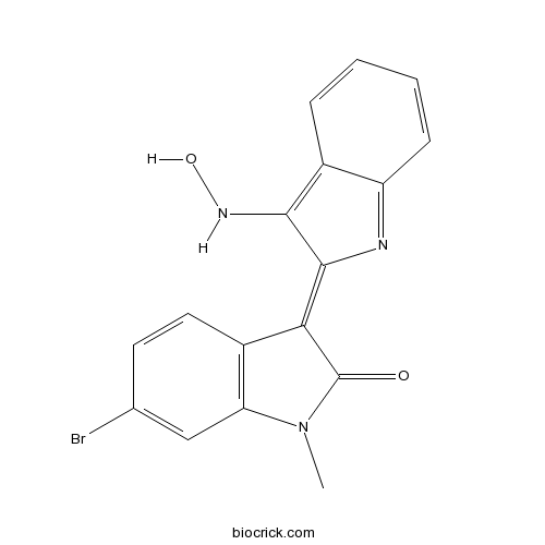
MeBIOCDK/Cyclin/ GSK-3/p25/AhR agonist CAS# 667463-95-8 |

- MK-4827
Catalog No.:BCC1761
CAS No.:1038915-60-4
- BMN-673 8R,9S
Catalog No.:BCC1422
CAS No.:1207456-00-5
- XAV-939
Catalog No.:BCC1120
CAS No.:284028-89-3
- PJ34
Catalog No.:BCC1865
CAS No.:344458-19-1
- ABT-888 (Veliparib)
Catalog No.:BCC1267
CAS No.:912444-00-9
Quality Control & MSDS
3D structure
Package In Stock
Number of papers citing our products

| Cas No. | 667463-95-8 | SDF | Download SDF |
| PubChem ID | 24906281 | Appearance | Powder |
| Formula | C17H12BrN3O2 | M.Wt | 370.2 |
| Type of Compound | N/A | Storage | Desiccate at -20°C |
| Solubility | Soluble to 10 mM in DMSO | ||
| Chemical Name | 6-bromo-3-[3-(hydroxyamino)indol-2-ylidene]-1-methylindol-2-one | ||
| SMILES | CN1C2=C(C=CC(=C2)Br)C(=C3C(=C4C=CC=CC4=N3)NO)C1=O | ||
| Standard InChIKey | WWVVQXIBSZPELF-UHFFFAOYSA-N | ||
| Standard InChI | InChI=1S/C17H12BrN3O2/c1-21-13-8-9(18)6-7-11(13)14(17(21)22)16-15(20-23)10-4-2-3-5-12(10)19-16/h2-8,20,23H,1H3 | ||
| General tips | For obtaining a higher solubility , please warm the tube at 37 ℃ and shake it in the ultrasonic bath for a while.Stock solution can be stored below -20℃ for several months. We recommend that you prepare and use the solution on the same day. However, if the test schedule requires, the stock solutions can be prepared in advance, and the stock solution must be sealed and stored below -20℃. In general, the stock solution can be kept for several months. Before use, we recommend that you leave the vial at room temperature for at least an hour before opening it. |
||
| About Packaging | 1. The packaging of the product may be reversed during transportation, cause the high purity compounds to adhere to the neck or cap of the vial.Take the vail out of its packaging and shake gently until the compounds fall to the bottom of the vial. 2. For liquid products, please centrifuge at 500xg to gather the liquid to the bottom of the vial. 3. Try to avoid loss or contamination during the experiment. |
||
| Shipping Condition | Packaging according to customer requirements(5mg, 10mg, 20mg and more). Ship via FedEx, DHL, UPS, EMS or other couriers with RT, or blue ice upon request. | ||
| Description | Control analog of 6-bromoindirubin-3'-oxime (BIO). Displays minimal activity against CDK1/Cyclin B, GSK-3 α/β, and CDK5/p25 (IC50 values are 92.0, 44-100 and >100 μM respectively). Aryl hydrocarbon receptor (AhR) agonist; causes redistribution of AhR to the nucleus. |

MeBIO Dilution Calculator

MeBIO Molarity Calculator
| 1 mg | 5 mg | 10 mg | 20 mg | 25 mg | |
| 1 mM | 2.7012 mL | 13.5062 mL | 27.0124 mL | 54.0249 mL | 67.5311 mL |
| 5 mM | 0.5402 mL | 2.7012 mL | 5.4025 mL | 10.805 mL | 13.5062 mL |
| 10 mM | 0.2701 mL | 1.3506 mL | 2.7012 mL | 5.4025 mL | 6.7531 mL |
| 50 mM | 0.054 mL | 0.2701 mL | 0.5402 mL | 1.0805 mL | 1.3506 mL |
| 100 mM | 0.027 mL | 0.1351 mL | 0.2701 mL | 0.5402 mL | 0.6753 mL |
| * Note: If you are in the process of experiment, it's necessary to make the dilution ratios of the samples. The dilution data above is only for reference. Normally, it's can get a better solubility within lower of Concentrations. | |||||

Calcutta University

University of Minnesota

University of Maryland School of Medicine

University of Illinois at Chicago

The Ohio State University

University of Zurich

Harvard University

Colorado State University

Auburn University

Yale University

Worcester Polytechnic Institute

Washington State University

Stanford University

University of Leipzig

Universidade da Beira Interior

The Institute of Cancer Research

Heidelberg University

University of Amsterdam

University of Auckland

TsingHua University

The University of Michigan

Miami University

DRURY University

Jilin University

Fudan University

Wuhan University

Sun Yat-sen University

Universite de Paris

Deemed University

Auckland University

The University of Tokyo

Korea University
Control analog of 6-bromoindirubin-3'-oxime. Displays minimal activity against CDK1/Cyclin B, GSK-3 α/β, and CDK5/p25 (IC50 values are 92.0, 44-100 and >100 μM respectively). Aryl hydrocarbon receptor (AhR) agonist; causes redistribution of AhR to the nuc
- BIO-acetoxime
Catalog No.:BCC6076
CAS No.:667463-85-6
- GSK-3 Inhibitor IX (BIO)
Catalog No.:BCC4510
CAS No.:667463-62-9
- PG 931
Catalog No.:BCC6363
CAS No.:667430-81-1
- 5-Hydroxy-7-acetoxyflavone
Catalog No.:BCN4217
CAS No.:6674-40-4
- DBU
Catalog No.:BCC2840
CAS No.:6674-22-2
- 3-Nitro-L-tyrosine ethyl ester hydrochloride
Catalog No.:BCN1384
CAS No.:66737-54-0
- E-64
Catalog No.:BCC1222
CAS No.:66701-25-5
- 8-Bromo-7-(but-2-yn-1-yl)-3-methyl-1H-purine-2,6(3H,7H)-dione
Catalog No.:BCC8786
CAS No.:666816-98-4
- Polygalacin D
Catalog No.:BCN3861
CAS No.:66663-91-0
- Platycodin D2
Catalog No.:BCN3847
CAS No.:66663-90-9
- Esculentoside H
Catalog No.:BCN2795
CAS No.:66656-92-6
- 7-Hydroxyflavone
Catalog No.:BCN3673
CAS No.:6665-86-7
- NSC 109555 ditosylate
Catalog No.:BCC7540
CAS No.:66748-43-4
- Diosbulbin D
Catalog No.:BCN4218
CAS No.:66756-57-8
- 6,8-Diprenylorobol
Catalog No.:BCN4602
CAS No.:66777-70-6
- Esculin Sesquihydrate
Catalog No.:BCC8324
CAS No.:66778-17-4
- Platycodin A
Catalog No.:BCN7997
CAS No.:66779-34-8
- Impurity B of Calcitriol
Catalog No.:BCC1645
CAS No.:66791-71-7
- Syringaresinol-di-O-glucoside
Catalog No.:BCN2600
CAS No.:66791-77-3
- (+/-)-Forbesione
Catalog No.:BCN6423
CAS No.:667914-50-3
- Hernandezine
Catalog No.:BCN7793
CAS No.:6681-13-6
- Jatrorrhizine chloride
Catalog No.:BCN4956
CAS No.:6681-15-8
- Magnoflorine chloride
Catalog No.:BCN2405
CAS No.:6681-18-1
- Linagliptin (BI-1356)
Catalog No.:BCC2110
CAS No.:668270-12-0
Arterial Tortuosity: An Imaging Biomarker of Childhood Stroke Pathogenesis?[Pubmed:27006453]
Stroke. 2016 May;47(5):1265-70.
BACKGROUND AND PURPOSE: Arteriopathy is the leading cause of childhood arterial ischemic stroke. Mechanisms are poorly understood but may include inherent abnormalities of arterial structure. Extracranial dissection is associated with connective tissue disorders in adult stroke. Focal cerebral arteriopathy is a common syndrome where pathophysiology is unknown but may include intracranial dissection or transient cerebral arteriopathy. We aimed to quantify cerebral arterial tortuosity in childhood arterial ischemic stroke, hypothesizing increased tortuosity in dissection. METHODS: Children (1 month to 18 years) with arterial ischemic stroke were recruited within the Vascular Effects of Infection in Pediatric Stroke (VIPS) study with controls from the Calgary Pediatric Stroke Program. Objective, multi-investigator review defined diagnostic categories. A validated imaging software method calculated the mean arterial tortuosity of the major cerebral arteries using 3-dimensional time-of-flight magnetic resonance angiographic source images. Tortuosity of unaffected vessels was compared between children with dissection, transient cerebral arteriopathy, meningitis, moyamoya, cardioembolic strokes, and controls (ANOVA and post hoc Tukey). Trauma-related versus spontaneous dissection was compared (Student t test). RESULTS: One hundred fifteen children were studied (median, 6.8 years; 43% women). Age and sex were similar across groups. Tortuosity means and variances were consistent with validation studies. Tortuosity in controls (1.346+/-0.074; n=15) was comparable with moyamoya (1.324+/-0.038; n=15; P=0.998), meningitis (1.348+/-0.052; n=11; P=0.989), and cardioembolic (1.379+/-0.056; n=27; P=0.190) cases. Tortuosity was higher in both extracranial dissection (1.404+/-0.084; n=22; P=0.021) and transient cerebral arteriopathy (1.390+/-0.040; n=27; P=0.001) children. Tortuosity was not different between traumatic versus spontaneous dissections (P=0.70). CONCLUSIONS: In children with dissection and transient cerebral arteriopathy, cerebral arteries demonstrate increased tortuosity. Quantified arterial tortuosity may represent a clinically relevant imaging biomarker of vascular biology in pediatric stroke.
[A clinical study of cryptococcal meningitis--sequential changes of cryptococcal antigen titers].[Pubmed:12708008]
Kansenshogaku Zasshi. 2003 Mar;77(3):150-7.
We reviewed the clinical manifestations, sequential changes in cryptococcal antigen titers in serum and cerebrospinal fluid (CSF), and the antifungal drug susceptibility of Cryptococcus neoformans in three patients with cryptococcal meningitis between 1996 and 2000. Cryptococcal antigen titers were measured using the latex agglutination method with Pastrex Cryptococcus (Fuji MeBIO, Tokyo) and Serodirect Cryptococcus (Eiken Chemical, Tokyo). The underlying systemic diseases in the three patients were liver cirrhosis, non-Hodgkin's lymphoma associated with miliary tuberculosis, and malignant thymoma associated with systemic lupus erythymatosus. The CSF samples showed positive indian ink staining in two of the three patients and C. neoformans was cultured from all three. The cryptococcal antigen titers in serum were higher than those in the CSF. The serum and CSF cryptococcal antigen titers measured by Serodirect Cryptococcus were higher than those measured by Pastrex Cryptococcus. The maximum titers of antigen in serum and CSF measured by Serodirect Cryptococcus were greater than 1,024 in all three patients. The treatment regimens used for the three patients were amphotericin-B (AMPH-B) and flucytosine (5-FC), fluconazole (FLCZ) and intrathecal AMPH-B, FLCZ and 5-FC, and intrathecal AMPH-B, respectively. The antigen titers in serum and CSF decreased after treatment in all three patients. The antigen titers decreased slowly over 7.3 months in the most seriously ill patient who had non-Hodgkin's lymphoma associated with miliary tuberculosis. The time between the beginning of treatment and CSF cryptococal antigen titers falling to less than 8 was 1.7 to 7.3 months in the three patients, but the serum titers did not decrease to less than 8 during this period. The minimum inhibitory concentration was 0.06-0.25 microgram/ml for AMPH-B, 4-8 micrograms/ml for 5-FC, 2-8 micrograms/ml for FLCZ, 0.125-0.5 microgram/ml for miconazole and 0.03-0.125 microgram/ml for itraconazole. The measurement of sequential changes in cryptococcal antigen titers in serum and CSF was useful for evaluating the response to treatment.
The binding of biotin analogues by streptavidin: a Raman spectroscopic study.[Pubmed:9639110]
Biospectroscopy. 1998;4(3):197-208.
Raman spectra of anhydrous complexes of streptavidin (Strep) with biotin (Bio) and some Bio analogues [Biotin methyl ester (MeBIO), desthiobiotin (DEBio), 2'-iminobiotin (IMBio), and diaminobiotin (DABio)] were recorded. The vibrational results indicate that the interaction with some of these ligands is able to modify the overall structure of the protein and this binding results in a decrease in the beta-sheet content and an increase in the alpha-helix content. To further confirm the conformational changes of the protein structure due to Bio analogue binding, the curve-fitting analysis of the amide I Raman band of neat Strep and of the complexes were performed. The intensity ratio of the components due to the beta-sheet and alpha-helix conformations decreased in the Strep-MeBIO, Strep-IMBio (pH 11), and Strep-Bio systems, whereas in all the other systems the changes were not significant. This behavior differs from that of Avi bound to the same ligands and suggests that Strep and Avi differ in their binding selectivity. A good correlation was found between the secondary structure percentages of the Avi and of the Strep complexes and deltaG(o). On the basis of this linear relationship, the vibrational results allow for an acceptable evaluation of the dissociation constants of the Strep complexes, not previously reported in the literature. The present results indicate a correlation between the type of interaction and the effects of the protein-substrate bonding on the overall structure of the proteins. The amino acid residues in the binding site appear to be positioned in a such a way as to provide a precise fit of Bio. Even slight change in the substrate structure causes a weakness in the strength of the binding. The vibrational results confirm that both the imidazolidinone and the thiophan rings are important in the Strep-Bio interactions, but the former is more responsible for the high affinity of the binding. One of the Tyr residues is hydrogen bound with the ureido ring and another Tyr could be involved in the binding pocket. Trp residues do not directly bind the ligand and probably stabilize other binding site residues which in turn interact directly with Bio.
Tendon-driven continuum robot for neuroendoscopy: validation of extended kinematic mapping for hysteresis operation.[Pubmed:26476639]
Int J Comput Assist Radiol Surg. 2016 Apr;11(4):589-602.
PURPOSE: The hysteresis operation is an outstanding issue in tendon-driven actuation--which is used in robot-assisted surgery--as it is incompatible with kinematic mapping for control and trajectory planning. Here, a new tendon-driven continuum robot, designed to fit existing neuroendoscopes, is presented with kinematic mapping for hysteresis operation. METHODS: With attention to tension in tendons as a salient factor of the hysteresis operation, extended forward kinematic mapping (FKM) has been developed. In the experiment, the significance of every component in the robot for the hysteresis operation has been investigated. Moreover, the prediction accuracy of postures by the extended FKM has been determined experimentally and compared with piecewise constant curvature assumption. RESULTS: The tendons were the most predominant factor affecting the hysteresis operation of the robot. The extended FKM including friction in tendons predicted the postures in the hysteresis operation with improved accuracy (2.89 and 3.87 mm for the single and the antagonistic-tendons layouts, respectively). The measured accuracy was within the target value of 5 mm for planning of neuroendoscopic resection of intraventricle tumors. CONCLUSION: The friction in tendons was the most predominant factor for the hysteresis operation in the robot. The extended FKM including this factor can improve prediction accuracy of the postures in the hysteresis operation. The trajectory of the new robot can be planned within target value for the neuroendoscopic procedure by using the extended FKM.
Independent actions on cyclin-dependent kinases and aryl hydrocarbon receptor mediate the antiproliferative effects of indirubins.[Pubmed:15077192]
Oncogene. 2004 May 27;23(25):4400-12.
Indirubin, a bis-indole obtained from various natural sources, is responsible for the reported antileukemia activity of a Chinese Medicinal recipe, Danggui Longhui Wan. However, its molecular mechanism of action is still not well understood. In addition to inhibition of cyclin-dependent kinases and glycogen synthase kinase-3, indirubins have been reported to activate the aryl hydrocarbon receptor (AhR), a cotranscriptional factor. Here, we confirm the interaction of AhR and indirubin using a series of indirubin derivatives and show that their binding modes to AhR and to protein kinases are unrelated. As reported for other AhR ligands, binding of indirubins to AhR leads to its nuclear translocation. Furthermore, the apparent survival of AhR-/- and +/+ cells, as measured by the MTT assay, is equally sensitive to the kinase-inhibiting indirubins. Thus, the cytotoxic effects of indirubins are AhR-independent and more likely to be linked to protein kinase inhibition. In contrast, a dramatic cytostatic effect, as measured by actual cell counts and associated with a sharp G1 phase arrest, is induced by 1-methyl-indirubins, a subfamily of AhR-active but kinase-inactive indirubins. As shown for TCDD (dioxin), this effect appears to be mediated through the AhR-dependent expression of p27(KIP1). Altogether these results suggest that AhR activation, rather than kinase inhibition, is responsible for the cytostatic effects of some indirubins. In contrast, kinase inhibition, rather than AhR activation, represents the main mechanism underlying the cytotoxic properties of this class of promising antitumor molecules.
Structural basis for the synthesis of indirubins as potent and selective inhibitors of glycogen synthase kinase-3 and cyclin-dependent kinases.[Pubmed:14761195]
J Med Chem. 2004 Feb 12;47(4):935-46.
Pharmacological inhibitors of glycogen synthase kinase-3 (GSK-3) and cyclin-dependent kinases have a promising potential for applications against several neurodegenerative diseases such as Alzheimer's disease. Indirubins, a family of bis-indoles isolated from various natural sources, are potent inhibitors of several kinases, including GSK-3. Using the cocrystal structures of various indirubins with GSK-3beta, CDK2 and CDK5/p25, we have modeled the binding of indirubins within the ATP-binding pocket of these kinases. This modeling approach provided some insight into the molecular basis of indirubins' action and selectivity and allowed us to forecast some improvements of this family of bis-indoles as kinase inhibitors. Predicted molecules, including 6-substituted and 5,6-disubstituted indirubins, were synthesized and evaluated as CDK and GSK-3 inhibitors. Control, kinase-inactive indirubins were obtained by introduction of a methyl substitution on N1.
GSK-3-selective inhibitors derived from Tyrian purple indirubins.[Pubmed:14700633]
Chem Biol. 2003 Dec;10(12):1255-66.
Gastropod mollusks have been used for over 2500 years to produce the "Tyrian purple" dye made famous by the Phoenicians. This dye is constituted of mixed bromine-substituted indigo and indirubin isomers. Among these, the new natural product 6-bromoindirubin and its synthetic, cell-permeable derivative, 6-bromoindirubin-3'-oxime (BIO), display remarkable selective inhibition of glycogen synthase kinase-3 (GSK-3). Cocrystal structure of GSK-3beta/BIO and CDK5/p25/indirubin-3'-oxime were resolved, providing a detailed view of indirubins' interactions within the ATP binding pocket of these kinases. BIO but not 1-methyl-BIO, its kinase inactive analog, also inhibited the phosphorylation on Tyr276/216, a GSK-3alpha/beta activation site. BIO but not 1-methyl-BIO reduced beta-catenin phosphorylation on a GSK-3-specific site in cellular models. BIO but not 1-methyl-BIO closely mimicked Wnt signaling in Xenopus embryos. 6-bromoindirubins thus provide a new scaffold for the development of selective and potent pharmacological inhibitors of GSK-3.


