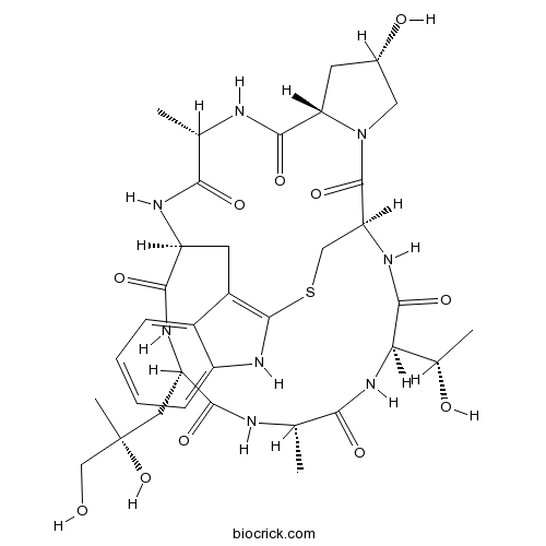
PhalloidinPromotes actin polymerization CAS# 17466-45-4 |

- LY2835219
Catalog No.:BCC1113
CAS No.:1231930-82-7
- Roscovitine (Seliciclib,CYC202)
Catalog No.:BCC1105
CAS No.:186692-46-6
- Nu 6027
Catalog No.:BCC1154
CAS No.:220036-08-8
- SNS-032 (BMS-387032)
Catalog No.:BCC1152
CAS No.:345627-80-7
- AT7519 Hydrochloride
Catalog No.:BCC1376
CAS No.:902135-91-5
Quality Control & MSDS
3D structure
Package In Stock
Number of papers citing our products

| Cas No. | 17466-45-4 | SDF | Download SDF |
| PubChem ID | 441542 | Appearance | Powder |
| Formula | C35H48N8O11S | M.Wt | 788.87 |
| Type of Compound | N/A | Storage | Desiccate at -20°C |
| Solubility | Soluble to 1 mg/ml in water | ||
| Sequence | ATCXAWL (Modifications: Cyclic Cys-3-Trp-6, X=Hyp, D-Thr-2) | ||
| SMILES | CC1C(=O)NC2CC3=C(NC4=CC=CC=C34)SCC(C(=O)N5CC(CC5C(=O)N1)O)NC(=O)C(NC(=O)C(NC(=O)C(NC2=O)CC(C)(CO)O)C)C(C)O | ||
| Standard InChIKey | KPKZJLCSROULON-QKGLWVMZSA-N | ||
| Standard InChI | InChI=1S/C35H48N8O11S/c1-15-27(47)38-22-10-20-19-7-5-6-8-21(19)41-33(20)55-13-24(34(53)43-12-18(46)9-25(43)31(51)37-15)40-32(52)26(17(3)45)42-28(48)16(2)36-30(50)23(39-29(22)49)11-35(4,54)14-44/h5-8,15-18,22-26,41,44-46,54H,9-14H2,1-4H3,(H,36,50)(H,37,51)(H,38,47)(H,39,49)(H,40,52)(H,42,48)/t15-,16-,17-,18-,22-,23-,24-,25-,26+,35+/m0/s1 | ||
| General tips | For obtaining a higher solubility , please warm the tube at 37 ℃ and shake it in the ultrasonic bath for a while.Stock solution can be stored below -20℃ for several months. We recommend that you prepare and use the solution on the same day. However, if the test schedule requires, the stock solutions can be prepared in advance, and the stock solution must be sealed and stored below -20℃. In general, the stock solution can be kept for several months. Before use, we recommend that you leave the vial at room temperature for at least an hour before opening it. |
||
| About Packaging | 1. The packaging of the product may be reversed during transportation, cause the high purity compounds to adhere to the neck or cap of the vial.Take the vail out of its packaging and shake gently until the compounds fall to the bottom of the vial. 2. For liquid products, please centrifuge at 500xg to gather the liquid to the bottom of the vial. 3. Try to avoid loss or contamination during the experiment. |
||
| Shipping Condition | Packaging according to customer requirements(5mg, 10mg, 20mg and more). Ship via FedEx, DHL, UPS, EMS or other couriers with RT, or blue ice upon request. | ||
| Description | Promotes actin polymerization. Decreases dissociation rate constant for actin subunits from filament ends; lowers critical concentration for polymerization. |

Phalloidin Dilution Calculator

Phalloidin Molarity Calculator

Calcutta University

University of Minnesota

University of Maryland School of Medicine

University of Illinois at Chicago

The Ohio State University

University of Zurich

Harvard University

Colorado State University

Auburn University

Yale University

Worcester Polytechnic Institute

Washington State University

Stanford University

University of Leipzig

Universidade da Beira Interior

The Institute of Cancer Research

Heidelberg University

University of Amsterdam

University of Auckland

TsingHua University

The University of Michigan

Miami University

DRURY University

Jilin University

Fudan University

Wuhan University

Sun Yat-sen University

Universite de Paris

Deemed University

Auckland University

The University of Tokyo

Korea University
- Talnetant
Catalog No.:BCC1981
CAS No.:174636-32-9
- SB-222200
Catalog No.:BCC1926
CAS No.:174635-69-9
- SB 218795
Catalog No.:BCC7037
CAS No.:174635-53-1
- SDZ 220-040
Catalog No.:BCC6992
CAS No.:174575-40-7
- SDZ 220-581
Catalog No.:BCC1939
CAS No.:174575-17-8
- 2-Allylphenol
Catalog No.:BCC8518
CAS No.:1745-81-9
- alpha-Spinasterol glucoside
Catalog No.:BCN1120
CAS No.:1745-36-4
- Tipranavir
Catalog No.:BCC2002
CAS No.:174484-41-4
- Sanggenol A
Catalog No.:BCN3602
CAS No.:174423-30-4
- Amiloride HCl dihydrate
Catalog No.:BCC5068
CAS No.:17440-83-4
- Riluzole
Catalog No.:BCC3849
CAS No.:1744-22-5
- Tsugaric acid A
Catalog No.:BCN2980
CAS No.:174391-64-1
- AN-2690
Catalog No.:BCC1360
CAS No.:174671-46-6
- CH 275
Catalog No.:BCC5913
CAS No.:174688-78-9
- 2-Amino-6-methoxybenzothiazole
Catalog No.:BCC8542
CAS No.:1747-60-0
- Ginsenoside Rh4
Catalog No.:BCN3503
CAS No.:174721-08-5
- Carabrone
Catalog No.:BCN1121
CAS No.:1748-81-8
- Fmoc-Hyp(Bzl)-OH
Catalog No.:BCC3255
CAS No.:174800-02-3
- 3-Amino-4-methoxybenzamide
Catalog No.:BCC8612
CAS No.:17481-27-5
- Rabdoketone B
Catalog No.:BCN6598
CAS No.:174819-51-3
- Parishin B
Catalog No.:BCN3812
CAS No.:174972-79-3
- Parishin C
Catalog No.:BCN3813
CAS No.:174972-80-6
- Tonabersat
Catalog No.:BCC2009
CAS No.:175013-84-0
- NQDI 1
Catalog No.:BCC2404
CAS No.:175026-96-7
Impact of C24:0 on actin-microtubule interaction in human neuronal SK-N-BE cells: evaluation by FRET confocal spectral imaging microscopy after dual staining with rhodamine-phalloidin and tubulin tracker green.[Pubmed:26214025]
Funct Neurol. 2015 Jan-Mar;30(1):33-46.
Disorganization of the cytoskeleton of neurons has major consequences on the transport of neurotransmitters via the microtubule network. The interaction of cytoskeleton proteins (actin and tubulin) was studied in neuronal SK-N-BE cells treated with tetracosanoic acid (C24:0), which is cytotoxic and increased in Alzheimer's disease patients. When SK-N-BE cells were treated with C24:0, mitochondrial dysfunctions and a non-apoptotic mode of cell death were observed. Fluorescence microscopy revealed shrunken cells with perinuclear condensation of actin and tubulin. Impact of C24:0 on actin-microtubule interaction in human neuronal SK-N-BE cells: evaluation by FRET confocal spectral imaging microscopy after dual staining with rhodamine-Phalloidin and tubulin tracker green After staining with rhodamine-Phalloidin and with an antibody raised against alpha-/beta-tubulin, modifications of F-actin and alpha-/beta-tubulin levels were detected by flow cytometry. Lower levels of alpha-tubulin were found by Western blotting. In C24:0-treated cells, spectral analysis and fluorescence recovery after photobleaching (FRAP) measured by confocal microscopy proved the existence of fluorescence resonance energy transfer (FRET) when actin and tubulin were stained with tubulin tracker and rhodamine-Phalloidin demonstrating actin and tubulin co-localization/interaction. In control cells, no FRET was observed. Our data demonstrate quantitative changes in actin and tubulin, and modified interactions between actin and tubulin in SK-N-BE cells treated with C24:0. They also show that FRET confocal imaging microscopy is an interesting method for specifying the impact of cytotoxic compounds on cytoskeleton proteins.
Actin-Dynamics in Plant Cells: The Function of Actin-Perturbing Substances: Jasplakinolide, Chondramides, Phalloidin, Cytochalasins, and Latrunculins.[Pubmed:26498789]
Methods Mol Biol. 2016;1365:243-61.
This chapter gives an overview of the most common F-actin-perturbing substances that are used to study actin dynamics in living plant cells in studies on morphogenesis, motility, organelle movement, or when apoptosis has to be induced. These substances can be divided into two major subclasses: F-actin-stabilizing and -polymerizing substances like jasplakinolide and chondramides and F-actin-severing compounds like chytochalasins and latrunculins. Jasplakinolide was originally isolated form a marine sponge, and can now be synthesized and has become commercially available, which is responsible for its wide distribution as membrane-permeable F-actin-stabilizing and -polymerizing agent, which may even have anticancer activities. Cytochalasins, derived from fungi, show an F-actin-severing function and many derivatives are commercially available (A, B, C, D, E, H, J), also making it a widely used compound for F-actin disruption. The same can be stated for latrunculins (A, B), derived from red sea sponges; however the mode of action is different by binding to G-actin and inhibiting incorporation into the filament. In the case of swinholide a stable complex with actin dimers is formed resulting also in severing of F-actin. For influencing F-actin dynamics in plant cells only membrane permeable drugs are useful in a broad range. We however introduce also the phallotoxins and synthetic derivatives, as they are widely used to visualize F-actin in fixed cells. A particular uptake mechanism has been shown for hepatocytes, but has also been described in siphonal giant algae. In the present chapter the focus is set on F-actin dynamics in plant cells where alterations in cytoplasmic streaming can be particularly well studied; however methods by fluorescence applications including Phalloidin and antibody staining as well as immunofluorescence-localization of the inhibitor drugs are given.
Staining Fission Yeast Filamentous Actin with Fluorescent Phalloidin Conjugates.[Pubmed:27250943]
Cold Spring Harb Protoc. 2016 Jun 1;2016(6). pii: 2016/6/pdb.prot091033.
The Schizosaccharomyces pombe filamentous (F)-actin cytoskeleton drives cell growth, morphogenesis, endocytosis, and cytokinesis. The protocol described here reveals the distribution of F-actin in fixed cells through the use of fluorescently conjugated Phalloidin. Simultaneous staining of cell wall landmarks (with calcofluor) and chromatin (with 4',6-diamidino-2-phenylindole, or DAPI) makes this rapid staining procedure highly effective for staging cell cycle progression, monitoring morphogenetic abnormalities, and assessing the impact of environmental and genetic changes on the integrity of the F-actin cytoskeleton.
CLSM Analysis of the Phalloidin-Stained Muscle System of the Nemertean Proboscis and Rhynchocoel.[Pubmed:26654037]
Zoolog Sci. 2015 Dec;32(6):547-60.
The proboscis and rhynchocoel musculature of 56 nemertean species was studied using Phalloidin labelling and confocal laser scanning microscopy. Six types of muscle layers are found in the anterior proboscis of the nemerteans: inner circular, inner diagonal, inner longitudinal, outer diagonal, outer circular, and outer longitudinal. Only the inner circular and inner longitudinal muscle layers are present in all the nemerteans studied. Ten types of arrangement of the proboscis musculature are described. Three primary types ('palaeotype', 'heterotype', and 'hoplotype') correspond to the three nemertean supergroups (Palaeonemertea, Heteronemertea, and Hoplonemertea). The evolutionary transformations of the initial 'palaeotype' proboscis proceeded in two primary ways: increasing bilateral symmetry (Callinera, Hubrechtella, and most of Heteronemertea) and increasing polyradial symmetry (Baseodiscidae, Oxypolellinae, and Hoplonemertea). The musculature of the middle portion of the proboscis differs among the three groups with armature: Palaeonemertea (genus Callinera), Polystilifera, and Monostilifera. The musculature of the stylet apparatus of the monostiliferous nemerteans is more complicated than that of the polystiliferous nemerteans, and consists of four muscle components--basal and anterior sphincters, radial and longitudinal musculature. Among the studied monostiliferans, the different components of the stylet musculature are developed to varying degrees. In addition, data on the structure of the rhynchocoel with interwoven musculature are provided. The taxonomic significance and phylogenetic interpretation of the proboscis and rhynchocoel musculature is discussed.
Phalloidin perturbs the interaction of human non-muscle myosin isoforms 2A and 2C1 with F-actin.[Pubmed:21295570]
FEBS Lett. 2011 Mar 9;585(5):767-71.
Phalloidin and fluorescently labeled Phalloidin analogs are established reagents to stabilize and mark actin filaments for the investigation of acto-myosin interactions. In the present study, we employed transient and steady-state kinetic measurements as well as in vitro motility assays to show that Phalloidin perturbs the productive interaction of human non-muscle myosin-2A and -2C1 with filamentous actin. Phalloidin binding to F-actin results in faster dissociation of the complex formed with non-muscle myosin-2A and -2C1, reduced actin-activated ATP turnover, and slower velocity of actin filaments in the in vitro motility assay. In contrast, Phalloidin binding to F-actin does not affect the interaction with human non-muscle myosin isoform 2B and Dictyostelium myosin-2 and myosin-5b.
Phalloidin enhances actin assembly by preventing monomer dissociation.[Pubmed:6746738]
J Cell Biol. 1984 Aug;99(2):529-35.
Incubation of the isolated acrosomal bundles of Limulus sperm with skeletal muscle actin results in assembly of actin onto both ends of the bundles. These cross-linked bundles of actin filaments taper, thus allowing one to distinguish directly the preferred end for actin assembly from the nonpreferred end; the preferred end is thinner. Incubation with actin in the presence of equimolar Phalloidin in 100 mM KCl, 1 mM MgCl2 and 0.5 mM ATP at pH 7.5 resulted in a slightly smaller association rate constant at the preferred end than in the absence of the drug (3.36 +/- 0.14 X 10(6) M-1 s-1 vs. 2.63 +/- 0.22 X 10(6) M-1 s-1, control vs. experimental). In the presence of Phalloidin, the dissociation rate constant at the preferred end was reduced from 0.317 +/- 0.097 s-1 to essentially zero. Consequently, the critical concentration at the preferred end dropped from 0.10 microM to zero in the presence of the drug. There was no detectable change in the rate constant of association at the nonpreferred end in the presence of Phalloidin (0.256 +/- 0.015 X 10(6) M-1 s-1 vs. 0.256 +/- 0.043 X 10(6) M-1 s-1, control vs. experimental); however, the dissociation rate constant was reduced from 0.269 +/- 0.043 s-1 to essentially zero. Thus, the critical concentration at the nonpreferred end changed from 1.02 microM to zero in the presence of Phalloidin. Dilution-induced depolymerization at both the preferred and nonpreferred ends was prevented in the presence of Phalloidin. Thus, Phalloidin enhances actin assembly by lowering the critical concentration at both ends of actin filaments, a consequence of reducing the dissociation rate constants at each end.
Phalloidin-induced actin polymerization in the cytoplasm of cultured cells interferes with cell locomotion and growth.[Pubmed:341163]
Proc Natl Acad Sci U S A. 1977 Dec;74(12):5613-7.
Phalloidin, the toxic drug from the mushroom Amanita phalloides, was injected into the cytoplasm of tissue culture cells and the changes in intracellular actin distribution were followed by immunofluorescence microscopy with actin antibody. At low concentrations, Phalloidin recruits the non- or less highly polymerized forms of cytoplasmic actin into stable "islands" of aggregated actin polymers and does not interfere with the preexisting thick bundles of microfilaments (stress fibers). Differential focusing shows that these islands of Phalloidin-induced actin polymers occur at a level in the cytoplasm that is above the submembranous bundles of microfilaments present on the adhesive side of the cells. The pattern of cytoplasmic microtubules remains unaffected by the injection of Phalloidin; however, filamin, a protein usually associated with actin in the cytoplasm, is also recruited into the islands. At higher Phalloidin concentrations, contraction of the cell is observed. These results are discussed in the light of previous biochemical studies by Wieland and Faulstich and their coworkers [for a review see Wieland, T. (1977) Naturwissenschaften 64, 303-309] on the in vitro interaction of Phalloidin with muscle actin, which have documented that Phalloidin reacts stoichiometrically with actin, promotes actin polymerization, and stabilizes actin polymers. In addition, we show that microinjection of Phalloidin interferes in a concentration-dependent manner with cell locomotion and cell growth. These results indicate that a well-balanced controlled reversible equilibrium between different polymerization states of actin may be a necessary requirement for cell locomotion and may also influence other cellular functions such as growth.


