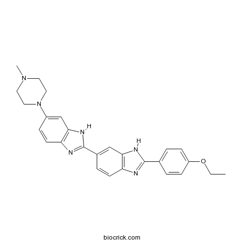
Hoechst 33342Blue fluorescent dyes CAS# 23491-52-3 |

- Hoechst 33342 analog 2
Catalog No.:BCC1631
CAS No.:106050-84-4
- Hoechst 33342
Catalog No.:BCC1629
CAS No.:23491-52-3
- Hoechst 33258 analog 2
Catalog No.:BCC1625
CAS No.:23491-54-5
- Hoechst 33258 analog 5
Catalog No.:BCC1627
CAS No.:23491-55-6
- Hoechst 34580
Catalog No.:BCC1632
CAS No.:23555-00-2
- Hoechst 33258 analog
Catalog No.:BCC1624
CAS No.:258843-62-8
Quality Control & MSDS
3D structure
Package In Stock
Number of papers citing our products

| Cas No. | 23491-52-3 | SDF | Download SDF |
| PubChem ID | 1464 | Appearance | Powder |
| Formula | C27H31Cl3N6O | M.Wt | 561.9 |
| Type of Compound | N/A | Storage | Desiccate at -20°C |
| Synonyms | bisBenzimide H 33342; HOE 33342 | ||
| Solubility | Soluble to 100 mM in water and to 50 mM in DMSO | ||
| Chemical Name | 2-(4-ethoxyphenyl)-6-[6-(4-methylpiperazin-1-yl)-1H-benzimidazol-2-yl]-1H-benzimidazole | ||
| SMILES | CCOC1=CC=C(C=C1)C2=NC3=C(N2)C=C(C=C3)C4=NC5=C(N4)C=C(C=C5)N6CCN(CC6)C | ||
| Standard InChIKey | PRDFBSVERLRRMY-UHFFFAOYSA-N | ||
| Standard InChI | InChI=1S/C27H28N6O/c1-3-34-21-8-4-18(5-9-21)26-28-22-10-6-19(16-24(22)30-26)27-29-23-11-7-20(17-25(23)31-27)33-14-12-32(2)13-15-33/h4-11,16-17H,3,12-15H2,1-2H3,(H,28,30)(H,29,31) | ||
| General tips | For obtaining a higher solubility , please warm the tube at 37 ℃ and shake it in the ultrasonic bath for a while.Stock solution can be stored below -20℃ for several months. We recommend that you prepare and use the solution on the same day. However, if the test schedule requires, the stock solutions can be prepared in advance, and the stock solution must be sealed and stored below -20℃. In general, the stock solution can be kept for several months. Before use, we recommend that you leave the vial at room temperature for at least an hour before opening it. |
||
| About Packaging | 1. The packaging of the product may be reversed during transportation, cause the high purity compounds to adhere to the neck or cap of the vial.Take the vail out of its packaging and shake gently until the compounds fall to the bottom of the vial. 2. For liquid products, please centrifuge at 500xg to gather the liquid to the bottom of the vial. 3. Try to avoid loss or contamination during the experiment. |
||
| Shipping Condition | Packaging according to customer requirements(5mg, 10mg, 20mg and more). Ship via FedEx, DHL, UPS, EMS or other couriers with RT, or blue ice upon request. | ||
| Description | Cell permeable fluorescent DNA stain; binds minor groove of AT-rich regions. Used to quantify DNA in viable cells. |

Hoechst 33342 Dilution Calculator

Hoechst 33342 Molarity Calculator
| 1 mg | 5 mg | 10 mg | 20 mg | 25 mg | |
| 1 mM | 1.7797 mL | 8.8984 mL | 17.7968 mL | 35.5935 mL | 44.4919 mL |
| 5 mM | 0.3559 mL | 1.7797 mL | 3.5594 mL | 7.1187 mL | 8.8984 mL |
| 10 mM | 0.178 mL | 0.8898 mL | 1.7797 mL | 3.5594 mL | 4.4492 mL |
| 50 mM | 0.0356 mL | 0.178 mL | 0.3559 mL | 0.7119 mL | 0.8898 mL |
| 100 mM | 0.0178 mL | 0.089 mL | 0.178 mL | 0.3559 mL | 0.4449 mL |
| * Note: If you are in the process of experiment, it's necessary to make the dilution ratios of the samples. The dilution data above is only for reference. Normally, it's can get a better solubility within lower of Concentrations. | |||||

Calcutta University

University of Minnesota

University of Maryland School of Medicine

University of Illinois at Chicago

The Ohio State University

University of Zurich

Harvard University

Colorado State University

Auburn University

Yale University

Worcester Polytechnic Institute

Washington State University

Stanford University

University of Leipzig

Universidade da Beira Interior

The Institute of Cancer Research

Heidelberg University

University of Amsterdam

University of Auckland

TsingHua University

The University of Michigan

Miami University

DRURY University

Jilin University

Fudan University

Wuhan University

Sun Yat-sen University

Universite de Paris

Deemed University

Auckland University

The University of Tokyo

Korea University
Description: IC50 Value: N/A Hoechst stains are part of a family of blue fluorescent dyes used to stain DNA. These Bis-benzimides were originally developed by Hoechst AG, which numbered all their compounds so that the dye Hoechst 33342 is the 33342nd compound made by the company. There are three related Hoechst stains: Hoechst 33258, Hoechst 33342, and Hoechst 34580. The dyes Hoechst 33258 and Hoechst 33342 are the ones most commonly used and they have similarexcitation/emission spectra. Both dyes are excited by ultraviolet light at around 350 nm, and both emit blue/cyan fluorescent light around anemission maximum at 461 nm. Unbound dye has its maximum fluorescence emission in the 510-540 nm range. Hoechst dyes are soluble in water and in organic solvents such as dimethyl formamide or dimethyl sulfoxide. Concentrations can be achieved of up to 10 mg/mL. Aqueous solutions are stable at 2-6 °C for at least six months when protected from light. For long-term storage the solutions are instead frozen at ≤-20 °C. The dyes bind to the minor groove of double-stranded DNA with a preference for sequences rich in adenine andthymine. Although the dyes can bind to all nucleic acids, AT-rich double-stranded DNA strands enhance fluorescence considerably. Hoechst dyes are cell-permeable and can bind to DNA in live or fixed cells. Therefore, these stains are often called supravital, which means that cells survive a treatment with these compounds. Cells that express specific ATP-binding cassette transporter proteins can also actively transport these stains out of their cytoplasm. in vitro: N/A in vivo: N/A Clinical trial: N/A
- Hoechst 33258
Catalog No.:BCC1623
CAS No.:23491-45-4
- 2-Amino-5-mercapto-1,3,4-thiadiazole
Catalog No.:BCC8536
CAS No.:2349-67-9
- U 99194 maleate
Catalog No.:BCC7029
CAS No.:234757-41-6
- 2-Palmitoylglycerol
Catalog No.:BCC7289
CAS No.:23470-00-0
- trans-Khellactone
Catalog No.:BCN6920
CAS No.:23458-04-0
- Decursinol
Catalog No.:BCN2638
CAS No.:23458-02-8
- alpha-Spinasterone
Catalog No.:BCN5086
CAS No.:23455-44-9
- Trenbolone cyclohexylmethylcarbonate
Catalog No.:BCC9185
CAS No.:23454-33-3
- Alternariol monomethyl ether
Catalog No.:BCN7384
CAS No.:23452-05-3
- Physcion 1-glucoside
Catalog No.:BCN8170
CAS No.:23451-01-6
- Irisolidone
Catalog No.:BCN8496
CAS No.:2345-17-7
- Swertianolin
Catalog No.:BCN2759
CAS No.:23445-00-3
- Hoechst 33258 analog 2
Catalog No.:BCC1625
CAS No.:23491-54-5
- Hoechst 33258 analog 5
Catalog No.:BCC1627
CAS No.:23491-55-6
- Peimine
Catalog No.:BCN1094
CAS No.:23496-41-5
- Axillaridine
Catalog No.:BCN2060
CAS No.:23506-96-9
- MEN 11270
Catalog No.:BCC6094
CAS No.:235082-52-7
- Humulon
Catalog No.:BCC8186
CAS No.:23510-81-8
- 8-Gingerol
Catalog No.:BCN5921
CAS No.:23513-08-8
- 6-Gingerol
Catalog No.:BCN1030
CAS No.:23513-14-6
- 10-Gingerol
Catalog No.:BCN5922
CAS No.:23513-15-7
- (-)-licarin A
Catalog No.:BCN5087
CAS No.:23518-30-1
- Vomifoliol
Catalog No.:BCN5088
CAS No.:23526-45-6
- 5-Aza-2'-deoxycytidine
Catalog No.:BCN2169
CAS No.:2353-33-5
Cell Cycle Analysis of CML Stem Cells Using Hoechst 33342 and Propidium Iodide.[Pubmed:27581138]
Methods Mol Biol. 2016;1465:47-57.
Chronic myeloid leukemia (CML) is a myeloproliferative disease with an expansion of white blood cells. The current treatments for CML are shown not to be long-term effective because of CML stem cells' insensitivity to tyrosine kinase inhibitors. Therefore, studying more about CML stem cells is essential to understand the pathways of CML stem cell development and proliferation and finally lead to effective treatments to eliminate CML stem cells and eradicate CML. This chapter describes two methods to analyze cell cycle of CML stem cells. The rare population of CML stem cells can be identified by staining with cell surface markers, and then DNA-binding dyes Hoechst 33342 and propidium iodide (PI) are added to stain the DNA content which is changed when cells go through different phases of the cell cycle. Samples are run through the flow cytometer to be analyzed based on different absorbance and emission wavelengths of different florescent colors.
Analyzing Cell Death by Nuclear Staining with Hoechst 33342.[Pubmed:27587774]
Cold Spring Harb Protoc. 2016 Sep 1;2016(9). pii: 2016/9/pdb.prot087205.
The nuclei of healthy cells are generally spherical, and the DNA is evenly distributed. During apoptosis the DNA becomes condensed, but this process does not occur during necrosis. Nuclear condensation can therefore be used to distinguish apoptotic cells from healthy cells or necrotic cells. Dyes that bind to DNA, such as Hoechst 33342 or 4',6-diamidino-2-phenylindole (DAPI), can be used to observe nuclear condensation. These dyes fluoresce at 461 nm when excited by ultraviolet light and can therefore be visualized using conventional fluorescent microscopes equipped with light sources that emit light at approximately 350 nm and filter sets that permit the transmission of light at approximately 460 nm. This protocol describes staining and visualization of cells stained with Hoechst 33342, but it can be adapted for staining with DAPI or other dyes.
Requirement of ABC transporter inhibition and Hoechst 33342 dye deprivation for the assessment of side population-defined C6 glioma stem cell metabolism using fluorescent probes.[Pubmed:27814696]
BMC Cancer. 2016 Nov 4;16(1):847.
BACKGROUND: Elucidating the precise properties of cancer stem cells (CSCs) is indispensable for the development of effective therapies against tumors, because CSCs are key drivers of tumor development, metastasis and relapse. We previously reported that the Hoechst 33342 dye-low staining side population (SP) method can enrich for CSCs in the C6 glioma cell line, and that the positively stained main population (MP) cells are non-CSCs. Presence of cancer stem-like SP cells is reported in various types of cancer. Although altered cellular energy metabolism is a hallmark of cancer, very little has been studied on the applicability of fluorescent probes for the understanding of CSC energy metabolism. METHODS: The metabolic status of C6 SP and MP cells are evaluated by CellROX, MitoTracker Green (MTG) and JC-1 for cellular oxidative stress, mitochondrial amount, and mitochondrial membrane potential, respectively. RESULTS: SP cells were found to exhibit significantly lower fluorescent intensities of CellROX and MTG than MP cells. However, inhibition of ATP binding cassette (ABC) transporters by verapamil enhanced the intensities of these probes in SP cells to the levels similar to those in MP cells, indicating that SP cells expel the probes outside of the cells through ABC transporters. Next, SP cells were stained with JC-1 dye which exhibits membrane potential dependent accumulation in mitochondrial matrix, followed by formation of aggregates. The mitochondrial membrane potential indicated by the aggregates of JC-1 was 5.0-fold lower in SP cells than MP cells. Inhibition of ABC transporters enhanced the fluorescent intensities of the JC-1 aggregates in both SP and MP cells, the former of which was still 2.2-fold lower than the latter. This higher JC-1 signal in MP cells was further found to be due to the Hoechst 33342 dye existing in MP cells. When SP and MP cells were recultured to deprive the intracellular Hoechst 33342 dye and then stained with JC-1 in the presence of verapamil, the intensities of JC-1 aggregates in such SP and MP cells became comparable. CONCLUSION: Inhibiting ABC transporters and depriving Hoechst 33342 dye are required for the accurate assessment of side population-defined C6 glioma stem cell metabolism using fluorescent probes.
Discovery of DNA dyes Hoechst 34580 and 33342 as good candidates for inhibiting amyloid beta formation: in silico and in vitro study.[Pubmed:27511370]
J Comput Aided Mol Des. 2016 Aug;30(8):639-50.
Combining Lipinski's rule with the docking and steered molecular dynamics simulations and using the PubChem data base of about 1.4 million compounds, we have obtained DNA dyes Hoechst 34580 and Hoechst 33342 as top-leads for the Alzheimer's disease. The binding properties of these ligands to amyloid beta (Abeta) fibril were thoroughly studied by in silico and in vitro experiments. Hoechst 34580 and Hoechst 33342 prefer to locate near hydrophobic regions with binding affinity mainly governed by the van der Waals interaction. By the Thioflavin T assay, it was found that the inhibition constant IC50 approximately 0.86 and 0.68 muM for Hoechst 34580 and Hoechst 33342, respectively. This result qualitatively agrees with the binding free energy estimated using the molecular mechanic-Poisson Boltzmann surface area method and all-atom simulations with the AMBER-f99SB-ILDN force field and water model TIP3P. In addition, DNA dyes have the high capability to cross the blood brain barrier. Thus, both in silico and in vitro experiments have shown that Hoechst 34580 and 33342 are good candidates for treating the Alzheimer's disease by inhibiting Abeta formation.
Assignment of DNA binding sites for 4',6-diamidine-2-phenylindole and bisbenzimide (Hoechst 33258). A comparative footprinting study.[Pubmed:2449244]
Biochim Biophys Acta. 1988 Feb 28;949(2):158-68.
DNA binding sites for the minor groove-binding ligands DAPI (4',6-diamidine-2-phenylindole) and Hoechst 33258 (bisbenzimide) have been analysed using DNAase I and micrococcal nuclease footprinting techniques. Both drugs appear to bind to AT-rich regions containing at least four such basepairs. Hoechst 33258 seems to bind relatively poorly to nucleotide sequences containing the alternating step TpA. However, in contrast to DAPI, it can more readily accommodate the presence of guanosine residues at the end of the binding site. We compare the DNA binding sites for DAPI and Hoechst 33258 with those determined for the related minor groove-binding ligands, berenil, netropsin and distamycin A, under comparable conditions, and discuss the importance of using different footprinting probes when analysing drug-DNA interactions.
Simultaneous quantitation of Hoechst 33342 and immunofluorescence on viable cells using a fluorescence activated cell sorter.[Pubmed:7028425]
Cytometry. 1980 Sep;1(2):136-42.
The cytochemical stain Hoechst 33342 has been used to quantify DNA in viable cells and has been used under nonsaturating conditions to discriminate between lymphoid cell types. In order to correlate the quantitative emission from Hoechst 33342 with cell surface antigens, a fluorescence activated cell sorter was modified to simultaneously detect emission from the UV excited Hoechst dye and fluorescein attached to the cell surface by immunofluorescence techniques. A special set of laser mirrors was installed in an argon ion laser so that all the lines from 351-488 nm could be used to illuminate the cells. Appropriate emission filters were used to separate the light emitted by Hoechst 33342 from the fluorescein. An electronic cross-over circuit was used to compensate for special overlap between the two dyes. Analysis of murine lymph node cells stained both with Hoechst 33342 under nonsaturating conditions and anti-Thy 1.2 indicated that the cells that stained dimly with the Hoechst dye expressed the Thy 1.2 marker while the cells that were brightly stained with Hoechst 33342 lacked this differentiation antigen. The correlation of cell surface myeloma protein with cell cycle on an in vitro cell line indicated that the heterogeneity of cell surface antigen expression could not be accounted for solely by variations occurring during the cell cycle.


