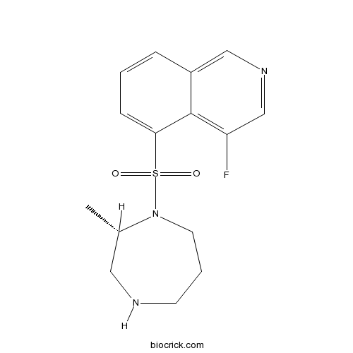
K-115 free baseCAS# 223645-67-8 |

- AM630
Catalog No.:BCC1353
CAS No.:164178-33-0
- Nepicastat
Catalog No.:BCC1795
CAS No.:173997-05-2
- Otenabant
Catalog No.:BCC1828
CAS No.:686344-29-6
- CP-945598 HCl
Catalog No.:BCC1082
CAS No.:686347-12-6
Quality Control & MSDS
3D structure
Package In Stock
Number of papers citing our products

| Cas No. | 223645-67-8 | SDF | Download SDF |
| PubChem ID | 9863672 | Appearance | Powder |
| Formula | C15H18FN3O2S | M.Wt | 323.39 |
| Type of Compound | N/A | Storage | Desiccate at -20°C |
| Synonyms | Ripasudil free base | ||
| Solubility | Soluble in DMSO | ||
| Chemical Name | 4-fluoro-5-[[(2S)-2-methyl-1,4-diazepan-1-yl]sulfonyl]isoquinoline | ||
| SMILES | CC1CNCCCN1S(=O)(=O)C2=CC=CC3=CN=CC(=C32)F | ||
| Standard InChIKey | QSKQVZWVLOIIEV-NSHDSACASA-N | ||
| Standard InChI | InChI=1S/C15H18FN3O2S/c1-11-8-17-6-3-7-19(11)22(20,21)14-5-2-4-12-9-18-10-13(16)15(12)14/h2,4-5,9-11,17H,3,6-8H2,1H3/t11-/m0/s1 | ||
| General tips | For obtaining a higher solubility , please warm the tube at 37 ℃ and shake it in the ultrasonic bath for a while.Stock solution can be stored below -20℃ for several months. We recommend that you prepare and use the solution on the same day. However, if the test schedule requires, the stock solutions can be prepared in advance, and the stock solution must be sealed and stored below -20℃. In general, the stock solution can be kept for several months. Before use, we recommend that you leave the vial at room temperature for at least an hour before opening it. |
||
| About Packaging | 1. The packaging of the product may be reversed during transportation, cause the high purity compounds to adhere to the neck or cap of the vial.Take the vail out of its packaging and shake gently until the compounds fall to the bottom of the vial. 2. For liquid products, please centrifuge at 500xg to gather the liquid to the bottom of the vial. 3. Try to avoid loss or contamination during the experiment. |
||
| Shipping Condition | Packaging according to customer requirements(5mg, 10mg, 20mg and more). Ship via FedEx, DHL, UPS, EMS or other couriers with RT, or blue ice upon request. | ||

K-115 free base Dilution Calculator

K-115 free base Molarity Calculator
| 1 mg | 5 mg | 10 mg | 20 mg | 25 mg | |
| 1 mM | 3.0922 mL | 15.4612 mL | 30.9224 mL | 61.8448 mL | 77.306 mL |
| 5 mM | 0.6184 mL | 3.0922 mL | 6.1845 mL | 12.369 mL | 15.4612 mL |
| 10 mM | 0.3092 mL | 1.5461 mL | 3.0922 mL | 6.1845 mL | 7.7306 mL |
| 50 mM | 0.0618 mL | 0.3092 mL | 0.6184 mL | 1.2369 mL | 1.5461 mL |
| 100 mM | 0.0309 mL | 0.1546 mL | 0.3092 mL | 0.6184 mL | 0.7731 mL |
| * Note: If you are in the process of experiment, it's necessary to make the dilution ratios of the samples. The dilution data above is only for reference. Normally, it's can get a better solubility within lower of Concentrations. | |||||

Calcutta University

University of Minnesota

University of Maryland School of Medicine

University of Illinois at Chicago

The Ohio State University

University of Zurich

Harvard University

Colorado State University

Auburn University

Yale University

Worcester Polytechnic Institute

Washington State University

Stanford University

University of Leipzig

Universidade da Beira Interior

The Institute of Cancer Research

Heidelberg University

University of Amsterdam

University of Auckland

TsingHua University

The University of Michigan

Miami University

DRURY University

Jilin University

Fudan University

Wuhan University

Sun Yat-sen University

Universite de Paris

Deemed University

Auckland University

The University of Tokyo

Korea University
K-115 free base a specific inhibitor of ROCK, with IC50s of 19 and 51 nM for ROCK2 and ROCK1, respectively.
In Vitro:K-115 free base is a potent inhibitor of ROCK, with IC50s of 19 and 51 nM for ROCK2 and ROCK1, respectively. K-115 also shows less potent inhibitory activities against CaMKIIα, PKACα and PKC, with IC50s of 370 nM, 2.1 μM and 27 μM, respectively[1]. K-115 (1, 10 μM) induces cytoskeletal changes, including retraction and cell rounding and reduced actin bundles of cultured trabecular meshwork (TM) cells. K-115 (5 μM) sifnificantly reduces transendothelial electrical resistance (TEER), and increases FITC-dextran permeability in Schlemm’s canal endothelial (SCE) cell monolayers[2].
In Vivo:K-115 reduces intraocular pressure (IOP) in a concentration-dependent manner at concentrations between 0.1% and 0.4% in monkey eyes and 0.0625% to 0.5% in rabbit eyes, respectively[1]. K-115 (1 mg/kg, p.o. daily) shows a neuroprotective effect on retinal ganglion cells (RGCs) after nerve crush (NC). K-115 also inhibits the oxidative stress induced by axonal injury in mice. K-115 suppresses the time-dependent production of ROS in RGCs after NC injury[3].
References:
[1]. Isobe T, et al. Effects of K-115, a rho-kinase inhibitor, on aqueous humor dynamics in rabbits. Curr Eye Res. 2014 Aug;39(8):813-22.
[2]. Kaneko Y, et al. Effects of K-115 (Ripasudil), a novel ROCK inhibitor, on trabecular meshwork and Schlemm's canal endothelial cells. Sci Rep. 2016 Jan 19;6:19640.
[3]. Yamamoto K, et al. The novel Rho kinase (ROCK) inhibitor K-115: a new candidate drug for neuroprotective treatment in glaucoma. Invest Ophthalmol Vis Sci. 2014 Oct 2;55(11):7126-36.
- Tiadinil
Catalog No.:BCC8070
CAS No.:223580-51-6
- CPA inhibitor
Catalog No.:BCC1500
CAS No.:223532-02-3
- YM 58483
Catalog No.:BCC7817
CAS No.:223499-30-7
- ONO 4817
Catalog No.:BCC2375
CAS No.:223472-31-9
- GLP-2 (human)
Catalog No.:BCC5891
CAS No.:223460-79-5
- FG2216
Catalog No.:BCC6402
CAS No.:223387-75-5
- Polygalacic acid
Catalog No.:BCN5898
CAS No.:22338-71-2
- Grandifloric acid
Catalog No.:BCN4669
CAS No.:22338-69-8
- Grandiflorenic acid
Catalog No.:BCN4670
CAS No.:22338-67-6
- 9-Hydroxy-alpha-lapachone
Catalog No.:BCN5060
CAS No.:22333-58-0
- Methyl ferulate
Catalog No.:BCN4023
CAS No.:22329-76-6
- Platycodigenin
Catalog No.:BCN3183
CAS No.:22327-82-8
- Collagen proline hydroxylase inhibitor-1
Catalog No.:BCC1495
CAS No.:223663-32-9
- Collagen proline hydroxylase inhibitor
Catalog No.:BCC1494
CAS No.:223666-07-7
- Mirabegron (YM178)
Catalog No.:BCC3814
CAS No.:223673-61-8
- Eupatilin
Catalog No.:BCN2336
CAS No.:22368-21-4
- 4-Methoxysalicylic acid
Catalog No.:BCN7783
CAS No.:2237-36-7
- Sodium Monensin
Catalog No.:BCC5319
CAS No.:22373-78-0
- NPE-caged-HPTS
Catalog No.:BCC5950
CAS No.:223759-19-1
- Bestatin trifluoroacetate
Catalog No.:BCC3909
CAS No.:223763-80-2
- Pedalitin
Catalog No.:BCN3954
CAS No.:22384-63-0
- Serratenediol
Catalog No.:BCN5061
CAS No.:2239-24-9
- 3,8-Di-O-methylellagic acid
Catalog No.:BCN5062
CAS No.:2239-88-5
- Aristola-1(10),8-dien-2-one
Catalog No.:BCN7608
CAS No.:22391-34-0


