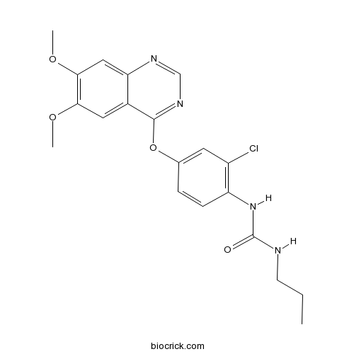
KRN 633VEGFR inhibitor,ATP-competitive CAS# 286370-15-8 |

- Elacridar
Catalog No.:BCC1546
CAS No.:143664-11-3
- Elacridar hydrochloride
Catalog No.:BCC1547
CAS No.:143851-98-3
Quality Control & MSDS
3D structure
Package In Stock
Number of papers citing our products

| Cas No. | 286370-15-8 | SDF | Download SDF |
| PubChem ID | 9549295 | Appearance | Powder |
| Formula | C20H21ClN4O4 | M.Wt | 416.86 |
| Type of Compound | N/A | Storage | Desiccate at -20°C |
| Solubility | DMSO : ≥ 8 mg/mL (19.19 mM) *"≥" means soluble, but saturation unknown. | ||
| Chemical Name | 1-[2-chloro-4-(6,7-dimethoxyquinazolin-4-yl)oxyphenyl]-3-propylurea | ||
| SMILES | CCCNC(=O)NC1=C(C=C(C=C1)OC2=NC=NC3=CC(=C(C=C32)OC)OC)Cl | ||
| Standard InChIKey | VPBYZLCHOKSGRX-UHFFFAOYSA-N | ||
| Standard InChI | InChI=1S/C20H21ClN4O4/c1-4-7-22-20(26)25-15-6-5-12(8-14(15)21)29-19-13-9-17(27-2)18(28-3)10-16(13)23-11-24-19/h5-6,8-11H,4,7H2,1-3H3,(H2,22,25,26) | ||
| General tips | For obtaining a higher solubility , please warm the tube at 37 ℃ and shake it in the ultrasonic bath for a while.Stock solution can be stored below -20℃ for several months. We recommend that you prepare and use the solution on the same day. However, if the test schedule requires, the stock solutions can be prepared in advance, and the stock solution must be sealed and stored below -20℃. In general, the stock solution can be kept for several months. Before use, we recommend that you leave the vial at room temperature for at least an hour before opening it. |
||
| About Packaging | 1. The packaging of the product may be reversed during transportation, cause the high purity compounds to adhere to the neck or cap of the vial.Take the vail out of its packaging and shake gently until the compounds fall to the bottom of the vial. 2. For liquid products, please centrifuge at 500xg to gather the liquid to the bottom of the vial. 3. Try to avoid loss or contamination during the experiment. |
||
| Shipping Condition | Packaging according to customer requirements(5mg, 10mg, 20mg and more). Ship via FedEx, DHL, UPS, EMS or other couriers with RT, or blue ice upon request. | ||
| Description | KRN 633 is an ATP-competitive inhibitor of VEGFR1, 2 and 3 with IC50 values of 170 nM, 160 nM and 125 nM,respectively. | |||||
| Targets | VEGFR3 | VEGFR2 | VEGFR1 | |||
| IC50 | 125 nM | 160 nM | 170 nM | |||

KRN 633 Dilution Calculator

KRN 633 Molarity Calculator
| 1 mg | 5 mg | 10 mg | 20 mg | 25 mg | |
| 1 mM | 2.3989 mL | 11.9944 mL | 23.9889 mL | 47.9777 mL | 59.9722 mL |
| 5 mM | 0.4798 mL | 2.3989 mL | 4.7978 mL | 9.5955 mL | 11.9944 mL |
| 10 mM | 0.2399 mL | 1.1994 mL | 2.3989 mL | 4.7978 mL | 5.9972 mL |
| 50 mM | 0.048 mL | 0.2399 mL | 0.4798 mL | 0.9596 mL | 1.1994 mL |
| 100 mM | 0.024 mL | 0.1199 mL | 0.2399 mL | 0.4798 mL | 0.5997 mL |
| * Note: If you are in the process of experiment, it's necessary to make the dilution ratios of the samples. The dilution data above is only for reference. Normally, it's can get a better solubility within lower of Concentrations. | |||||

Calcutta University

University of Minnesota

University of Maryland School of Medicine

University of Illinois at Chicago

The Ohio State University

University of Zurich

Harvard University

Colorado State University

Auburn University

Yale University

Worcester Polytechnic Institute

Washington State University

Stanford University

University of Leipzig

Universidade da Beira Interior

The Institute of Cancer Research

Heidelberg University

University of Amsterdam

University of Auckland

TsingHua University

The University of Michigan

Miami University

DRURY University

Jilin University

Fudan University

Wuhan University

Sun Yat-sen University

Universite de Paris

Deemed University

Auckland University

The University of Tokyo

Korea University
KRN 633 is a selective inhibitor of VEGFR-1, VEGFR-2 and VEGFR-3 with IC50 value of 170 nM, 160 nM and 125 nM [1].
Vascular endothelial growth factor receptor (VEGFR) is a protein and plays an important role in tumor angiogenesis by cooperating with its ligand VEGF [1].
KRN 633 is a potent VEGFR inhibitor. When tested with HUVECs, KRN 633 inhibited the cell proliferation that mediated by VEGF with the IC50 value of 14.9 nmol/L and suppressed the capillary tube formation by ~50% at the dose of 10 nmol/L [1].
In mid-pregnant mice model, KRN633 was used at the dose of 5 mg/kg once daily from embryonic day 13.5 until the day of delivery and the effect on vascular growth was slightly delayed on postnatal day 4 (P4) and on P8 it was observed that KRN633 resulted in the decreased numbers of central arteries and veins and abnormal branching of the central arteries [2]. When tested with athymic mouse xenograft HT29 cells model, administration of KRN633 inhibited tumor growth as ~90% from the initial tumor volume rangs from 500-667 mm3, while had less effect Du145 xenograft mouse models by inhibiting tumor angiogenesis and vascular permeability [1].
References:
[1]. Nakamura, K., et al., KRN633: A selective inhibitor of vascular endothelial growth factor receptor-2 tyrosine kinase that suppresses tumor angiogenesis and growth. Mol Cancer Ther, 2004. 3(12): p. 1639-49.
[2]. Morita, A., et al., Treatment of mid-pregnant mice with KRN633, an inhibitor of vascular endothelial growth factor receptor tyrosine kinase, induces abnormal retinal vascular patterning in their newborn pups. Birth Defects Res B Dev Reprod Toxicol, 2014. 101(4): p. 293-9.
- S 14506 hydrochloride
Catalog No.:BCC7174
CAS No.:286369-38-8
- Erythristemine
Catalog No.:BCN5184
CAS No.:28619-41-2
- 8-Prenylkaempferol
Catalog No.:BCN3311
CAS No.:28610-31-3
- Isoanhydroicaritin
Catalog No.:BCN3879
CAS No.:28610-30-2
- Orientin
Catalog No.:BCN4984
CAS No.:28608-75-5
- CCG-1423
Catalog No.:BCC5581
CAS No.:285986-88-1
- BIRB 796 (Doramapimod)
Catalog No.:BCC2535
CAS No.:285983-48-4
- 22-Dehydroclerosteryl acetate
Catalog No.:BCN5183
CAS No.:28594-00-5
- Docosyl caffeate
Catalog No.:BCN5182
CAS No.:28593-92-2
- Eicosanyl caffeate
Catalog No.:BCN7209
CAS No.:28593-90-0
- Persicogenin
Catalog No.:BCN7744
CAS No.:28590-40-1
- 7-Geranyloxy-6-methoxycoumarin
Catalog No.:BCN5181
CAS No.:28587-43-1
- Nigericin sodium salt
Catalog No.:BCC7915
CAS No.:28643-80-3
- Meloscandonine
Catalog No.:BCN5186
CAS No.:28645-27-4
- L-838,417
Catalog No.:BCC7617
CAS No.:286456-42-6
- Multicaulisin
Catalog No.:BCN7840
CAS No.:286461-76-5
- Euphorbiasteroid
Catalog No.:BCN2781
CAS No.:28649-59-4
- Epoxylathyrol
Catalog No.:BCN5382
CAS No.:28649-60-7
- 6'-Amino-3',4'-(methylenedioxy)acetophenone
Catalog No.:BCC8760
CAS No.:28657-75-2
- Fesoterodine Fumarate
Catalog No.:BCC4584
CAS No.:286930-03-8
- Ezatiostat hydrochloride
Catalog No.:BCC4259
CAS No.:286942-97-0
- NCX 4040
Catalog No.:BCC7944
CAS No.:287118-97-2
- PD 180970
Catalog No.:BCC3894
CAS No.:287204-45-9
- MA 2029
Catalog No.:BCC7983
CAS No.:287206-61-5
Regression of retinal capillaries following N-methyl-D-aspartate-induced neurotoxicity in the neonatal rat retina.[Pubmed:25284371]
J Neurosci Res. 2015 Feb;93(2):380-90.
Degeneration of retinal capillaries occurs following N-methyl-D-aspartate (NMDA)-induced retinal neurotoxicity, and the degree of capillary degeneration decreases in an age-dependent manner. To determine the role of vascular endothelial growth factor (VEGF) in the high susceptibility of capillaries to neuronal damage during the early postnatal stage, this study compares the vascular regression patterns between NMDA-treated retinas and retinas treated with N-[2-chloro-4-{(6,7-dimethoxy-4-quinazolinyl)oxy}phenyl]-N'-propylurea (KRN633), a VEGF receptor tyrosine kinase inhibitor, in neonatal rats. Two days after a single intravitreal injection of NMDA (200 nmol/eye) on postnatal day (P) 7, substantial retinal neuron loss and delayed expansion of the retinal vascular bed were observed. The reduction in the capillary density in the central retina reached statistical significance 4 days after NMDA treatment. In retinas of rats injected subcutaneously with KRN633 (10 mg/kg) on P7 and P8, simplified vasculature attributable to capillary regression and prevention of endothelial cell growth were seen on P9, whereas no visible changes in the morphology of the retinal layers were observed. The degree of capillary degeneration in NMDA-treated retinas was less than that in KRN633-treated retinas. No apparent changes in immunoreactivities for VEGF were found 2 days after NMDA treatment. These results indicate that neuronal cell loss in the retina precedes retinal capillary degeneration following NMDA treatment, and VEGF-dependent immature capillaries might be more susceptible to NMDA-induced neuronal damage.
Treatment of mid-pregnant mice with KRN633, an inhibitor of vascular endothelial growth factor receptor tyrosine kinase, induces abnormal retinal vascular patterning in their newborn pups.[Pubmed:24831925]
Birth Defects Res B Dev Reprod Toxicol. 2014 Aug;101(4):293-9.
We previously reported that treatment of mid-pregnant mice with KRN633, a vascular endothelial growth factor receptor tyrosine kinase inhibitor, caused fetal growth restriction resulting from diminished vascularization in the placenta and fetal organs. In this study, we examined how the treatment of mid-pregnant mice with KRN633 affects the development and morphology of vascular components (endothelial cells, pericytes, and basement membrane) in the retinas of their newborn pups. Pregnant mice were treated with KRN633 (5 mg/kg) once daily from embryonic day 13.5 until the day of delivery. Vascular components were examined using immunohistochemistry with specific markers for each component. Radial vascular growth in the retina was slightly delayed until postnatal day 4 (P4) in the newborn pups of KRN633-treated mothers. On P8, compared with the pups of control mothers, the pups of KRN633-treated mothers exhibited decreased numbers of central arteries and veins and abnormal branching of the central arteries. No apparent differences in pericytes or basement membrane were observed between the pups of control and KRN633-treated mothers. These results suggest that a critical period for determining retinal vascular patterning is present at the earliest stages of retinal vascular development, and that the impaired vascular endothelial growth factor signaling during this period induces abnormal architecture in the retinal vascular network.
Effects of mTOR inhibition on normal retinal vascular development in the mouse.[Pubmed:25446323]
Exp Eye Res. 2014 Dec;129:127-34.
We aimed to determine the role of age-related changes in the mammalian target of rapamycin (mTOR) activity in endothelial cell growth during retinal vascular development in mice. Mice were administered the mTOR inhibitor rapamycin as follows: (i) for 6 days from postnatal day 0 (P0) to P5, (ii) for 2 days on P6 and P7, and (iii) for 2 days on P12 and P13. For comparison, we examined the effects of KRN633, an inhibitor of vascular endothelial growth factor (VEGF) receptor tyrosine kinase, on retinal vascular development. The retinal vasculature and phosphorylated ribosomal protein S6 (pS6), a downstream indicator of mTOR activity, were evaluated using immunohistochemistry. Vascularization was delayed and capillary density was reduced in mice administered rapamycin from P0 to P5 compared to the vehicle-treated mice. Rapamycin administration on P6 and P7 decreased the vascular density but did not significantly delay the radial vascular growth. Rapamycin administration on P12 and P13 did not significantly affect the retinal superficial blood vessels. Immunoreactivity for pS6 was detected in both endothelial cells in the vascular front and non-vascular cells in the retinal parenchyma, and rapamycin markedly diminished the pS6 immunoreactivity. KRN633 administration on P0 and P1 completely inhibited retinal vascularization. The effects of KRN633 on retinal blood vessels decreased in magnitude in an age-dependent manner. These results suggest that the mTOR pathway in endothelial cells activated by VEGF contributes to physiologic vascular development, and that the mTOR pathway in endothelial cells is modulated in a postnatal age-dependent manner.
Short-term treatment with VEGF receptor inhibitors induces retinopathy of prematurity-like abnormal vascular growth in neonatal rats.[Pubmed:26500193]
Exp Eye Res. 2016 Feb;143:120-31.
Retinal arterial tortuosity and venous dilation are hallmarks of plus disease, which is a severe form of retinopathy of prematurity (ROP). In this study, we examined whether short-term interruption of vascular endothelial growth factor (VEGF) signals leads to the formation of severe ROP-like abnormal retinal blood vessels. Neonatal rats were treated subcutaneously with the VEGF receptor (VEGFR) tyrosine kinase inhibitors, KRN633 (1, 5, or 10 mg/kg) or axitinib (10 mg/kg), on postnatal day (P) 7 and P8. The retinal vasculatures were examined on P9, P14, or P21 in retinal whole-mounts stained with an endothelial cell marker. Prevention of vascular growth and regression of some preformed capillaries were observed on P9 in retinas of rats treated with KRN633. However, on P14 and P21, density of capillaries, tortuosity index of arterioles, and diameter of veins significantly increased in KRN633-treated rats, compared to vehicle (0.5% methylcellulose)-treated animals. Similar observations were made with axitinib-treated rats. Expressions of VEGF and VEGFR-2 were enhanced on P14 in KRN633-treated rat retinas. The second round of KRN633 treatment on P11 and P12 completely blocked abnormal retinal vascular growth on P14, but thereafter induced ROP-like abnormal retinal blood vessels by P21. These results suggest that an interruption of normal retinal vascular development in neonatal rats as a result of short-term VEGFR inhibition causes severe ROP-like abnormal retinal vascular growth in a VEGF-dependent manner. Rats treated postnatally with VEGFR inhibitors could serve as an animal model for studying the mechanisms underlying the development of plus disease.
Effects of pre- and post-natal treatment with KRN633, an inhibitor of vascular endothelial growth factor receptor tyrosine kinase, on retinal vascular development and patterning in mice.[Pubmed:24462631]
Exp Eye Res. 2014 Mar;120:127-37.
The impaired function of angiogenic factors, including vascular endothelial growth factor (VEGF), during pregnancy is associated with preeclampsia and intrauterine growth restriction. To determine how the attenuation of VEGF signals during retinal vascular development affects retinal vascular growth and patterns, we examined the effects of pre- and post-natal treatment of mice with KRN633, a VEGF receptor tyrosine kinase inhibitor, on retinal vascular development and structure. Delays in retinal vascular development were observed in the pups of mother mice that were treated daily with KRN633 (5 mg/kg/day) from embryonic day 13.5 until the day of delivery. A more marked delay was seen in pups treated with the inhibitor (5 mg/kg/day) on the day of birth and on the following day. Pups treated postnatally with KRN633 showed abnormal retinal vascular patterns, such as highly dense capillary networks and decreased numbers of central arteries and veins. The high-density vascular networks in KRN633-treated pups showed a greater sensitivity to VEGF signaling inhibition than the normal vascular networks in vehicle-treated pups. Compared to vehicle-treated pups, more severe hypoxia and stronger VEGF mRNA expression were observed in avascular areas in KRN633-treated pups. These results suggest that a short-term loss of VEGF function at the earliest stages of vascular development suppresses vascular growth, leading to abnormal vascular patterning, at least in part via mechanisms involving VEGF in the mouse retina.


