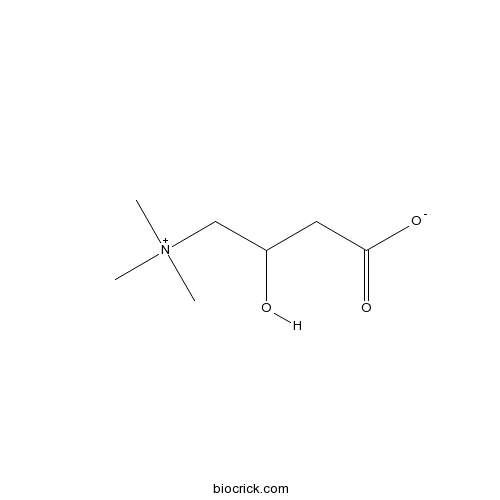
L-Carnitine inner saltCAS# 541-15-1 |

Quality Control & MSDS
3D structure
Package In Stock
Number of papers citing our products

| Cas No. | 541-15-1 | SDF | Download SDF |
| PubChem ID | 288 | Appearance | White cryst. |
| Formula | C7H15NO3 | M.Wt | 161.20 |
| Type of Compound | Miscellaneous | Storage | Desiccate at -20°C |
| Synonyms | Levocarnitine | ||
| Solubility | H2O : ≥ 50 mg/mL (310.17 mM) *"≥" means soluble, but saturation unknown. | ||
| Chemical Name | 3-hydroxy-4-(trimethylazaniumyl)butanoate | ||
| SMILES | C[N+](C)(C)CC(CC(=O)[O-])O | ||
| Standard InChIKey | PHIQHXFUZVPYII-UHFFFAOYSA-N | ||
| Standard InChI | InChI=1S/C7H15NO3/c1-8(2,3)5-6(9)4-7(10)11/h6,9H,4-5H2,1-3H3 | ||
| General tips | For obtaining a higher solubility , please warm the tube at 37 ℃ and shake it in the ultrasonic bath for a while.Stock solution can be stored below -20℃ for several months. We recommend that you prepare and use the solution on the same day. However, if the test schedule requires, the stock solutions can be prepared in advance, and the stock solution must be sealed and stored below -20℃. In general, the stock solution can be kept for several months. Before use, we recommend that you leave the vial at room temperature for at least an hour before opening it. |
||
| About Packaging | 1. The packaging of the product may be reversed during transportation, cause the high purity compounds to adhere to the neck or cap of the vial.Take the vail out of its packaging and shake gently until the compounds fall to the bottom of the vial. 2. For liquid products, please centrifuge at 500xg to gather the liquid to the bottom of the vial. 3. Try to avoid loss or contamination during the experiment. |
||
| Shipping Condition | Packaging according to customer requirements(5mg, 10mg, 20mg and more). Ship via FedEx, DHL, UPS, EMS or other couriers with RT, or blue ice upon request. | ||
| Description | L-carnitine is constituent of striated muscle and liver, it is used therapeutically to stimulate gastric and pancreatic secretions and in the treatment of hyperlipoproteinemias. L-Carnitine inner salt regulates the PTEN/Akt/mTOR signaling pathway, and enhances axonal plasticity while concurrently ameliorating oxidative stress and increasing oligodendrocyte myelination of axons, thereby improving WMLs and cognitive impairment in a rat chronic hypoperfusion model. |
| Targets | Akt | mTOR | PTEN |
| In vitro | L-carnitine exposure and mitochondrial function in human neuronal cells.[Pubmed: 24005823]Neurochem Res. 2013 Nov;38(11):2336-41.L-Carnitine inner salt is a naturally occurring substance required in mammalian energy metabolism that functions by facilitating long-chain fatty acid entry into cellular mitochondria, thereby delivering substrate for oxidation and subsequent energy production. It has been purposed that L-Carnitine inner salt may improve and preserve cognitive performance, and may lead to better cognitive aging through the life span, and several controlled human clinical trials with L-Carnitine inner salt support the hypothesis that this substance has the ability to improve cognitive function.
We further hypothesized that, since L-Carnitine inner salt is an important co-factor of mammalian mitochondrial energy metabolism, acute administration of L-Carnitine inner salt to human tissue culture cells should result in detectable increases in mitochondrial function.
|
| In vivo | L-Carnitine intake prevents irregular feeding-induced obesity and lipid metabolism disorder.[Pubmed: 25445284]Gene. 2015 Jan 10;554(2):148-54.L-Carnitine inner salt supplementation has been used to reduce obesity caused by high-fat diet, which is beneficial for lowering blood and hepatic lipid levels, and for ameliorating fatty liver. However, whether L-Carnitine inner salt may affect irregular feeding-induced obesity and lipid metabolism disorder is still largely unknown.
|
| Kinase Assay | Characterization of L-carnitine transport into rat skeletal muscle plasma membrane vesicles.[Pubmed: 10727937]Eur J Biochem. 2000 Apr;267(7):1985-94.
|
| Animal Research | L-carnitine enhances axonal plasticity and improves white-matter lesions after chronic hypoperfusion in rat brain.[Pubmed: 25465043]J Cereb Blood Flow Metab. 2015 Mar;35(3):382-91.Chronic cerebral hypoperfusion causes white-matter lesions (WMLs) with oxidative stress and cognitive impairment. However, the biologic mechanisms that regulate axonal plasticity under chronic cerebral hypoperfusion have not been fully investigated.
|

L-Carnitine inner salt Dilution Calculator

L-Carnitine inner salt Molarity Calculator
| 1 mg | 5 mg | 10 mg | 20 mg | 25 mg | |
| 1 mM | 6.2035 mL | 31.0174 mL | 62.0347 mL | 124.0695 mL | 155.0868 mL |
| 5 mM | 1.2407 mL | 6.2035 mL | 12.4069 mL | 24.8139 mL | 31.0174 mL |
| 10 mM | 0.6203 mL | 3.1017 mL | 6.2035 mL | 12.4069 mL | 15.5087 mL |
| 50 mM | 0.1241 mL | 0.6203 mL | 1.2407 mL | 2.4814 mL | 3.1017 mL |
| 100 mM | 0.062 mL | 0.3102 mL | 0.6203 mL | 1.2407 mL | 1.5509 mL |
| * Note: If you are in the process of experiment, it's necessary to make the dilution ratios of the samples. The dilution data above is only for reference. Normally, it's can get a better solubility within lower of Concentrations. | |||||

Calcutta University

University of Minnesota

University of Maryland School of Medicine

University of Illinois at Chicago

The Ohio State University

University of Zurich

Harvard University

Colorado State University

Auburn University

Yale University

Worcester Polytechnic Institute

Washington State University

Stanford University

University of Leipzig

Universidade da Beira Interior

The Institute of Cancer Research

Heidelberg University

University of Amsterdam

University of Auckland

TsingHua University

The University of Michigan

Miami University

DRURY University

Jilin University

Fudan University

Wuhan University

Sun Yat-sen University

Universite de Paris

Deemed University

Auckland University

The University of Tokyo

Korea University
L-carnitine is constituent of striated muscle and liver. It is used therapeutically to stimulate gastric and pancreatic secretions and in the treatment of hyperlipoproteinemias. Target: Others L-Carnitine is an endogenous molecule involved in fatty acid metabolism, biosynthesized within the human body using amino acids: L-lysine and L-methionine, as substrates. L-Carnitine can also be found in many foods, but red meats, such as beef and lamb, are the best choices for adding carnitine into the diet [1]. Administering L-carnitine (510 mg/day) to patients with the disease. L-carnitine treatment significantly improved the total time for dozing off during the daytime, calculated from the sleep logs, compared with that of placebo-treated periods. L-carnitine efficiently increased serum acylcarnitine levels, and reduced serum triglycerides concentration [2]. L-carnitine and its derivatives show promise in the treatment of chronic conditions and diseases associated with mitochondrial dysfunction but further translational studies are needed to fully explore their potential [3].
References:
[1]. Marcovina, S.M., et al., Translating the basic knowledge of mitochondrial functions to metabolic therapy: role of L-carnitine. Transl Res, 2013. 161(2): p. 73-84.
[2]. Pekala, J., et al., L-carnitine--metabolic functions and meaning in humans life. Curr Drug Metab, 2011. 12(7): p. 667-78.
[3]. Miyagawa, T., et al., Effects of oral L-carnitine administration in narcolepsy patients: a randomized, double-blind, cross-over and placebo-controlled trial. PLoS One, 2013. 8(1): p. e53707.
- 2-(1-Hydroxy-1-methylethyl)-4-methoxy-7H-furo[3,2-g][1]benzopyran-7-one
Catalog No.:BCN1422
CAS No.:54087-32-0
- Isoastilbin
Catalog No.:BCN5719
CAS No.:54081-48-0
- Palosuran
Catalog No.:BCC4311
CAS No.:540769-28-6
- Tofacitinib (CP-690550) Citrate
Catalog No.:BCC2189
CAS No.:540737-29-9
- Etonogestrel
Catalog No.:BCC5230
CAS No.:54048-10-1
- Albendazole Oxide
Catalog No.:BCC4757
CAS No.:54029-12-8
- Amifampridine
Catalog No.:BCC5185
CAS No.:54-96-6
- Pentylenetetrazole
Catalog No.:BCC7453
CAS No.:54-95-5
- Isoniazid
Catalog No.:BCC9003
CAS No.:54-85-3
- Cinanserin hydrochloride
Catalog No.:BCC6653
CAS No.:54-84-2
- Pilocarpine HCl
Catalog No.:BCC4702
CAS No.:54-71-7
- Idoxuridine
Catalog No.:BCC4666
CAS No.:54-42-2
- Decamethonium Bromide
Catalog No.:BCC4568
CAS No.:541-22-0
- Isovaleramide
Catalog No.:BCC4668
CAS No.:541-46-8
- Muscone
Catalog No.:BCN6275
CAS No.:541-91-3
- 15-Hydroxydehydroabietic acid
Catalog No.:BCN5720
CAS No.:54113-95-0
- 9-Benzylcarbazole-3-carboxaldehyde
Catalog No.:BCC8800
CAS No.:54117-37-2
- Apoptosis Inhibitor
Catalog No.:BCC1143
CAS No.:54135-60-3
- Neoisoastilbin
Catalog No.:BCN6532
CAS No.:54141-72-9
- Flecainide acetate
Catalog No.:BCC1578
CAS No.:54143-56-5
- Vicriviroc Malate
Catalog No.:BCC1230
CAS No.:541503-81-5
- Apilimod
Catalog No.:BCC5286
CAS No.:541550-19-0
- 1-Monopalmitin
Catalog No.:BCN7749
CAS No.:542-44-9
- Cimaterol
Catalog No.:BCC6647
CAS No.:54239-37-1
Characterization of L-carnitine transport into rat skeletal muscle plasma membrane vesicles.[Pubmed:10727937]
Eur J Biochem. 2000 Apr;267(7):1985-94.
Transport of L-carnitine into skeletal muscle was investigated using rat sarcolemmal membrane vesicles. In the presence of an inwardly directed sodium chloride gradient, L-carnitine transport showed a clear overshoot. The uptake of L-carnitine was increased, when vesicles were preloaded with potassium. When sodium was replaced by lithium or cesium, and chloride by nitrate or thiocyanate, transport activities were not different from in the presence of sodium chloride. However, L-carnitine transport was clearly lower in the presence of sulfate or gluconate, suggesting potential-dependent transport. An osmolarity plot revealed a positive slope and a significant intercept, indicating transport of L-carnitine into the vesicle lumen and binding to the vesicle membrane. Displacement experiments revealed that approximately 30% of the L-carnitine associated with the vesicles was bound to the outer and 30% to the inner surface of the vesicle membrane, whereas 40% was unbound inside the vesicle. Saturable transport could be described by Michaelis-Menten kinetics with an apparent Km of 13.1 microM and a Vmax of 2.1 pmol.(mg protein-1).s-1. L-Carnitine transport could be trans-stimulated by preloading the vesicles with L-carnitine but not with the carnitine precursor butyrobetaine, and was cis-inhibited by L-palmitoylcarnitine, L-isovalerylcarnitine, and glycinebetaine. On comparing carnitine transport into rat kidney brush-border membrane vesicles and OCTN2, a sodium-dependent high-affinity human carnitine transporter, cloned recently from human kidney also expressed in muscle, the Km values are similar but driving forces, pattern of inhibition and stereospecificity are different. This suggests the existence of more than one carnitine carrier in skeletal muscle.
L-Carnitine intake prevents irregular feeding-induced obesity and lipid metabolism disorder.[Pubmed:25445284]
Gene. 2015 Jan 10;554(2):148-54.
L-Carnitine supplementation has been used to reduce obesity caused by high-fat diet, which is beneficial for lowering blood and hepatic lipid levels, and for ameliorating fatty liver. However, whether l-carnitine may affect irregular feeding-induced obesity and lipid metabolism disorder is still largely unknown. In the present study, we developed a time-delayed pattern of eating, and investigated the effects of l-carnitine on the irregular eating induced adiposity in mice. After an experimental period of 8 weeks with l-carnitine supplementation, l-carnitine significantly inhibited body weight increase and epididymal fat weight gain induced by the time-delayed feeding. In addition, l-carnitine administration decreased levels of serum alanine aminotransferase (GPT), glutamic oxalacetic transaminase (GOT) and triglyceride (TG), which were significantly elevated by the irregular feeding. Moreover, mice supplemented with l-carnitine did not display glucose intolerance-associated hallmarks, which were found in the irregular feeding-induced obesity. Furthermore, quantitative real-time polymerase chain reaction (qRT-PCR) analysis indicated that l-carnitine counteracted the negative alterations of lipid metabolic gene expression (fatty acid synthase, 3-hydroxy-3-methyl-glutaryl coenzyme A reductase, cholesterol 7alpha-hydroxylase, carnitine/acylcarnitine translocase) in the liver and fat of mice caused by the irregular feeding. Therefore, our results suggest that the time-delayed pattern of eating can induce adiposity and lipid metabolic disorders, while l-carnitine supplementation might prevent these negative symptoms.
L-carnitine enhances axonal plasticity and improves white-matter lesions after chronic hypoperfusion in rat brain.[Pubmed:25465043]
J Cereb Blood Flow Metab. 2015 Mar;35(3):382-91.
Chronic cerebral hypoperfusion causes white-matter lesions (WMLs) with oxidative stress and cognitive impairment. However, the biologic mechanisms that regulate axonal plasticity under chronic cerebral hypoperfusion have not been fully investigated. Here, we investigated whether L-carnitine, an antioxidant agent, enhances axonal plasticity and oligodendrocyte expression, and explored the signaling pathways that mediate axonal plasticity in a rat chronic hypoperfusion model. Adult male Wistar rats subjected to ligation of the bilateral common carotid arteries (LBCCA) were treated with or without L-carnitine. L-carnitine-treated rats exhibited significantly reduced escape latency in the Morris water maze task at 28 days after chronic hypoperfusion. Western blot analysis indicated that L-carnitine increased levels of phosphorylated high-molecular weight neurofilament (pNFH), concurrent with a reduction in phosphorylated phosphatase tensin homolog deleted on chromosome 10 (PTEN), and increased phosphorylated Akt and mammalian target of rapamycin (mTOR) at 28 days after chronic hypoperfusion. L-carnitine reduced lipid peroxidation and oxidative DNA damage, and enhanced oligodendrocyte marker expression and myelin sheath thickness after chronic hypoperfusion. L-carnitine regulates the PTEN/Akt/mTOR signaling pathway, and enhances axonal plasticity while concurrently ameliorating oxidative stress and increasing oligodendrocyte myelination of axons, thereby improving WMLs and cognitive impairment in a rat chronic hypoperfusion model.
L-carnitine exposure and mitochondrial function in human neuronal cells.[Pubmed:24005823]
Neurochem Res. 2013 Nov;38(11):2336-41.
L-Carnitine is a naturally occurring substance required in mammalian energy metabolism that functions by facilitating long-chain fatty acid entry into cellular mitochondria, thereby delivering substrate for oxidation and subsequent energy production. It has been purposed that L-carnitine may improve and preserve cognitive performance, and may lead to better cognitive aging through the life span, and several controlled human clinical trials with L-carnitine support the hypothesis that this substance has the ability to improve cognitive function. We further hypothesized that, since L-carnitine is an important co-factor of mammalian mitochondrial energy metabolism, acute administration of L-carnitine to human tissue culture cells should result in detectable increases in mitochondrial function. Cultures of SH-SY-5Y human neuroblastoma and 1321N1 human astrocytoma cells grown in 96-well cell culture plates were acutely administered L-carnitine hydrochloride, and then, mitochondrial function was assayed using the colorimetric 2,3-bis[2-methoxy-4-nitro-5-sulfophenyl]-2H-tetrazolium-5-carboxyanilide inner salt cell assay kit in a VERSAmax tunable microplate reader. Significant increases in mitochondrial function were observed when human neuroblastoma or human astrocytoma cells were exposed to 100 nM (20 mug L-carnitine hydrochloride/L) to 100 muM (20 mg L-carnitine hydrochloride/L) concentrations of L-carnitine hydrochloride in comparison to unexposed cells, whereas no significant positive effects were observed at lower or higher concentrations of L-carnitine hydrochloride. The results of the present study provide insights for how L-carnitine therapy may significantly improve human neuronal function, but we recommend that future studies further explore different derivatives of L-carnitine compounds in different in vitro cell-based systems using different markers of mitochondrial function.


