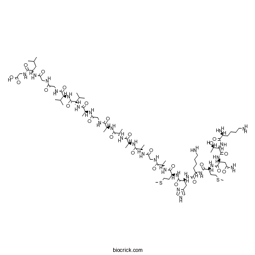
Prion Protein 106-126 (human)Prion protein fragment CAS# 148439-49-0 |

- CP 31398 dihydrochloride
Catalog No.:BCC2406
CAS No.:1217195-61-3
- Tenovin-1
Catalog No.:BCC2239
CAS No.:380315-80-0
- PRIMA-1
Catalog No.:BCC2413
CAS No.:5608-24-2
- Pifithrin-α (PFTα)
Catalog No.:BCC2241
CAS No.:63208-82-2
- NSC 319726
Catalog No.:BCC2242
CAS No.:71555-25-4
- PhiKan 083
Catalog No.:BCC2411
CAS No.:880813-36-5
Quality Control & MSDS
3D structure
Package In Stock
Number of papers citing our products

| Cas No. | 148439-49-0 | SDF | Download SDF |
| PubChem ID | 71312203 | Appearance | Powder |
| Formula | C80H138N26O24S2 | M.Wt | 1912.26 |
| Type of Compound | N/A | Storage | Desiccate at -20°C |
| Solubility | Soluble to 1 mg/ml in water | ||
| Sequence | KTNMKHMAGAAAAGAVVGGLG | ||
| SMILES | CC(C)CC(C(=O)NCC(=O)O)NC(=O)CNC(=O)CNC(=O)C(C(C)C)NC(=O)C(C(C)C)NC(=O)C(C)NC(=O)CNC(=O)C(C)NC(=O)C(C)NC(=O)C(C)NC(=O)C(C)NC(=O)CNC(=O)C(C)NC(=O)C(CCSC)NC(=O)C(CC1=CNC=N1)NC(=O)C(CCCCN)NC(=O)C(CCSC)NC(=O)C(CC(=O)N)NC(=O)C(C(C)O)NC(=O)C(CCCCN)N | ||
| Standard InChIKey | XPZWWTIIKSODDO-MBNDGZRNSA-N | ||
| Standard InChI | InChI=1S/C80H138N26O24S2/c1-38(2)28-53(72(122)90-36-61(113)114)98-60(112)33-86-57(109)32-89-78(128)62(39(3)4)105-79(129)63(40(5)6)104-70(120)44(10)93-59(111)35-87-65(115)41(7)94-68(118)45(11)96-69(119)46(12)95-67(117)43(9)92-58(110)34-88-66(116)42(8)97-73(123)51(22-26-131-14)100-76(126)54(29-48-31-85-37-91-48)102-74(124)50(21-17-19-25-82)99-75(125)52(23-27-132-15)101-77(127)55(30-56(84)108)103-80(130)64(47(13)107)106-71(121)49(83)20-16-18-24-81/h31,37-47,49-55,62-64,107H,16-30,32-36,81-83H2,1-15H3,(H2,84,108)(H,85,91)(H,86,109)(H,87,115)(H,88,116)(H,89,128)(H,90,122)(H,92,110)(H,93,111)(H,94,118)(H,95,117)(H,96,119)(H,97,123)(H,98,112)(H,99,125)(H,100,126)(H,101,127)(H,102,124)(H,103,130)(H,104,120)(H,105,129)(H,106,121)(H,113,114)/t41-,42-,43-,44-,45-,46-,47+,49-,50-,51-,52-,53-,54-,55-,62-,63-,64-/m0/s1 | ||
| General tips | For obtaining a higher solubility , please warm the tube at 37 ℃ and shake it in the ultrasonic bath for a while.Stock solution can be stored below -20℃ for several months. We recommend that you prepare and use the solution on the same day. However, if the test schedule requires, the stock solutions can be prepared in advance, and the stock solution must be sealed and stored below -20℃. In general, the stock solution can be kept for several months. Before use, we recommend that you leave the vial at room temperature for at least an hour before opening it. |
||
| About Packaging | 1. The packaging of the product may be reversed during transportation, cause the high purity compounds to adhere to the neck or cap of the vial.Take the vail out of its packaging and shake gently until the compounds fall to the bottom of the vial. 2. For liquid products, please centrifuge at 500xg to gather the liquid to the bottom of the vial. 3. Try to avoid loss or contamination during the experiment. |
||
| Shipping Condition | Packaging according to customer requirements(5mg, 10mg, 20mg and more). Ship via FedEx, DHL, UPS, EMS or other couriers with RT, or blue ice upon request. | ||
| Description | Prion peptide fragment that shares many physiochemical features with PrPSc. Exhibits neurotoxicity caused by amplification of PrPC-associated signaling responses and induces NF-κB-mediated apoptosis in the mouse neuroblastoma cell line N2a. Forms β-sheet-rich, insoluble, protease-resistant fibrils and is used as a model to study prion diseases in vitro. |

Prion Protein 106-126 (human) Dilution Calculator

Prion Protein 106-126 (human) Molarity Calculator

Calcutta University

University of Minnesota

University of Maryland School of Medicine

University of Illinois at Chicago

The Ohio State University

University of Zurich

Harvard University

Colorado State University

Auburn University

Yale University

Worcester Polytechnic Institute

Washington State University

Stanford University

University of Leipzig

Universidade da Beira Interior

The Institute of Cancer Research

Heidelberg University

University of Amsterdam

University of Auckland

TsingHua University

The University of Michigan

Miami University

DRURY University

Jilin University

Fudan University

Wuhan University

Sun Yat-sen University

Universite de Paris

Deemed University

Auckland University

The University of Tokyo

Korea University
- (+)-Matairesinol
Catalog No.:BCN7021
CAS No.:148409-36-3
- Docetaxel Trihydrate
Catalog No.:BCC1535
CAS No.:148408-66-6
- Secoisolariciresinol Diglucoside
Catalog No.:BCN1212
CAS No.:148244-82-0
- H-Dap-OH.HCl
Catalog No.:BCC3186
CAS No.:1482-97-9
- UNC 0642
Catalog No.:BCC8014
CAS No.:1481677-78-4
- (±)-Epibatidine
Catalog No.:BCC6750
CAS No.:148152-66-3
- trans-2-Tridecene-1,13-dioic acid
Catalog No.:BCN3667
CAS No.:14811-82-6
- Ac-Lys(Fmoc)-OH
Catalog No.:BCC2679
CAS No.:148101-51-3
- Fmoc-Lys(Dnp)-OH
Catalog No.:BCC3519
CAS No.:148083-64-1
- Talc
Catalog No.:BCC4730
CAS No.:14807-96-6
- Calcineurin Autoinhibitory Peptide
Catalog No.:BCC2456
CAS No.:148067-21-4
- 25-Hydroxycycloart-23-en-3-one
Catalog No.:BCN1657
CAS No.:148044-47-7
- L-732,138
Catalog No.:BCC6821
CAS No.:148451-96-1
- JMV 390-1
Catalog No.:BCC5922
CAS No.:148473-36-3
- MNS
Catalog No.:BCC3943
CAS No.:1485-00-3
- GRK2i
Catalog No.:BCC6048
CAS No.:148505-03-7
- Pregabalin
Catalog No.:BCN2175
CAS No.:148553-50-8
- 3,5-Dihydroxyergosta-7,22-dien-6-one
Catalog No.:BCN1658
CAS No.:14858-07-2
- 3-O-Methylquercetin tetraacetate
Catalog No.:BCN1659
CAS No.:1486-69-7
- 3-O-Methylquercetin
Catalog No.:BCN1660
CAS No.:1486-70-0
- Fmoc-Prolinol
Catalog No.:BCC2710
CAS No.:148625-77-8
- GR 127935 hydrochloride
Catalog No.:BCC7081
CAS No.:148642-42-6
- L-733,060 hydrochloride
Catalog No.:BCC5707
CAS No.:148687-76-7
- SB 204070
Catalog No.:BCC5752
CAS No.:148688-01-1
Conformational properties of peptide fragments homologous to the 106-114 and 106-126 residues of the human prion protein: a CD and NMR spectroscopic study.[Pubmed:15678187]
Org Biomol Chem. 2005 Feb 7;3(3):490-7.
Two peptide fragments, corresponding to the amino acid residues 106-126 (PrP[Ac-106-126-NH(2)]) and 106-114 (PrP[Ac-106-114-NH(2)]) of the human prion protein have been synthesised in the acetylated and amide form at their N- and C-termini, respectively. The conformational preferences of PrP[Ac-106-126-NH(2)] and PrP[Ac-106-114-NH(2)] were investigated using CD and NMR spectroscopy. CD results showed that PrP[Ac-106-126-NH(2)] mainly adopts an alpha-helical conformation in TFE-water mixture and in SDS micelles, while a predominantly random structure is observed in aqueous solution. The shorter PrP[Ac-106-114-NH(2)] fragment showed similar propensities when investigated under the same experimental conditions as those employed for PrP[Ac-106-126-NH(2)]. From CD experiments at different SDS concentrations, an alpha-helix/beta-sheet conformational transition was only observed in the blocked PrP[Ac-106-126-NH(2)] sequence. The NMR analysis confirmed the helical nature of PrP[Ac-106-126-NH(2)] in the presence of SDS micelles. The shorter PrP[Ac-106-114-NH(2)] manifested a similar behaviour. The results as a whole suggest that both hydrophobic effects and electrostatic interactions play a significant role in the formation and stabilisation of ordered secondary structures in PrP[Ac-106-126-NH(2)].
PACAP protects neuronal PC12 cells from the cytotoxicity of human prion protein fragment 106-126.[Pubmed:12095620]
FEBS Lett. 2002 Jul 3;522(1-3):65-70.
Misfolding of the prion protein yields amyloidogenic isoforms, and it shows exacerbating neuronal damage in neurodegenerative disorders including prion diseases. Pituitary adenylate cyclase-activating polypeptide (PACAP) and vasoactive intestinal peptide (VIP) potently stimulate neuritogenesis and survival of neuronal cells in the central nervous system. Here, we tested these neuropeptides on neurotoxicity in PC12 cells induced by the prion protein fragment 106-126 [PrP (106-126)]. Concomitant application of neuropeptide with PrP(106-126) (5x10(-5) M) inhibited the delayed death of neuron-like PC12 cells. In particular, PACAP27 inhibited the neurotoxicity of PrP(106-126) at low concentrations (>10(-15) M), characterized by the deactivation of PrP(106-126)-stimulated caspase-3. The neuroprotective effect of PACAP27 was antagonized by the selective PKA inhibitor, H89, or the MAP kinase inhibitor, U0126. These results suggest that PACAP27 attenuates PrP(106-126)-induced delayed neurotoxicity in PC12 cells by activating both PKA and MAP kinases mediated by PAC1 receptor.
Core structure of amyloid fibrils formed by residues 106-126 of the human prion protein.[Pubmed:19278656]
Structure. 2009 Mar 11;17(3):417-26.
Peptides comprising residues 106-126 of the human prion protein (PrP) exhibit many features of the full-length protein. PrP(106-126) induces apoptosis in neurons, forms fibrillar aggregates, and can mediate the conversion of native cellular PrP (PrP(C)) to the scrapie form (PrP(Sc)). Despite a wide range of biochemical and biophysical studies on this peptide, including investigation of its propensity for aggregation, interactions with cell membranes, and PrP-like toxicity, the structure of amyloid fibrils formed by PrP(106-126) remains poorly defined. In this study we use solid-state nuclear magnetic resonance to define the secondary and quaternary structure of PrP(106-126) fibrils. Our results reveal that PrP(106-126) forms in-register parallel beta sheets, stacked in an antiparallel fashion within the mature fibril. The close intermolecular contacts observed in the fibril core provide a rational for the sequence-dependent behavior of PrP(106-126), and provide a basis for further investigation of its biological properties.
Isolation of human neuronal cells resistant to toxicity by the prion protein peptide 106-126.[Pubmed:12214058]
J Alzheimers Dis. 2001 Apr;3(2):169-180.
Prion diseases or transmissible spongiform encephalopathies, are neurodegenerative disorders that are genetic, sporadic, or infectious. The pathogenetic event common to all prion disorders is the conformational transformation of the cellular prion protein (PrP^C) to the scrapie form (PrP^Sc), that deposits in the brain parenchyma and induces neuronal death. Infectious prion disorders are caused by exogenously introduced PrP^Sc that acts as a template in the conversion of endogenous PrP^C to nascent PrP^Sc, and subsequently the process becomes autocatalytic. To understand the process of cellular uptake of PrP^Sc and its mechanism of cellular toxicity, previous studies have used a PrP fragment spanning residues 106-126 (PrP^Tx) that is toxic to primary neurons in culture, and mimics PrP^Sc in its biophysical properties [9,11,14]. Several possible mechanisms of cell death by PrP^Tx have been proposed [2,3,10,11,18], but the existing data are unclear. To identify the biochemical pathways of neurotoxicity by this fragment, we have isolated mutant neuroblastoma and NT-2 cells that are resistant to toxicity by PrP^Tx. We show that these cells bind and internalize PrP^Tx in a temperature dependent fashion, and the peptide accumulates in intracellular compartments, probably lysosomes, where it has an unusually long half-life. The PrP^Tx-resistant phenotype of the cells reported in this study could result from aberrant binding or internalization of the peptide, or due to an abnormality in the downstream pathway(s) of neuronal toxicity. The PrP^Tx-resistant cells are therefore a useful tool for evaluating the cellular and biochemical pathways that lead to cell death by this peptide, and will provide insight into the mechanism(s) of neurotoxicity by PrP^Sc.
p75(NTR) activation of NF-kappaB is involved in PrP106-126-induced apoptosis in mouse neuroblastoma cells.[Pubmed:18602709]
Neurosci Res. 2008 Sep;62(1):9-14.
Neuronal death is a pathological hallmark of prion diseases. Synthetic prion peptide PrP106-126 can convert PrP(C) into protease-resistant aggregates, which can cause neurotoxicity in vivo and in vitro. Various cell surface proteins can participate in the infection process of prions. p75(NTR) can interact with PrP106-126 and has a neurotoxic effect on neurons. However, for p75(NTR) lacking intrinsic catalytic activity domain in cytoplasm, p75(NTR) -associated signaling molecular and the signaling events in cytoplasm in p75(NTR)-mediated apoptosis responding to PrP106-126 remain still unknown. Thus p75(NTR) -associated NF-kappaB signaling pathway was investigated in this study. Herein PrP106-126-induced apoptosis in mouse neuroblastoma cell line N2a, PrP106-126 significantly up-regulated p75(NTR) expression on mRNA and protein levels. For the first time we found that PrP106-126 induced activation of NF-kappaB by Western blot assay, and blocking the interaction of p75(NTR) with PrP106-126 by p75(NTR) polyclonal antibody sc-6189 or pretreatment with inhibitor NF-kappaB SN50 reduced the activation of NF-kappaB and attenuated the apoptotic effect by PrP106-126. This study offers a possible interpretation that NF-kappaB signaling pathway was activated by the interaction of PrP106-126 with p75(NTR), and NF-kappaB activity showed the pro-apoptotic effect in PrP106-126-induced apoptosis in N2a cells. Involvement of NF-kappaB signaling pathway in p75(NTR)-mediated apoptosis may partially account for the PrP106-126-induced neurotoxicity in N2a cells.
Overstimulation of PrPC signaling pathways by prion peptide 106-126 causes oxidative injury of bioaminergic neuronal cells.[Pubmed:16864581]
J Biol Chem. 2006 Sep 22;281(38):28470-9.
Transmissible spongiform encephalopathies, also called prion diseases, are characterized by neuronal loss linked to the accumulation of PrP(Sc), a pathologic variant of the cellular prion protein (PrP(C)). Although the molecular and cellular bases of PrP(Sc)-induced neuropathogenesis are not yet fully understood, increasing evidence supports the view that PrP(Sc) accumulation interferes with PrP(C) normal function(s) in neurons. In the present work, we exploit the properties of PrP-(106-126), a synthetic peptide encompassing residues 106-126 of PrP, to investigate into the mechanisms sustaining prion-associated neuronal damage. This peptide shares many physicochemical properties with PrP(Sc) and is neurotoxic in vitro and in vivo. We examined the impact of PrP-(106-126) exposure on 1C11 neuroepithelial cells, their neuronal progenies, and GT1-7 hypothalamic cells. This peptide triggers reactive oxygen species overflow, mitogen-activated protein kinase (ERK1/2), and SAPK (p38 and JNK1/2) sustained activation, and apoptotic signals in 1C11-derived serotonergic and noradrenergic neuronal cells, while having no effect on 1C11 precursor and GT1-7 cells. The neurotoxic action of PrP-(106-126) relies on cell surface expression of PrP(C), recruitment of a PrP(C)-Caveolin-Fyn signaling platform, and overstimulation of NADPH-oxidase activity. Altogether, these findings provide actual evidence that PrP-(106-126)-induced neuronal injury is caused by an amplification of PrP(C)-associated signaling responses, which notably promotes oxidative stress conditions. Distorsion of PrP(C) signaling in neuronal cells could hence represent a causal event in transmissible spongiform encephalopathy pathogenesis.
Neurotoxicity of a prion protein fragment.[Pubmed:8464494]
Nature. 1993 Apr 8;362(6420):543-6.
The cellular prion protein (PrPC) is a sialoglycoprotein of M(r) 33-35K that is expressed predominantly in neurons. In transmissible and genetic neurodegenerative disorders such as scrapie of sheep, spongiform encephalopathy of cattle and Creutzfeldt-Jakob or Gerstmann-Straussler-Scheinker diseases of humans, PrPC is converted into an altered form (termed PrPSc) which is distinguishable from its normal homologue by its relative resistance to protease digestion. PrPSc accumulates in the central nervous system of affected individuals, and its protease-resistant core aggregates extracellularly into amyloid fibrils. The process is accompanied by nerve cell loss, whose pathogenesis and molecular basis are not understood. We report here that neuronal death results from chronic exposure of primary rat hippocampal cultures to micromolar concentrations of a peptide corresponding to residues 106-126 of the amino-acid sequence deduced from human PrP complementary DNA. DNA fragmentation of degenerating neurons indicates that cell death occurred by apoptosis. The PrP peptide 106-126 has a high intrinsic ability to polymerize into amyloid-like fibrils in vitro. These findings indicate that cerebral accumulation of PrPSc and its degradation products may play a role in the nerve cell degeneration that occurs in prion-related encephalopathies.


