
Auramine OCAS# 2465-27-2 |
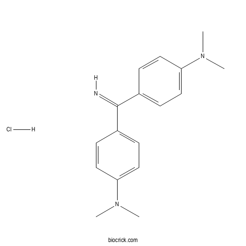
| Cas No. | 2465-27-2 | SDF | File under preparation. |
| PubChem ID | 17170.0 | Appearance | Powder |
| Formula | C17H22ClN3 | M.Wt | 303.8 |
| Type of Compound | Amines | Storage | Desiccate at -20°C |
| Solubility | Soluble in Chloroform,Dichloromethane,Ethyl Acetate,DMSO,Acetone,etc. | ||
| Chemical Name | 4-[4-(dimethylamino)benzenecarboximidoyl]-N,N-dimethylaniline;hydrochloride | ||
| SMILES | CN(C)C1=CC=C(C=C1)C(=N)C2=CC=C(C=C2)N(C)C.Cl | ||
| Standard InChIKey | KSCQDDRPFHTIRL-UHFFFAOYSA-N | ||
| Standard InChI | InChI=1S/C17H21N3.ClH/c1-19(2)15-9-5-13(6-10-15)17(18)14-7-11-16(12-8-14)20(3)4;/h5-12,18H,1-4H3;1H | ||
| General tips | For obtaining a higher solubility , please warm the tube at 37 ℃ and shake it in the ultrasonic bath for a while.Stock solution can be stored below -20℃ for several months. We recommend that you prepare and use the solution on the same day. However, if the test schedule requires, the stock solutions can be prepared in advance, and the stock solution must be sealed and stored below -20℃. In general, the stock solution can be kept for several months. Before use, we recommend that you leave the vial at room temperature for at least an hour before opening it. |
||
| About Packaging | 1. The packaging of the product may be reversed during transportation, cause the high purity compounds to adhere to the neck or cap of the vial.Take the vail out of its packaging and shake gently until the compounds fall to the bottom of the vial. 2. For liquid products, please centrifuge at 500xg to gather the liquid to the bottom of the vial. 3. Try to avoid loss or contamination during the experiment. |
||
| Shipping Condition | Packaging according to customer requirements(5mg, 10mg, 20mg and more). Ship via FedEx, DHL, UPS, EMS or other couriers with RT, or blue ice upon request. | ||
Quality Control & MSDS
3D structure
Package In Stock
Number of papers citing our products

Auramine O Dilution Calculator
Auramine O Molarity Calculator
| 1 mg | 5 mg | 10 mg | 20 mg | 25 mg | |
| 1 mM | 3.2916 mL | 16.4582 mL | 32.9164 mL | 65.8328 mL | 82.291 mL |
| 5 mM | 0.6583 mL | 3.2916 mL | 6.5833 mL | 13.1666 mL | 16.4582 mL |
| 10 mM | 0.3292 mL | 1.6458 mL | 3.2916 mL | 6.5833 mL | 8.2291 mL |
| 50 mM | 0.0658 mL | 0.3292 mL | 0.6583 mL | 1.3167 mL | 1.6458 mL |
| 100 mM | 0.0329 mL | 0.1646 mL | 0.3292 mL | 0.6583 mL | 0.8229 mL |
| * Note: If you are in the process of experiment, it's necessary to make the dilution ratios of the samples. The dilution data above is only for reference. Normally, it's can get a better solubility within lower of Concentrations. | |||||
- Cadalene
Catalog No.:BCX2814
CAS No.:483-78-3
- Glycocholic acid
Catalog No.:BCX2813
CAS No.:475-31-0
- 2-Deoxokanshone M
Catalog No.:BCX2250
CAS No.:3062873-62-2
- Z-isogeranic acid
Catalog No.:BCX2249
CAS No.:85916-85-4
- 6,11,12,16-Tetrahydroxy-5,8,11,13-abitetetraen-7-one
Catalog No.:BCX2248
CAS No.:1295650-67-7
- 3-(2-Hydroxyphenyl)propionic acid
Catalog No.:BCX2247
CAS No.:495-78-3
- Oxypeucedanin hydrate-3-ethyl ether
Catalog No.:BCX2246
CAS No.:90686-46-7
- 4'-Demethyldeoxypodophyllotoxin beta-D-glucopyranoside
Catalog No.:BCX2245
CAS No.:78178-42-4
- Dihydromelilotoside
Catalog No.:BCX2244
CAS No.:24696-05-7
- 7-Feruloylloganic acid
Catalog No.:BCX2243
CAS No.:178666-92-7
- rel-(7S,8S,7''R,8''R)-3,3'',4'',5''-hexamethoxy-7-O-7'',8,8''-lignan
Catalog No.:BCX2242
CAS No.:50394-05-3
- Neoligustilide
Catalog No.:BCX2241
CAS No.:179382-45-7
- 3-Phenylpropyl Acetate
Catalog No.:BCX2816
CAS No.:122-72-5
- Gweicurculactone
Catalog No.:BCX2817
CAS No.:123914-43-2
- Esculentoside L
Catalog No.:BCX2818
CAS No.:130364-33-9
- Rorifamide
Catalog No.:BCX2819
CAS No.:53078-91-4
- Stemoninine
Catalog No.:BCX2820
CAS No.:69772-72-1
- Dracorubin
Catalog No.:BCX2821
CAS No.:6219-63-2
- Astin J
Catalog No.:BCX2822
CAS No.:163136-31-0
- Kalopanaxsaponin G
Catalog No.:BCX2823
CAS No.:57539-70-5
- Kuwanon L
Catalog No.:BCX2824
CAS No.:88524-65-6
- Evodiamide
Catalog No.:BCX2825
CAS No.:116965-70-9
- Dihydrochelirubine
Catalog No.:BCX2826
CAS No.:28342-26-9
- Protodioscin
Catalog No.:BCX2827
CAS No.:60478-69-5
Progressive fluorescence amplification of molecular rotors via G-run-length engineering of G-rich DNA scaffolds.[Pubmed:42702446]
Anal Chim Acta. 2026 Nov 1;1421:346015.
BACKGROUND: G-quadruplexes (G4s) act as versatile scaffolds for biosensing, yet a generalizable strategy to systematically amplify the fluorescence of G-rich scaffold-bound molecular rotors remains lacking. RESULTS: Herein, we propose a G-run-length engineering strategy to enhance the "light-up" performance of fluorogenic ligands using Thioflavin T (ThT) as a model molecular rotor. Through rational design of a sequence library derived from EAD4 with variable G-run lengths (N = 2-6, where N denotes the number of consecutive guanines per G-tract), we show that increasing N progressively amplified ThT fluorescence by up to approximately 1000-fold under metal-free conditions. Mechanistic investigations-including induced circular dichroism, thermal melting analysis, Job plots, isothermal titration calorimetry and hydroxylation kinetics-support that G-run extension increases ligand-accessible binding environments and shields bound ThT from the aqueous phase, thereby restricting intramolecular rotation and suppressing non-radiative decay. The enhanced shielding likely arises from strengthened terminal stacking and/or possible cavity-associated binding, although the present data do not define a single G4 topology or a unique microscopic binding mode. The G-run-length-dependent enhancement trend was further observed in derivative libraries based on different G-rich DNA scaffolds and with other molecular rotors, including Thiazole Orange and Auramine O, suggesting functional generalizability. By contrast, macrocyclic planar ligands such as hemin and N-Methyl Mesoporphyrin IX, which are dominated by terminal G-tetrad association rather than intramolecular rotation restriction, did not show the same trend. SIGNIFICANCE: These findings establish G-run-length engineering as a practical strategy for regulating G-rich scaffold-fluorophore interactions and provide mechanistic guidance for nucleic-acid-based fluorescence signal amplification.
Construction of multivalent aptamer-based fluorogenic DNA nanostructures for label-free fluorescent biosensors.[Pubmed:42685561]
Talanta. 2026 Aug 30;312(Pt C):130535.
Fluorescent light-up aptamers (FLAPs) are attractive signaling tools due to their label-free fluorescence and programmability. However, DNA-based FLAP sensors are often constrained by low fluorogenic activation efficiency, limited intrinsic target responsiveness, and insufficient signal amplification. In this study, we integrated a DNA-based FLAP (DAP) with a three-branched DNA (TBD) scaffold to construct a multivalent FLAP nanostructure (DAP-TBD) that exhibited enhanced fluorogenic activation toward Auramine O (AO). Compared with monovalent DAP, the trivalent DAP-TBD showed a 3.3-fold increase in fluorogenic activation efficiency, indicating a clear multivalent effect. The system adopts a modular architecture in which DAP functions as a replaceable FLAP model. Using a target-triggered catalytic DNA assembly (CDA) circuit composed of three rationally designed hairpin DNAs, multiple DAP-TBD nanostructures are assembled isothermally, enabling target-triggered signal generation and amplification without the need for labeled probes. The CDA-based DAP-TBD system selectively detects target single-stranded DNA with a detection limit of 235 pM. Furthermore, coupling the DAP-TBD system with an exonuclease-assisted hairpin and an aptazyme enabled label-free detection of microRNA and adenosine triphosphate, demonstrating the versatility of the system as a signal sensor that can interface with diverse upstream transducer modules. The system requires no fluorophore or quencher labeling, relies on only three unmodified DNA strands, and offers programmability, modularity, and cost-effectiveness. Overall, this study presents a multivalent DNA-based FLAP nanostructure and provides a versatile strategy for developing label-free fluorescent biosensors.
Evaluating Pollen Viability and Tube Growth in Rice (Oryza sativa).[Pubmed:42642567]
Methods Mol Biol. 2026;3042:19-33.
Pollen maturity, viability, and pollen tube growth are essential indicators of male fertility in rice (Oryza sativa). This chapter presents standardized protocols to evaluate these traits using a range of staining methods, including iodine-potassium iodide (I(2)-KI), DAPI, calcofluor white, Auramine O, LM19/LM20 antibodies, and FM4-64. Pollen germination is assessed through in vitro assays on sucrose-based germination medium optimized for rice, while in vivo pollen tube growth is visualized with aniline blue staining. Given the rapid and short-lived nature of rice pollen under laboratory conditions, practical tips are included to enhance reproducibility. Together, these methods provide a reliable toolkit for the functional analysis of rice pollen in developmental, genetic, and breeding research.
Environmental Contaminant Auramine O, a Synthetic Dye Exposure Induces Multiorgan Toxicity via Inflammatory and Apoptotic Pathways in Sprague-Dawley Rats.[Pubmed:42551918]
J Appl Toxicol. 2026 Aug 4.
Auramine O (AO) is a widely used synthetic dye that poses serious environmental and health risks, yet the mechanisms underlying its multiorgan toxicity remain poorly understood. This investigation aims to assess the systemic toxicity of AO in Sprague-Dawley rats. The animals were divided into a control group, AO-exposed groups (5 and 10 mg/kg), and a positive control group receiving bisphenol A (50 mg/kg) for 90 days of exposure via oral gavage. Toxicity was assessed through body weight, serum biochemical markers, histopathology, gene expression (RT-PCR), immunohistochemistry, and HPLC-based quantification of AO in both urine and plasma. AO showed a quick systemic distribution to physiological organs and was primarily eliminated via the renal pathway. In addition, this study revealed a dose-dependent decrease in body weight. Biomarkers, such as bilirubin, albumin, glucose, creatinine, blood urea nitrogen (BUN), and creatine kinase levels, showed a significant elevation. In the brain homogenate, acetylcholinesterase level was notably reduced. Histopathological analysis of tissue samples revealed considerable damage to the liver (hepatic injury), kidney (tubular degeneration), heart (myocardial injury), and brain (neuronal degeneration). Gene expression analysis showed significant upregulation of inflammatory (tnf-alpha and nfkb) and apoptotic (caspase-3 and caspase-9) markers, and immunohistochemistry resulted in upregulated expression of nfkb. These results emphasize the potential health hazards linked to prolonged AO exposure, necessitating stringent environmental regulatory measures and further research into its long-term safety and potential therapeutic interventions.
A smear test aiding with an automated slide reader acquires equivalent sensitivity to MGIT960 culture in pulmonary tuberculosis diagnosis.[Pubmed:42360030]
Microbiol Spectr. 2026 Aug 4;14(8):e0397025.
Smear microscopy remains widely used for tuberculosis (TB) diagnosis due to its rapid turnaround and low cost, despite its relatively low sensitivity. With the advent of molecular tests, there is growing debate on whether the smear test remains relevant in clinical settings. An artificial intelligence (AI)-aided automated smear slide reader system (Zopomed, Zhengzhou, China) was evaluated at Beijing Chest Hospital. Sputum samples from suspected TB patients were used to prepare both Ziehl-Neelsen (ZN) and Auramine O-stained slides. These were scanned by the automated reader under predefined field-of-view settings. Manual light-emitting diode fluorescence microscopy (for Auramine O slides), MGIT960 culture, and Xpert MTB/RIF assay served as reference standards. Among 352 clinically confirmed TB patients, the detection rates were 18.75% by manual microscopy, 20.74% by automated ZN (1,000 fields), 32.67% by automated Auramine O (300 fields), 30.68% by MGIT960 culture, and 42.05% by Xpert. Expanding the field of view significantly enhanced detection rates for both staining methods, with Auramine O staining consistently outperforming ZN. The automated Auramine O method (300 fields) identified 78.70% of culture-positive and 66.89% of Xpert-positive TB cases. All methods maintained specificities exceeding 99% when tested on 158 patients with non-mycobacterial pulmonary diseases. AI-aided Auramine O smear testing represents a sensitive, rapid, and cost-effective approach to TB diagnosis. With the one-time investment in the automated system, this technique achieves sensitivity comparable to MGIT960 culture, offering a practical diagnostic solution for high-throughput clinical laboratories and resource-limited settings. IMPORTANCE: This study demonstrated that an AI-aided automated smear microscopy system, when used with Auramine O-stained slides scanning 300 fields, achieves a sensitivity comparable to MGIT960 culture, while offering the advantages of rapid, same-day results and significant cost-effectiveness; this one-time investment in equipment, which is compatible with routine staining protocols, provides a highly practical and scalable solution to enhance tuberculosis diagnosis in both high-throughput laboratories and resource-limited settings.
Revealing Temperature-Dependent Dynamic Crossover of Molecular Solutes in Thymol-Camphor-Based Type V Deep Eutectic Solvent via Optical Spectroscopy.[Pubmed:42272294]
J Phys Chem B. 2026 Jun 25;130(25):6333-6346.
Type V deep eutectic solvents (DESs) are composed exclusively of nonionic molecular species, in contrast to type I-IV DESs, which contain at least one ionic component. In the absence of ionic constituents, the cohesive energy of type V DESs is governed primarily by hydrogen bonding and van der Waals interactions. Such an intermolecular framework imparts distinct physicochemical properties, such as increased hydrophobicity and relatively low viscosity (compared to their ionic counterparts), substantially broadening the application potential of DESs. Owing to their fundamental distinction from ionic DESs and ionic liquids (ILs), it is imperative to understand how the nonionic nature of type V DESs influences their microstructure and dynamic behavior for their rational and strategic utilization. To address this, we examined the microenvironment of a thymol/camphor-based type V DES using three molecular probes: Coumarin-153, a neutral benchmark probe widely employed for investigating dynamics in solutions, and two fluorescent molecular rotors, Thioflavin T and Auramine O, whose fluorescence quantum yield is sensitive to the microviscosity of the local environment. Absorption and steady-state fluorescence measurements reveal that the local micropolarity experienced by Coumarin-153 in the DES is comparable to that of acetonitrile. A slight red-edge excitation shift observed in the steady-state fluorescence spectra with varying excitation wavelengths points to the presence of subtle spatial heterogeneity within the solvent. Time-resolved emission spectra (TRES) were recorded over the temperature range of 278-333 K and used to construct the solvent correlation function, which describes the solvent's reorganization in the excited state following the electronic excitation of Coumarin-153. The solvation dynamics were found to be bimodal, with a dominant, markedly slow component indicative of a highly sluggish solvent response. Correlating the solvation dynamics with the shear viscosity of the solvent reveals significant decoupling (p approximately 0.62, where p is the decoupling parameter), suggesting decoupling of the solvation dynamics from the viscosity of the medium. Furthermore, a dynamic crossover was observed at approximately 308 K or 35 degrees C, showing temperature-dependent modulation in the structural features of the thymol/camphor-based DES. In contrast, the rotational dynamics of Coumarin-153 closely follow the Stokes-Einstein-Debye hydrodynamic model (p approximately 0.8), indicating that despite heterogeneity, Coumarin-153 experiences an effectively homogeneous local environment. In the thymol/camphor-based DES, the temperature-dependent evolution of fluorescence quantum yield for both Thioflavin T and Auramine O reveals a nonuniform response across the studied range, manifesting as two distinct friction regimes separated by a well-defined crossover at 308 K. Microviscosity determination of both probes reveals decoupling of the torsional relaxation from the bulk viscosity of the solvent, which is complemented by the disparity in the activation energy values of viscous flow and nonradiative transition obtained by utilizing Arrhenius-type curves. This observation suggests that the photophysical behavior of molecular rotors like Thioflavin T and Auramine O is not governed solely by the bulk viscosity of the DES; rather, the local microstructure of the solvent plays a dominant role. These findings underscore how variations in local polarity and specific solute-solvent interactions drive preferential solvation, revealing the presence of microscopically heterogeneous domains within the nonionic, hydrogen-bonded framework of the DES.
Surface-confined CRISPR-Cas12a biosensor with metal-enhanced fluorescence for rapid and ultrasensitive detection of SARS-CoV-2 nucleocapsid protein.[Pubmed:42061344]
Biosens Bioelectron. 2026 Aug 15;306:118649.
CRISPR-Cas12a integrated with nanomaterials has formulated powerful biosensors for viral protein detection, addressing the urgent need for point-of-care diagnostics. However, existing platforms are hindered by either multi-step separation procedures or insufficient signal amplification, limiting their sensitivity and practicality. Here, we report a one-pot "on-off" biosensor that combines metal-enhanced fluorescence (MEF) and nanoscale spatial confinement by co-localizing both reporter substrates and the CRISPR-Cas12a system on gold-silica core-shell nanoparticles (Au@SiO(2) NPs), enabling rapid and ultrasensitive protein detection. Using SARS-CoV-2 nucleocapsid (N) protein as a model analyte, Au@SiO(2) NPs are co-functionalized with (i) ssDNA activators blocked by N protein-specific aptamers, (ii) light-up hairpin DNA (DAP) complexed with Auramine O (AO) as reporters, and (iii) short polyethylene glycol (PEG) spacers to mitigate steric hindrance. The nanoplatform displays an ultrabright "on-state" fluorescence, with an intensity >860-fold higher than that of free AO, enabled by the interaction with DAP and optimized fluorophore-metal spacing ( approximately 20 nm). Upon target binding, aptamer displacement exposes the activator to locally initiate Cas12a trans-cleavage, disrupting proximal DAP structure and its interaction with AO, thereby producing a distinct "off-state" signal. Within the linear detection range, the system demonstrates up to approximately 85% signal reduction relative to the initial signal and a signal-to-noise ratio (SNR) of 83.89, corresponding to a approximately 2.5-fold higher SNR than the solution-phase system. The platform attains a limit of detection at 67.2 fg/mL within 30 min, with excellent sensitivity, selectivity, stability, and recovery in bronchoalveolar lavage fluid. By combining MEF-driven signal amplification with surface-confined CRISPR-Cas12a trans-cleavage, this platform establishes an efficient strategy for sensitive N protein detection.
Ordered Macro-Microporous ZIF-8 Decorated with Nanoparticles for Highly Sensitive Detection of Auramine O in Tropical Fruits.[Pubmed:41972614]
Nanomaterials (Basel). 2026 Mar 25;16(7):398.
Herein, an electrochemical sensor is reported for the first time based on an ordered macro-microporous composite derived from metal-organic frameworks (MOFs) for the highly sensitive detection of Auramine O (AO), a Group 2B carcinogen. The hierarchical pore architecture, integrating an ordered macroporous network with a microporous ZIF-8 framework, enables the uniform dispersion of a high density of catalytically active sites. The interconnected macroporous channels facilitate efficient mass transport and rapid removal of reaction byproducts, effectively preventing pore blockage and ensuring stable sensing performance during repeated measurements. Owing to these structural advantages, the proposed sensor exhibits outstanding analytical performance toward AO detection, with a sensitivity of 0.4843 muA muM(-1), a detection limit of 0.168 muM (S/N = 3), and a wide linear range from 0.5 to 50 muM. Moreover, the sensor demonstrates excellent selectivity and reproducibility, maintaining reliable responses even in the presence of 100-fold excess common food constituents such as tartrazine and glucose. Real sample analysis further confirms its high accuracy and operational stability. Overall, the electrochemical sensor based on silver nanoparticle-decorated ordered macro-microporous ZIF-8 synthesized via in situ reduction shows great potential as a portable and on-site tool for rapid AO detection in food. More broadly, ordered macro-microporous MOF-derived materials represent a promising platform for advanced electrochemical sensor applications.
Engineering novel ceramic metal oxides with residual carbon for efficient sequestration of Auramine O from wastewater.[Pubmed:41917472]
Sci Rep. 2026 Mar 31;16(1):11643.
Auramine O is a hazardous cationic dye that threatens aquatic ecosystems by suppressing photosynthesis, and it poses health risks to humans through toxicity and bioaccumulation. Its chemical persistence allows it to remain in water bodies and enter food chains, which amplifies environmental and medical concerns. Here, we report a facile Pechini sol-gel route to synthesize CuO/MgCr(2)O(4)/C and CuO/MgCr(2)O(4)/MgO/C at 600 and 800 degrees C nanohybrids labeled MCC600 and MCC800, respectively, for efficient Auramine O removal. XRD confirms monoclinic CuO and cubic MgCr(2)O(4) in MCC600 and reveals additional cubic MgO in MCC800 with average crystallite sizes of 60.97 nm and 75.35 nm, respectively, which reflects thermally driven grain growth. EDX verifies C, O, Mg, Cr, and Cu, with carbon decreasing and chromium increasing at higher temperature due to enhanced organic burnout and phase development. FE-SEM shows cauliflower-like agglomerates with fine nanospheres for MCC600 and a more uniform granular texture for MCC800, while HR-TEM reveals ultrafine quasi-spherical clusters for MCC600 and larger faceted polyhedral grains for MCC800. The capacities reach 442.48 mg/g for MCC600 and 298.51 mg/g for MCC800. Kinetic and thermodynamic analyses demonstrate pseudo-first-order behavior with exothermic spontaneous and physical adsorption that follows the Langmuir isotherm. Nearly complete acid-driven regeneration and stable multicycle reuse are achieved, and high performance is retained in real wastewater, which confirms suitability for practical treatment.
Flow cytometric distinction of mature spores of Bacillus subtilis 168 using Auramine O.[Pubmed:41533672]
Biosci Biotechnol Biochem. 2026 Mar 20;90(4):576-584.
We developed a simple and effective method for distinguishing mature spores (MSs) of Bacillus subtilis 168 from mixed cell population using Auramine O (AuO) staining combined with flow cytometry (FCM) analysis. AuO preferentially stains MSs, enabling fluorescence-based discrimination. To optimize the method, B. subtilis 168 was stained with AuO and heated at different temperatures (25, 40, 55, or 70 degrees C) before FCM analysis. Among the tested conditions, heating for 30 min at 55 degrees C yielded the most distinct separation between MSs and other cell types based on fluorescence intensity. This approach combines the high-throughput capability of FCM with temperature-enhanced fluorescent staining to achieve efficient and accurate spore identification. This method is simple, rapid, and scalable, with potential applications in food safety testing and probiotic product manufacturing, where fast and reliable bacterial enumeration is essential.

Calcutta University

University of Minnesota

University of Maryland School of Medicine

University of Illinois at Chicago

The Ohio State University

University of Zurich

Harvard University

Colorado State University

Auburn University

Yale University

Worcester Polytechnic Institute

Washington State University

Stanford University

University of Leipzig

Universidade da Beira Interior

The Institute of Cancer Research

Heidelberg University

University of Amsterdam

University of Auckland

TsingHua University

The University of Michigan

Miami University

DRURY University

Jilin University

Fudan University

Wuhan University

Sun Yat-sen University

Universite de Paris

Deemed University

Auckland University

The University of Tokyo

Korea University