
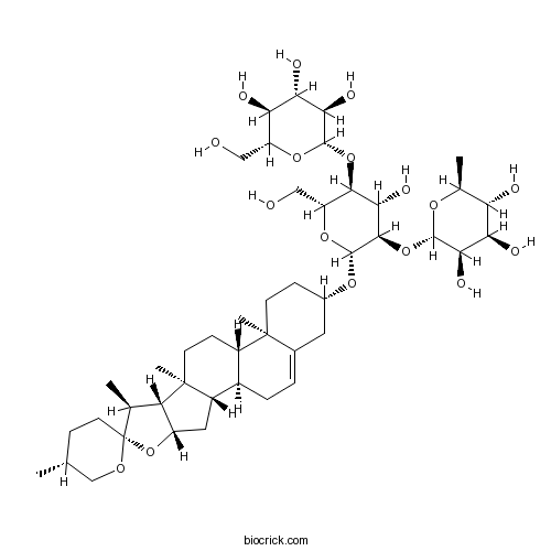
ProtodioscinCAS# 60478-69-5 |
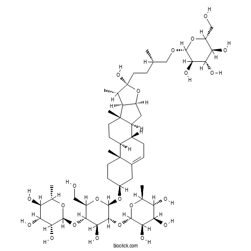
| Cas No. | 60478-69-5 | SDF | File under preparation. |
| PubChem ID | 441899.0 | Appearance | Powder |
| Formula | C51H84O22 | M.Wt | 1049.0 |
| Type of Compound | Steroid Saponins and its Sapogenins | Storage | Desiccate at -20°C |
| Solubility | Soluble in Chloroform,Dichloromethane,Ethyl Acetate,DMSO,Acetone,etc. | ||
| Chemical Name | (2S,3R,4R,5R,6S)-2-[(2R,3S,4S,5R,6R)-4-hydroxy-2-(hydroxymethyl)-6-[[(1S,2S,4S,6R,7S,8R,9S,12S,13R,16S)-6-hydroxy-7,9,13-trimethyl-6-[(3S)-3-methyl-4-[(2R,3R,4S,5S,6R)-3,4,5-trihydroxy-6-(hydroxymethyl)oxan-2-yl]oxybutyl]-5-oxapentacyclo[10.8.0.02,9.04,8.013,18]icos-18-en-16-yl]oxy]-5-[(2S,3R,4R,5R,6S)-3,4,5-trihydroxy-6-methyloxan-2-yl]oxyoxan-3-yl]oxy-6-methyloxane-3,4,5-triol | ||
| SMILES | CC1C2C(CC3C2(CCC4C3CC=C5C4(CCC(C5)OC6C(C(C(C(O6)CO)OC7C(C(C(C(O7)C)O)O)O)O)OC8C(C(C(C(O8)C)O)O)O)C)C)OC1(CCC(C)COC9C(C(C(C(O9)CO)O)O)O)O | ||
| Standard InChIKey | LVTJOONKWUXEFR-YFQJTNJMSA-N | ||
| Standard InChI | InChI=1S/C51H84O22/c1-20(19-65-45-39(60)38(59)35(56)30(17-52)69-45)9-14-51(64)21(2)32-29(73-51)16-28-26-8-7-24-15-25(10-12-49(24,5)27(26)11-13-50(28,32)6)68-48-44(72-47-41(62)37(58)34(55)23(4)67-47)42(63)43(31(18-53)70-48)71-46-40(61)36(57)33(54)22(3)66-46/h7,20-23,25-48,52-64H,8-19H2,1-6H3/t20-,21-,22-,23-,25-,26+,27-,28-,29-,30+,31+,32-,33-,34-,35+,36+,37+,38-,39+,40+,41+,42-,43+,44+,45+,46-,47-,48+,49-,50-,51+/m0/s1 | ||
| General tips | For obtaining a higher solubility , please warm the tube at 37 ℃ and shake it in the ultrasonic bath for a while.Stock solution can be stored below -20℃ for several months. We recommend that you prepare and use the solution on the same day. However, if the test schedule requires, the stock solutions can be prepared in advance, and the stock solution must be sealed and stored below -20℃. In general, the stock solution can be kept for several months. Before use, we recommend that you leave the vial at room temperature for at least an hour before opening it. |
||
| About Packaging | 1. The packaging of the product may be reversed during transportation, cause the high purity compounds to adhere to the neck or cap of the vial.Take the vail out of its packaging and shake gently until the compounds fall to the bottom of the vial. 2. For liquid products, please centrifuge at 500xg to gather the liquid to the bottom of the vial. 3. Try to avoid loss or contamination during the experiment. |
||
| Shipping Condition | Packaging according to customer requirements(5mg, 10mg, 20mg and more). Ship via FedEx, DHL, UPS, EMS or other couriers with RT, or blue ice upon request. | ||
Quality Control & MSDS
3D structure
Package In Stock
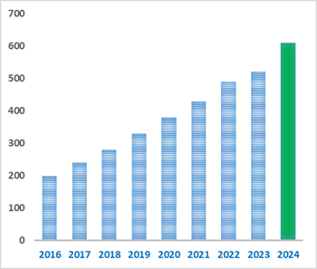
Number of papers citing our products

Protodioscin Dilution Calculator
Protodioscin Molarity Calculator
-
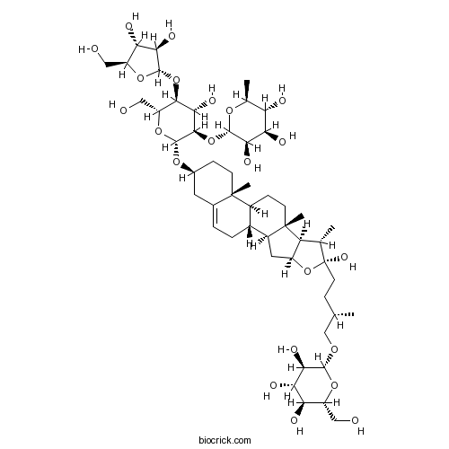 Parisaponin ICatalog No.: BCN2835CAS No.: 561007-63-4
-
 ProtodioscinCatalog No.: BCN6274CAS No.: 55056-80-9
-
 ProtodeltoninCatalog No.: BCN8611CAS No.: 94992-08-2
-
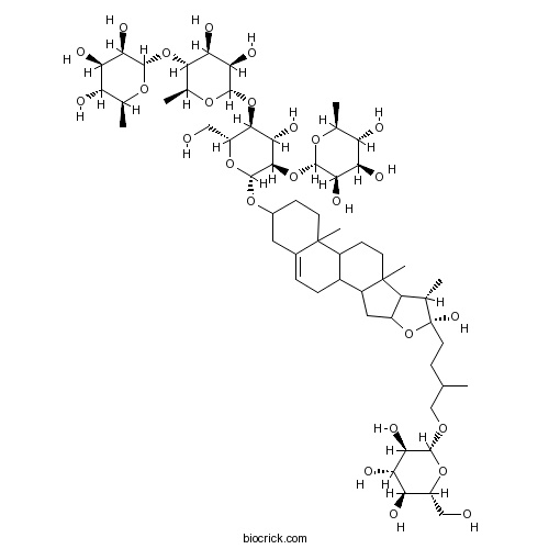 DichotominCatalog No.: BCN2836CAS No.: 53093-47-3
-
 Protogracillin(P)Catalog No.: BCC8352CAS No.: 54848-30-5
-
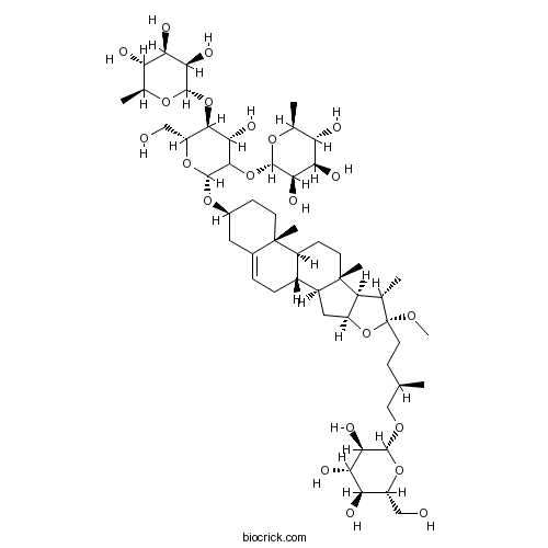 Methyl protodioscinCatalog No.: BCN6342CAS No.: 54522-52-0
-
 Polyphyllin GCatalog No.: BCN1054CAS No.: 76296-75-8
-
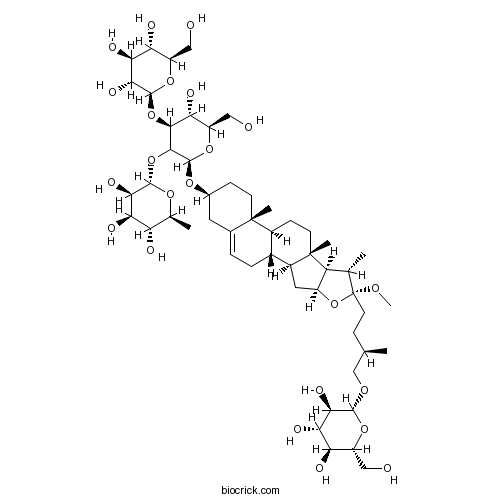 Methyl protogracillinCatalog No.: BCN8177CAS No.: 54522-53-1
-
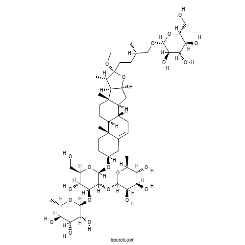 Taccafurostanoside BCatalog No.: BCX2275CAS No.: 98569-68-7
-
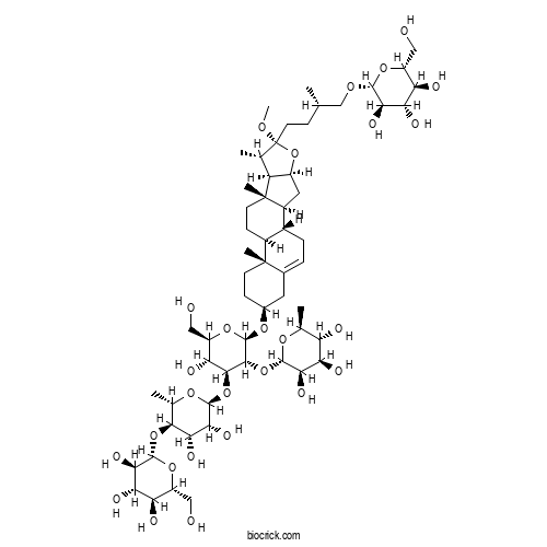 Taccafurostanoside ACatalog No.: BCX2274CAS No.: 290809-76-6
-
 Parisyunnanoside BCatalog No.: BCN2837CAS No.: 945865-37-2
-
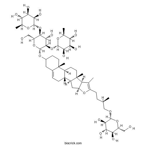 PseudoprotodioscinCatalog No.: BCN2827CAS No.: 102115-79-7
-
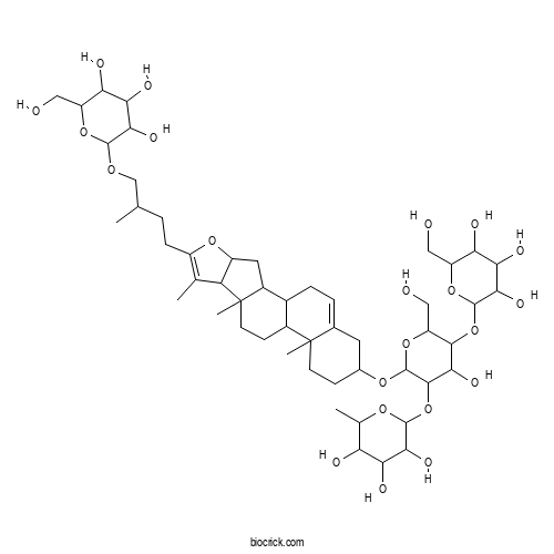 HuangjiangSu ACatalog No.: BCX1589CAS No.: 1026020-27-8
-
 Pseudoproto PbCatalog No.: BCN2838CAS No.: 102100-46-9
-
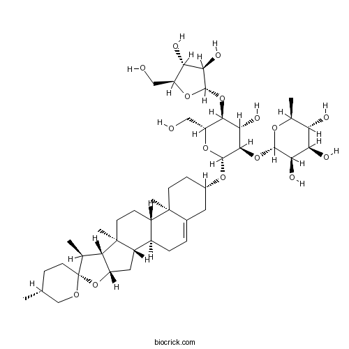 Polyphyllin DCatalog No.: BCN2401CAS No.: 50773-41-6
-
 DeltoninCatalog No.: BCC8933CAS No.: 55659-75-1
| 1 mg | 5 mg | 10 mg | 20 mg | 25 mg | |
| 1 mM | 0.9533 mL | 4.7664 mL | 9.5329 mL | 19.0658 mL | 23.8322 mL |
| 5 mM | 0.1907 mL | 0.9533 mL | 1.9066 mL | 3.8132 mL | 4.7664 mL |
| 10 mM | 0.0953 mL | 0.4766 mL | 0.9533 mL | 1.9066 mL | 2.3832 mL |
| 50 mM | 0.0191 mL | 0.0953 mL | 0.1907 mL | 0.3813 mL | 0.4766 mL |
| 100 mM | 0.0095 mL | 0.0477 mL | 0.0953 mL | 0.1907 mL | 0.2383 mL |
| * Note: If you are in the process of experiment, it's necessary to make the dilution ratios of the samples. The dilution data above is only for reference. Normally, it's can get a better solubility within lower of Concentrations. | |||||
- Dihydrochelirubine
Catalog No.:BCX2826
CAS No.:28342-26-9
- Evodiamide
Catalog No.:BCX2825
CAS No.:116965-70-9
- Kuwanon L
Catalog No.:BCX2824
CAS No.:88524-65-6
- Kalopanaxsaponin G
Catalog No.:BCX2823
CAS No.:57539-70-5
- Astin J
Catalog No.:BCX2822
CAS No.:163136-31-0
- Dracorubin
Catalog No.:BCX2821
CAS No.:6219-63-2
- Stemoninine
Catalog No.:BCX2820
CAS No.:69772-72-1
- Rorifamide
Catalog No.:BCX2819
CAS No.:53078-91-4
- Esculentoside L
Catalog No.:BCX2818
CAS No.:130364-33-9
- Gweicurculactone
Catalog No.:BCX2817
CAS No.:123914-43-2
- 3-Phenylpropyl Acetate
Catalog No.:BCX2816
CAS No.:122-72-5
- Auramine O
Catalog No.:BCX2815
CAS No.:2465-27-2
- Parthenin
Catalog No.:BCX2828
CAS No.:508-59-8
- (+)-(R)-Ribalinine
Catalog No.:BCX2829
CAS No.:62928-56-7
- Rorifone
Catalog No.:BCX2830
CAS No.:53078-90-3
- Esculentoside F
Catalog No.:BCX2831
CAS No.:95263-31-3
- Trans-resveratrol-3-O-β-D-glucuronide
Catalog No.:BCX2832
CAS No.:387372-17-0
- Molephantin
Catalog No.:BCX2833
CAS No.:50656-66-1
- Molephantinin
Catalog No.:BCX2834
CAS No.:56221-98-8
- 3-Butylidene-7-hydroxyphthalide
Catalog No.:BCX2835
CAS No.:93236-67-0
- (7S)-Hellicoside
Catalog No.:BCX2836
CAS No.:132278-04-7
- Cistanoside C
Catalog No.:BCX2837
CAS No.:94492-22-5
- β-Curcumene
Catalog No.:BCX2838
CAS No.:451-56-9
- (E)-Ligustilide
Catalog No.:BCX2839
CAS No.:81944-08-3
Protodioscin mitigates cyclophosphamide-induced renal toxicity in HEK-293 cells by regulating endoplasmic reticulum stress, antioxidant defense, and apoptotic pathways.[Pubmed:42766610]
PLoS One. 2026 Sep 21;21(9):e0357227.
Cyclophosphamide (CTX) is a widely used chemotherapeutic agent with well-established efficacy; however, its clinical use is limited by dose-dependent adverse effects, particularly nephrotoxicity. Protodioscin (Prot), a bioactive saponin derived from plants of the Dioscoreaceae family, has demonstrated notable antioxidant and anti-inflammatory properties. This study aimed to evaluate the protective effects of Prot against CTX-induced cytotoxicity in HEK-293 cells. Primary HEK-293 kidney cells were exposed to the IC50 concentration of CTX in the presence or absence of Prot (5 and 10 microM) for 24 hours. Cell viability was assessed using the MTT assay. Gene expression of Bax and Bcl-2 was measured by real-time PCR assay, while protein expression of Bax, cleaved caspase-3, Bcl-2, ATF6, PERK, GRP78, CHOP, p-eIF2alpha, p-PERK, and cleaved ATF6 were evaluated by Western blot analysis. Oxidative stress parameters, including CAT, SOD, GPx, GSH, MDA, and NO, were quantified using spectrophotometric assays, and kidney injury markers (KIM-1 and cystatin C) were measured by ELISA. Prot decreased Bax and caspase 3 expression and increased Bcl-2 expression in HEK-293 cells exposed to CTX, which was accompanied by reduced apoptosis. Treatment with 5 and 10 microM Prot significantly reduced the expression of ATF6, PERK, GRP78, CHOP, p-eIF2alpha, p-PERK and Cleaved- ATF6 proteins in CTX-exposed HEK-293 cells. Additionally, treatment with Prot significantly increased catalase, GPx, GSH, and SOD levels, while reducing KIM-1, cystatin C, MDA, and NO levels in HEK-293 cells subjected to CTX. These findings suggest that Prot exerts a protective role against CTX-induced renal injury in HEK-293 cells by modulating apoptotic pathways, attenuating endoplasmic reticulum stress, and improving antioxidant indices.
Protodioscin alleviates cyclophosphamide-mediated liver damage in mice by targeting Nrf2/HO-1, NF-kappaB/NLRP3 inflammasome, and mitochondrial apoptosis pathways.[Pubmed:42586447]
Toxicol Appl Pharmacol. 2026 Oct;515:117983.
Cyclophosphamide is a currently used antineoplastic drug to manage a number of malignant tumors. Hepatotoxicity is one of the most significant complications of cyclophosphamide therapy, which restricts its clinical application. Protodioscin, as a natural saponin, possesses multiple striking biological properties. This preclinical research was conducted to assess the ameliorative influences of Protodioscin against cyclophosphamide-triggered hepatotoxicity. Forty male mice were apportioned into five distinct groups: control (normal saline) group, cyclophosphamide (200 mg/kg, i.p.) group, Protodioscin (12.5, 25, and 50 mg/kg, i.p.) + cyclophosphamide groups. The mice received Protodioscin for 10 days and cyclophosphamide was administrated on day 5. The results exhibited that Protodioscin significantly attenuated the activities of ALT, AST, and ALP as well as the serum level of total bilirubin. Protodioscin remarkably inhibited ROS generation and upregulated the protein expression of NF-E2-related factor 2 (Nrf2) and heme oxygenase-1 (HO-1) to suppress hepatic oxidative stress as documented by an increase in SOD level and a reduction in MDA level. Furthermore, Protodioscin remarkably diminished the protein expression of nuclear factor kappaB (NF-kappaB) and NLRP3 as well as the mRNA expression of tumor necrosis factor alpha (TNF-alpha) and interleukin 1beta (IL-1beta) to inhibit hepatic inflammation. Protodioscin markedly downregulated the mRNA expression of Bax and caspase-3 and upregulated the mRNA expression of Bcl-2 to block hepatic apoptosis. Notably, Protodioscin treatment effectively improved the cyclophosphamide-induced pathological alterations in liver tissue. These data accentuate that Protodioscin can serve as a promising hepatoprotective compound to considerably prevent liver damage caused by cyclophosphamide.
[Quality evaluation of Asparagi Radix based on saponin fingerprinting and content determination].[Pubmed:42392713]
Zhongguo Zhong Yao Za Zhi. 2026 Apr;51(8):2258-2270.
This study established a fingerprinting method and a content determination method for Asparagi Radix saponins, providing methodological and data support for the quality control of Asparagi Radix. High-performance liquid chromatography coupled with evaporative light-scattering detection(HPLC-ELSD) was used to construct the fingerprint of Asparagi Radix and to determine the content of Protodioscin as a marker compound. A total of 49 batches of samples from different origins were analyzed and 8 common peaks were observed. These peaks were characterized by ultra-performance liquid chromatography coupled with LTQ-Orbitrap mass spectrometry(UPLC-LTQ-Orbitrap-MS) as protoneodioscin(1), Protodioscin(2), pseudoprotoneodioscin(3), pseudoProtodioscin(4), asparagoside F(5), 3-O-beta-D-glucopyranosyl-(1-->2)-beta-D-glucopyranosyl-26-O-beta-D-glucopyranosyl-(25S)-5beta-furosta-3beta,22alpha,26-triol(6), Asp Ⅳ'(7), and 26-O-beta-D-glucopyranosyl-furosta-20(22)-en-3beta,26-diol-3-O-[beta-D-glucopyranosyl-(1-->2)]-[alpha-L-rhamnopyranosyl-(1-->4)]-beta-D-glucopyranoside(8). According to the fingerprint characteristics, the samples were clustered into 3 categories. Category A included samples from Yunnan, Chongqing, and Guizhou, with 4 common peaks(peaks 1-4). Category B consisted mostly of samples from Guangxi and a few from Sichuan, showing 4 common peaks(peaks 2, 5, 6, and 7). Category C consisted exclusively of small specifications of samples from Neijiang, Sichuan, with common peaks 3, 5, 6, and 8. The content of Protodioscin in Asparagi Radix of different categories varied greatly. The content of Protodioscin in samples of category A was significantly higher than that in samples of category B, and this compound was not detected in samples of category C. On the basis of appearance, Asparagi Radix samples were classified into large and small specifications. According to the fingerprint and content determination results, Asparagi Radix samples were classified into three categories above. The medicinal properties of Asparagi Radix are reflected in the bitter saponins, and the edible properties are reflected in the oligosaccharides. Asparagi Radix samples of category C(without bitter saponins) are more suitable to be used as raw materials for edible and medicinal substances, while those of category A are more suitable to be used as medicinal materials.
A tri-target in silico analysis of Testolift: a nutraceutical formulation targeting aromatase, myostatin, and prolyl hydroxylase-2 in testosterone regulation and muscle performance.[Pubmed:42277171]
Sci Rep. 2026 Jun 11;16(1):26636.
Declining testosterone levels and associated impairments in muscle function, endurance, and metabolic health represent a growing global concern, while limitations associated with long-term testosterone replacement therapy have intensified interest in safer, mechanism-based nutraceutical approaches. In the present study, a systems-oriented multi-target computational framework integrating physicochemical profiling, ADMET prediction, molecular docking, and molecular dynamics (MD) simulations was employed to investigate the mechanistic basis of the Testolift formulation composed of Protodioscin, diosgenin, 5,7-dimethoxyflavone (DMF), and 5,7,4'-trimethoxyflavone (TMF). The phytochemicals were evaluated against three mechanistically interconnected targets namely aromatase (CYP19A1), myostatin, and prolyl hydroxylase-2 (PHD2), which are associated with testosterone regulation, muscle physiology, and oxygen-dependent metabolic adaptation. Protodioscin demonstrated extensive polar interactions and persistent hydrogen bonding within the CYP19A1 binding region, whereas diosgenin exhibited favorable lipophilicity and interaction characteristics consistent with steroidogenic signaling. DMF and TMF displayed the most favorable drug-like physicochemical profiles (QED = 0.74), high predicted intestinal absorption, and stable interactions with myostatin and PHD2. Molecular dynamics simulations performed over 150 ns confirmed stable ligand-protein complexes without major conformational destabilization. ADMET analysis further indicated complementary pharmacokinetic behavior together with an overall favorable predicted safety profile. Collectively, these findings support a coordinated multi-target nutraceutical framework in which structurally distinct phytochemicals modulate complementary peripheral pathways associated with testosterone balance, muscle anabolic regulation, and endurance-related metabolic adaptation, thereby supporting further experimental validation of the Testolift formulation.
Screening, validation, and mechanism study of antitumor components from Dioscorea nipponica Makino subsp. rosthornii based on spectrum-effect relationship analysis.[Pubmed:42137146]
Front Oncol. 2026 Apr 29;16:1836431.
OBJECTIVES: This study aimed to screen potential antitumor compounds from Dioscorea nipponica Makino subsp. rosthornii, validate them in cellular models, and investigate their mechanisms underlying the inhibition of proliferation in A549 cells. METHODS: The chemical constituents in 12 batches of D. nipponica Makino subsp. rosthornii were identified using ultra-performance liquid chromatography coupled with quadrupole time-of-flight mass spectrometry (UPLC-QTOF-MS). The antitumor activity of these samples was assessed in A549 cells through cell proliferation, wound healing, migration, and invasion assays. A multivariate regression spectrum-effect relationship model was developed, treating the peak areas of common chromatographic peaks as independent variables and the antitumor activity as the dependent variable. Potential antitumor components were identified based on this spectrum-effect correlation and subsequently validated. The protein expression levels of N-cadherin, MMP2, and E-cadherin in A549 cells were measured to investigate the underlying mechanisms. RESULTS: A comprehensive chemical analysis was conducted on 12 batches of D. nipponica Makino subsp. rosthornii from varied geographical origins using UPLC-QTOF-MS, resulting in the identification of 23 distinct chemical constituents. The extract derived from D. nipponica Makino subsp. rosthornii notably inhibited the proliferation, migration, and invasion of A549 cells. Spectrum-effect relationship analysis pinpointed three potential antitumor compounds: Protodioscin, diosgenin, and gracillin. Further mechanistic investigations demonstrated that these saponins inhibit the malignant phenotype of A549 cells through a dual regulatory mechanism. Specifically, they markedly reduced the expression of pro-metastatic proteins N-cadherin and MMP2, while simultaneously enhancing the expression of E-cadherin. CONCLUSION: This study elucidated the key active constituents responsible for the antitumor effects of D. nipponica Makino subsp. rosthornii, thereby providing scientific evidence to support its potential application in cancer therapy.
Methyl Protodioscin Promotes Ferroptosis of Prostate Cancer Cells by Facilitating Dissociation of RB1CC1 from the Detergent-Resistant Membranes and Its Nuclear Translocation.[Pubmed:41594579]
Biomolecules. 2025 Dec 25;16(1):38.
Methyl Protodioscin (MPD), a furostanol saponin found in the rhizomes of Dioscorea plants, has been shown to effectively inhibit proliferation of prostate cancer cells in vitro and in vivo. However, the mechanism underlying this inhibitory action remains unclear. To elucidate the mechanism, we used mass spectrometry to analyze protein rearrangements in detergent-resistant membranes (DRMs). Ferroptosis-related factors were identified in cells in vitro and in vivo. MPD induced the expression of acyl-CoA synthetase long chain family member 4 and reduced expression levels of glutathione peroxidase 4 and solute carrier family 7 member 11. Following MPD treatment, RB1-inducible coiled-coil 1 (RB1CC1) dissociated from DRMs and translocated from the cytoplasm to the nucleus. This translocation induced the expression of ferroptosis-related protein coiled-coil-helix-coiled-coil-helix domain containing 3, promoting ferroptosis in prostate cancer cells. As the nuclear translocation of RB1CC1 was promoted by the JNK signaling pathway, SP600125, a JNK inhibitor, prevented the MPD-induced RB1CC1 nuclear translocation. In summary, MPD induced the dissociation of RB1CC1 from DRMs and its subsequent nuclear translocation, contributing to ferroptosis of prostate cancer cells.
Decrypting the impact of ferroptosis and cuproptosis in development of pancreatic ductal adenocarcinoma.[Pubmed:41475070]
Comput Biol Chem. 2026 Apr;121:108867.
Pancreatic cancer (PanCa) is a devastating disease with very low survival rate. Ferroptosis and cuproptosis, regulated forms of cell death, resulting from the accumulation of iron and copper ions, respectively are gaining increasing attention for their roles in tumor development. These non-apoptotic cell death forms would be worth to explore in the context of PanCa, as it is a difficult disease to treat and often become resistant to apoptosis inducing conventional therapeutic modes. So, targeting the transcription factors (TFs) regulating both these cell death pathways are critical for pancreatic ductal adenocarcinoma (PDAC) therapeutics because they control the cellular mechanisms that execute these specialized, non-apoptotic forms of cell death. Therefore, using differentially expressed genes (DEGs) from RNA-Seq and TCGA data, we first rebuilt the protein-protein interaction network (PPIN) for PDAC (PPIN(PDAC)). We then identified 71 strongly connected clusters within the PPIN(PDAC). Functional enrichment of these clusters revealed that key cell death mechanisms are compromised in this cancer. Next, we discovered that 37 deregulated clusters of PDAC are regulated by 10 ferroptotic and 1 cuproptotic transcription factors (FerrTFs and CuprTF), respectively. NFE2L2 was found to be both FerrTF and CuprTF. Using various clinicopathological parameters for PDAC, we screened 4 critical TFs among the 10 TFs viz. NFE2L2, TP53, ZEB1, and HIF1A which might be critical for disease progression. Lastly, drugs epigallocatechin gallate (EGCG) and Protodioscin (PTN) were identified as promising therapeutic candidates against the 4 TFs using molecular docking and simulation studies. This preliminary investigation pinpoints possible TFs of ferroptosis and cuproptosis in PDAC and develops countermeasures against them.
Protodioscin enhances Methotrexate-induced senescence and senolytic activity in HepG2 liver cancer spheroids by modulating cell cycle regulators and the TGF-beta/p-Smad2-4 signaling pathway.[Pubmed:41430416]
Sci Rep. 2025 Dec 22;15(1):44296.
Protodioscin (PD) is a natural saponin with anti-inflammatory, anticancer, and metabolic regulatory properties. Senolytic agents, which selectively eliminate senescent cells, are gaining attention for enhancing cancer therapy. This study evaluated the senolytic potential of PD in 3D HepG2 spheroids subjected to Methotrexate (MTX)-induced senescence. Spheroids were generated using the hanging drop method with a 1:1 mixture of HepG2 medium and HFF1-conditioned medium. Senescence was induced with 6 microM MTX, followed by 24-h PD treatment at 1.6, 4, and 8.14 microM (10%, 25%, and 50% of IC(5)(0)). Cell viability was assessed via MTT; ROS, NO, and TGF-beta levels via ELISA; apoptosis via Annexin V/PI; and cell death via Calcein-AM. Expression of p16, p21, p27, p53, and p-Smad2/4 was measured by Western blot, and beta-Galactosidase activity was used as a senescence marker. PD dose-dependently reduced spheroid viability and, combined with MTX, significantly enhanced apoptosis, cell death, ROS, NO, beta-Gal activity, and expression of p16, p27, and p53, while downregulating p-Smad2/4 and TGF-beta compared to MTX treatment alone. These findings suggest that PD potentiates MTX-induced senescence through modulation of cell cycle regulators and inhibition of the TGF-beta/p-Smad2/4 pathway, highlighting its potential as a senolytic adjuvant in liver cancer therapy.
n-Butanol Fraction of Dioscorea dumetorum Tuber Aqueous Extract Exhibited Potent Antimalarial Activity in Plasmodium berghei-infected Mice.[Pubmed:41410906]
Acta Parasitol. 2025 Dec 18;71(1):9.
PURPOSE: Dioscorea dumetorum (locally known as Esuru Yoruba tribe of Nigeria) has been claimed to be used in folk medicine for the management of malaria without information on the anti-plasmodial rich fraction. It is against this background that the current study was designed to provide information on the anti-plasmodial rich fraction of Dioscorea dumetorium tubers. METHODS: The n-hexane (nHF), ethylacetate (EAF), and n-butanol (nBF) fractions obtained from D. dumetorum tubers were screened for anti-plasmodial activity. The most potent fraction, nBF, was further subjected to column chromatography and the resulting three sub-fractions (CCF1-CCF3) were equally screened for anti-plasmodial activity. The most potent sub-fraction, CCF1, was chemically profiled using HPLC. RESULTS: The results revealed that the nBF and CCF1 demonstrated the most profound inhibition (p < 0.05) of parasite growth by 49.13% and 40.80% respectively. The CCF1contained 11 chemical compounds with allantoin and Protodioscin being relatively the most abundant. The significant and dose-dependent suppression of parasitemia by the n-BF and its CCF1 in the murine model conferred potent anti-plasmodial activity on them. CONCLUSION: In conclusion, the nBF and CCF1 from the D. dumetorum tubers represents a promising and pharmacologically relevant source for the development of novel antimalarial therapies or lead compounds.
Identification of a chromatin-modifying gene-histone lysine N-methyl transferase (KMT2C/MLL3) as a potential immunomodulator oncogene in Indian pancreatic cancer patients.[Pubmed:41392150]
Eur J Med Res. 2025 Dec 14;31(1):99.
BACKGROUND: Due to lack of early biomarkers, pancreatic cancer (PanCa), often manifests late, with few treatment options and poor prognosis, Although epigenetic regulators-particularly lysine methyltransferases like KMT2C-are becoming increasingly linked to cancer biology, their function in PanCa is still poorly understood. OBJECTIVES: This study aims to investigate KMT2C's mutational and expression landscape in Indian PanCa patients, to explore it's possible role in carcinogenesis and immune modulation, and to assess its druggability through computational docking and dynamic simulations. METHODS: Clinical samples from Indian PanCa patients were used for differential expression and mutation studies. Differential expression, methylation, mutation, and immune cell infiltration profiling were also conducted using public databases (TCGA, GEO, CPTAC, and CCLE). Regulatory networks, scRNA-seq analysis, and protein-protein interaction networks were mapped. To evaluate ligand binding to KMT2C, molecular docking and 100-ns molecular dynamics simulations were used. RESULTS: KMT2C was overexpressed and exhibited a significantly higher mutational frequency (62.5%) in Indian PanCa samples in contrast with Western cohorts. Its role in immune suppression was implicated by positive correlations observed between KMT2C expression and several. immune-checkpoint receptor expression and regulatory T-cell infiltration. KMT2C was connected by functional enrichment to inositol phosphate metabolism and chromatin remodelling. The therapeutic potential of Protodioscin was suggested by its strong binding affinity to KMT2C and the formation of stable interactions confirmed by MD simulations. CONCLUSION: This study suggests KMT2C to be a putative oncogene in Indian PanCa patients in contrast with Western PanCa patients, with immunomodulatory effects and therapeutic potential, implicating its role as a promising biomarker, requiring additional clinical validation, emphasizing the necessity of ethnically informed precision oncology.
Standardized fenugreek seed extract alleviates menopausal sleep disturbances via GABAA, melatonin, and adenosine receptors in ovariectomized (OVX) rodents.[Pubmed:41221726]
Food Funct. 2025 Nov 24;16(23):9174-9190.
Fenugreek seed (Trigonella foenum-graecum L.), a dietary component traditionally used in food and herbal medicine, is known for its wide range of health-promoting properties. However, its effects on menopause-associated sleep disturbances remain largely unexplored. This study investigated the potential of a standardized fenugreek seed extract (FSE), enriched in trigonelline (1.72%), Protodioscin (6.17%), and 4-hydroxy-L-isoleucine (2.55%), to alleviate sleep impairments in ovariectomized (OVX) mice, a model of menopause-related symptoms. FSE administration significantly improved OVX-induced sleep disturbances by increasing the sleep duration in a pentobarbital-induced hypnotic test. Mechanistic investigations showed that FSE activated GABAA and melatonin receptor 1A (MTNR1A), confirmed by antagonist treatment with picrotoxin, flumazenil, and luzindole. Electroencephalogram (EEG) analysis revealed enhanced delta activity during NREM sleep, indicating improved sleep quality. FSE restored neurochemical and hormonal balance by elevating GABA and dopamine levels while reducing cortisol. It also increased the expression of estrogen receptors ERalpha, ERbeta, and GPR30, without altering systemic estradiol levels. Furthermore, the adenosine A2A receptor (ADORA2A) was significantly elevated. Administration of trigonelline, Protodioscin, and 4-hydroxy-L-isoleucine, either individually or in combination, replicated the sleep-promoting effects of FSE, supporting their role in the overall bioactivity. This study highlights FSE and its bioactive compounds as promising functional food ingredients for improving menopausal sleep disorders by modulating GABAergic, melatonergic, and adenosinergic signaling pathways, as well as estrogen receptor expression and neurohormonal homeostasis.
Saponins from Dioscorea nipponinca Makino rehabilitate UVB-induced skin photoaging by inhibiting oxidative stress and apoptosis via targeting STAT3.[Pubmed:41175580]
Phytomedicine. 2025 Nov 25;148:157452.
BACKGROUND: Skin photoaging is one of the main causes of disability, and there are few effective methods for the treatment. In recent years, some studies have demonstrated that natural saponins have bioactivity against skin photoaging. However, the pharmacological data, targets and molecular mechanisms are limited. Dioscorea nipponinca Makino processes various pharmacological actions. To date, there have been no reports on the actions of it against skin photoaging. PURPOSE: Total saponins from D. nipponinca Makino (TSDN) were isolated for investigating the activity and mechanisms against skin photoaging. METHODS: UPLC-MS/MS was used for chemical identification and determination. The skin photoaging model was constructed by UVB irradiation on HaCaT cells and C57/BL mice. The activity of TSDN was evaluated by biochemical and pathological analyses. The potential targets of the components in TSDN were predicted by the Swiss Target Prediction database. The binding relationships between the components and the target were verified by molecular docking, molecular dynamics simulation, cell thermal shift assay (CETSA) and surface plasmon resonance (SPR). The molecular mechanism underlying the anti-photoaging of TSND was further investigated by gene overexpression, immunofluorescence and western blotting tests. RESULTS: TSDN, primarily containing diosgenin, dioscin, trillin, pseudoproto -dioscin and Protodioscin, exhibited significant protective effects against skin photoaging by attenuating UVB-induced oxidative stress and apoptosis. STAT3 was the target of TSDN against skin photoaging. After overexpression of STAT3, TSDN lost its ability to resist skin photoaging, which was also no longer able to adjust the signal. CONCLUSION: TSDN showed the potential to rehabilitate UVB-induced skin photoaging by inhibiting oxidative stress and apoptosis via targeting of STAT3.
Targeting LC3B/MCL-1 expression by Protodioscin induces autophagy and apoptosis in human glioblastoma cells in vitro and in vivo.[Pubmed:41049425]
Int J Med Sci. 2025 Aug 22;22(15):3828-3838.
Protodioscin (PD), a natural steroidal saponin extracted from Dioscorea and Tribulus terrestris, has been shown to exhibit anticancer, antimetastatic, and pro-autophagic and apoptotic activities in various malignant tumor cells. However, its antitumor potential and molecular mechanisms in glioblastoma remain unclear. This study aimed to investigate the effects of PD on apoptosis and autophagy in human glioblastoma cells using both in vitro and in vivo models. Our results suggested that PD significantly inhibited the proliferation, induced mitochondrial dysfunction and apoptosis of human GBM8401 and M059K cells, as evidenced by the activation of cleaved-PARP (c-PARP) and MCL-1 expression. However, PD also enhanced autophagic activity, as indicated by the upregulation of LC3B expression. Silencing LC3 expression using siRNA markedly attenuated PD-induced autophagy and apoptosis, these results demonstrated the crucial regulatory role of LC3 in mediating cell death pathways. In an in vivo GBM8401 xenograft model, PD treatment significantly suppressed GBM8401 tumor growth without affecting body weight or causing organ toxicity in PD-treated mice. Immunohistochemical analysis further suggested that PD reduced the expression of the Ki67 expression in glioblastoma tumor tissues and molecular docking finding that PD strong interaction with LC3B and MCL-1. In summary, PD induces both apoptosis and autophagy in glioblastoma cells through LC3B/MCL-1 modulation, demonstrating strong antitumor potential against human glioblastoma. These findings suggest that PD may serve as a novel therapeutic strategy and a promising candidate for glioblastoma treatment.

Calcutta University

University of Minnesota

University of Maryland School of Medicine

University of Illinois at Chicago

The Ohio State University

University of Zurich

Harvard University

Colorado State University

Auburn University

Yale University

Worcester Polytechnic Institute

Washington State University

Stanford University

University of Leipzig

Universidade da Beira Interior

The Institute of Cancer Research

Heidelberg University

University of Amsterdam

University of Auckland

TsingHua University

The University of Michigan

Miami University

DRURY University

Jilin University

Fudan University

Wuhan University

Sun Yat-sen University

Universite de Paris

Deemed University

Auckland University

The University of Tokyo

Korea University