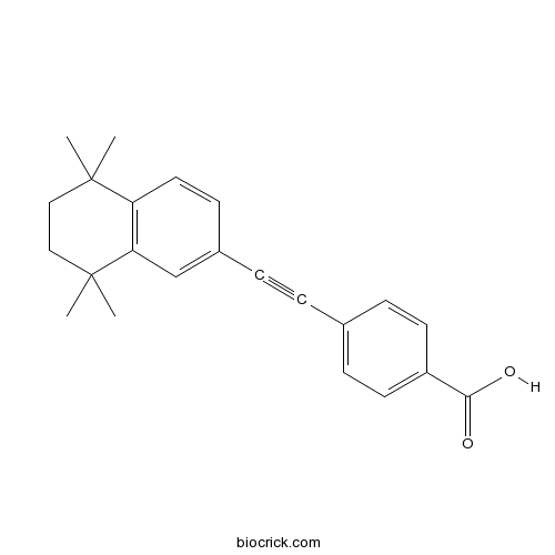
EC 23Synthetic retinoid; induces neural differentiation of hESCs CAS# 104561-41-3 |

- Metiamide
Catalog No.:BCC1742
CAS No.:34839-70-8
- Famotidine
Catalog No.:BCC4529
CAS No.:76824-35-6
Quality Control & MSDS
3D structure
Package In Stock
Number of papers citing our products

| Cas No. | 104561-41-3 | SDF | Download SDF |
| PubChem ID | 10314719 | Appearance | Powder |
| Formula | C23H24O2 | M.Wt | 332.44 |
| Type of Compound | N/A | Storage | Desiccate at -20°C |
| Solubility | Soluble to 100 mM in DMSO and to 10 mM in ethanol | ||
| Chemical Name | 4-[2-(5,5,8,8-tetramethyl-6,7-dihydronaphthalen-2-yl)ethynyl]benzoic acid | ||
| SMILES | CC1(CCC(C2=C1C=CC(=C2)C#CC3=CC=C(C=C3)C(=O)O)(C)C)C | ||
| Standard InChIKey | OQVLOWLEEHYBJH-UHFFFAOYSA-N | ||
| Standard InChI | InChI=1S/C23H24O2/c1-22(2)13-14-23(3,4)20-15-17(9-12-19(20)22)6-5-16-7-10-18(11-8-16)21(24)25/h7-12,15H,13-14H2,1-4H3,(H,24,25) | ||
| General tips | For obtaining a higher solubility , please warm the tube at 37 ℃ and shake it in the ultrasonic bath for a while.Stock solution can be stored below -20℃ for several months. We recommend that you prepare and use the solution on the same day. However, if the test schedule requires, the stock solutions can be prepared in advance, and the stock solution must be sealed and stored below -20℃. In general, the stock solution can be kept for several months. Before use, we recommend that you leave the vial at room temperature for at least an hour before opening it. |
||
| About Packaging | 1. The packaging of the product may be reversed during transportation, cause the high purity compounds to adhere to the neck or cap of the vial.Take the vail out of its packaging and shake gently until the compounds fall to the bottom of the vial. 2. For liquid products, please centrifuge at 500xg to gather the liquid to the bottom of the vial. 3. Try to avoid loss or contamination during the experiment. |
||
| Shipping Condition | Packaging according to customer requirements(5mg, 10mg, 20mg and more). Ship via FedEx, DHL, UPS, EMS or other couriers with RT, or blue ice upon request. | ||
| Description | Photostable synthetic retinoid; analog of ATRA. Displays similar activity to ATRA when tested on the embryonal carcinoma stem cell model TERA2.cl.SP12. Potently induces neural differentiation in human pluripotent embryonic stem cells. |

EC 23 Dilution Calculator

EC 23 Molarity Calculator
| 1 mg | 5 mg | 10 mg | 20 mg | 25 mg | |
| 1 mM | 3.0081 mL | 15.0403 mL | 30.0806 mL | 60.1612 mL | 75.2015 mL |
| 5 mM | 0.6016 mL | 3.0081 mL | 6.0161 mL | 12.0322 mL | 15.0403 mL |
| 10 mM | 0.3008 mL | 1.504 mL | 3.0081 mL | 6.0161 mL | 7.5202 mL |
| 50 mM | 0.0602 mL | 0.3008 mL | 0.6016 mL | 1.2032 mL | 1.504 mL |
| 100 mM | 0.0301 mL | 0.1504 mL | 0.3008 mL | 0.6016 mL | 0.752 mL |
| * Note: If you are in the process of experiment, it's necessary to make the dilution ratios of the samples. The dilution data above is only for reference. Normally, it's can get a better solubility within lower of Concentrations. | |||||

Calcutta University

University of Minnesota

University of Maryland School of Medicine

University of Illinois at Chicago

The Ohio State University

University of Zurich

Harvard University

Colorado State University

Auburn University

Yale University

Worcester Polytechnic Institute

Washington State University

Stanford University

University of Leipzig

Universidade da Beira Interior

The Institute of Cancer Research

Heidelberg University

University of Amsterdam

University of Auckland

TsingHua University

The University of Michigan

Miami University

DRURY University

Jilin University

Fudan University

Wuhan University

Sun Yat-sen University

Universite de Paris

Deemed University

Auckland University

The University of Tokyo

Korea University
- Bayogenin 3-O-beta-D-glucopyranoside
Catalog No.:BCN7868
CAS No.:104513-86-2
- Testosterone acetate
Catalog No.:BCC9165
CAS No.:1045-69-8
- RVX-208
Catalog No.:BCC4475
CAS No.:1044870-39-4
- 3'-Methylflavokawin
Catalog No.:BCN3990
CAS No.:1044743-35-2
- Typhaneoside
Catalog No.:BCN4994
CAS No.:104472-68-6
- L803-mts
Catalog No.:BCC5889
CAS No.:1043881-55-5
- RU-SKI 43
Catalog No.:BCC5441
CAS No.:1043797-53-0
- Tetrahydroxysqualene
Catalog No.:BCN5858
CAS No.:1043629-23-7
- Bisoprolol fumarate
Catalog No.:BCC4344
CAS No.:104344-23-2
- IRAK inhibitor 6
Catalog No.:BCC1658
CAS No.:1042672-97-8
- Famciclovir
Catalog No.:BCC4780
CAS No.:104227-87-4
- IRAK inhibitor 1
Catalog No.:BCC1654
CAS No.:1042224-63-4
- CAY10603
Catalog No.:BCC5542
CAS No.:1045792-66-2
- p-Menthan-3-one
Catalog No.:BCN3850
CAS No.:10458-14-7
- Caffeic acid phenethyl ester
Catalog No.:BCN2695
CAS No.:104594-70-9
- CGS 15943
Catalog No.:BCC7157
CAS No.:104615-18-1
- Pramipexole dihydrochloride
Catalog No.:BCN2181
CAS No.:104632-25-9
- Pramipexole
Catalog No.:BCC4467
CAS No.:104632-26-0
- Dexpramipexole dihydrochloride
Catalog No.:BCC1528
CAS No.:104632-27-1
- Dexpramipexole
Catalog No.:BCC1527
CAS No.:104632-28-2
- H-Ile-NH2.HCl
Catalog No.:BCC2962
CAS No.:10466-56-5
- 7,3'-Di-O-methylorobol
Catalog No.:BCN6831
CAS No.:104668-88-4
- Iriflophenone 3-C-beta-D-glucopyranoside
Catalog No.:BCN1635
CAS No.:104669-02-5
- Lupiwighteone
Catalog No.:BCN4045
CAS No.:104691-86-3
Molecular, kinetic, thermodynamic, and structural analyses of Mycobacterium tuberculosis hisD-encoded metal-dependent dimeric histidinol dehydrogenase (EC 1.1.1.23).[Pubmed:21672513]
Arch Biochem Biophys. 2011 Aug 15;512(2):143-53.
The emergence of drug-resistant strains of Mycobacterium tuberculosis, the major causative agent of tuberculosis (TB), and the deadly HIV-TB co-infection have led to an urgent need for the development of new anti-TB drugs. The histidine biosynthetic pathway is present in bacteria, archaebacteria, lower eukaryotes and plants, but is absent in mammals. Disruption of the hisD gene has been shown to be essential for M. tuberculosis survival. Here we present cloning, expression and purification of recombinant hisD-encoded histidinol dehydrogenase (MtHisD). N-terminal amino acid sequencing and electrospray ionization mass spectrometry analyses confirmed the identity of homogeneous MtHisD. Analytical gel filtration, metal requirement analysis, steady-state kinetics and isothermal titration calorimetry data showed that homodimeric MtHisD is a metalloprotein that follows a Bi Uni Uni Bi Ping-Pong mechanism. pH-rate profiles and a three-dimensional model of MtHisD allowed proposal of amino acid residues involved in either catalysis or substrate(s) binding.
Viral screening of couples undergoing partner donation in assisted reproduction with regard to EU Directives 2004/23/EC, 2006/17/EC and 2006/86/EC: what is the evidence for repeated screening?[Pubmed:20956268]
Hum Reprod. 2010 Dec;25(12):3058-65.
BACKGROUND: This paper concerns the requirements of the EU Tissue and Cells Directives with regard to the biological screening of donors of reproductive cells which are to be used for partner donation. METHODS: We review the evidence regarding the risks of transmission of blood-borne viruses [hepatitis B (HBV), hepatitis C (HCV) and human immunodeficiency virus (HIV)] in the assisted reproductive technology (ART) setting. We document the experience in seven Irish ART clinics since the introduction of the legislation. RESULTS: Even among those known to be HBV-, HCV- or HIV-positive, when current best practice ART procedures are employed for gamete and embryo processing, cross-contamination in the ART facility or horizontal or vertical transmission to a partner or neonate has never been documented. When samples are processed and high-security straws are used for cryopreservation, transmission of virus and cross-contamination in storage have not been reported. CONCLUSIONS: While initial screening of those about to embark on ART treatment is good practice, we can find no medical or scientific evidence to support re-screening prior to each treatment cycle for individuals undergoing partner donation in ART. It would seem more appropriate to focus on risk reduction using a combination of initial baseline screening (with a reduced frequency of re-testing), appropriate sample processing and best possible containment systems for cryostorage.
"Eurocode International Blood Labeling System" enables unique identification of all biological products from human origin in accordance with the European Directive 2004/23/EC.[Pubmed:20563859]
Cell Tissue Bank. 2010 Nov;11(4):345-52.
Due to their limited availability and compatibility, biological products must be exchanged between medical institutions. In addition to a number of national systems and agreements which strive to implement a unique identification and classification of blood products, the ISBT 128 was developed in 1994, followed by the Eurocode in 1998. In contrast to other coding systems, these both make use of primary identifiers as stipulated by the document ISO/IEC 15418 of the International Organization for Standardization (ISO), and thus provide a unique international code. Due to their flexible data structures, which make use of secondary identifiers, both systems are able to integrate additional biological products and their producers. Tissue and cells also constitute a comparable risk to the recipient as that of blood products in terms of false labeling and the danger of infection. However, in contrast to blood products, the exchange of tissue and cells is much more intensively pursued at the international level. This fact is recognised by Directives 2004/23/EC and 2006/86/EC of the European Union (EU), which demand a standardized coding system for cells and tissue throughout the EU. The 2008 workshop agreement of the European Committee for Standardization (CEN) was unique identification by means of a Key Code consisting of country code corresponding to ISO 3166-1, as well as competent authority and tissue establishment. As agreed at the meeting of the Working Group on the European Coding System for Human Tissues and Cells of the Health and Consumers Directorate-General of the European Commission (DG SANCO) held on 19 May 2010 in Brussels, this Key Code could also be used with existing coding systems to provide unique identification and allow EU traceability of all materials from one donation event. Today Eurocode already uses country codes according to ISO 3166-1, and thus the proposed Key Code can be integrated into the current Eurocode data structure and does not need to be introduced separately. The Eurocode product classification for all products is based on its own unique coding system, which can be accessed over the internet by all users who are not themselves members of Eurocode. In summary, it can be said that the standardized single coding system for tissues and cells requires only unique sections in the data structure such the Key Code to fulfil the requirements of the EU Directive. Thus, various systems currently in place in different EU member states can continue to operate if the Key Code as suggested by the EU is integrated into them. The classification and description of each product characteristic is currently being discussed by the DG SANCO Working Group on the European Coding System for Human Tissues and Cells. Following intensive scrutiny in light of the stipulations laid out in EU Directives 2004/23/EC and 2006/86/EC as well as the CEN/ISSS workshop agreements, the Germany Federal Ministry for Health and organisations representing German tissue establishments under the responsibility of the German Society of Transfusion Medicine and Immunohematology, Working Party "Tissue preparations" proposed in 2009 that Eurocode be adopted for the donor identification and product coding of tissue and cells in Germany. The technical details for implementation have already been completed and are presented in the current article.
Cathepsin E (EC 3.4.23.34)--a review.[Pubmed:22252749]
Folia Histochem Cytobiol. 2011;49(4):547-57.
Cathepsin E belongs to the third class of enzymes - hydrolases, a subclass of peptide bond hydrolases and a sub-subclass of endopeptidases with aspartic catalytic sites. Cathepsin E is an endopeptidase with substrate specificity similar to that of cathepsin D. In a human organism, cathepsin E occurs in: erythrocytes, thymus, dendritic cells, epithelial M cells, microglia cells, Langerhans cells, lymphocytes, epithelium of gastrointestinal tract, urinary bladder, lungs, osteoclasts, spleen and lymphatic nodes. In human cells, loci of the gene of pre-procathepsin E are located on chromosome 1 in the region 1231-32. The catalytic site of cathepsin E is two residues of aspartic acid - Asp96 and Asn281, occurring in amino acid triads with sequences DTG96-98 and DTG281-283. To date, no particular role of cathepsin E in the metabolism of proteins in normal tissues has been found. However, it is known that there are many documented pathological conditions in which overexpression of cathepsin E occurs.
Synthetic retinoids: structure-activity relationships.[Pubmed:19821467]
Chemistry. 2009 Nov 2;15(43):11430-42.
Retinoid signalling pathways are involved in numerous processes in cells, particularly those mediating differentiation and apoptosis. The endogenous ligands that bind to the retinoid receptors, namely all-trans-retinoic acid (ATRA) and 9-cis-retinoic acid, are prone to double-bond isomerisation and to oxidation by metabolic enzymes, which can have significant and deleterious effects on their activities and selectivities. Many of these problems can be overcome through the use of synthetic retinoids, which are often much more stable, as well as being more active. Modification of their molecular structures can result in retinoids that act as antagonists, rather than agonists, or exhibit a large degree of selectivity for particular retinoid-receptor isotypes. Several such selective retinoids are likely to be of value as pharmaceutical agents with reduced toxicities, particularly in cancer therapy, as reagents for controlling cell differentiation, and as tools for elucidating the precise roles that specific retinoid signalling pathways play within cells.
Proteomic profiling of the stem cell response to retinoic acid and synthetic retinoid analogues: identification of major retinoid-inducible proteins.[Pubmed:19381361]
Mol Biosyst. 2009 May;5(5):458-71.
The natural retinoid, all-trans retinoic acid (ATRA), is widely used to direct the in vitro differentiation of stem cells. However, substantial degradation and isomerisation of ATRA in response to UV-vis light has serious implications with regard to experimental reproducibility and standardisation. We present the novel application of proteomic biomarker profiling technology to stem cell lysates to rapidly compare the differentiation effects of ATRA with those of two stable synthetic retinoid analogues, EC19 and EC23, which have both been shown to induce differentiation in the embryonal carcinoma cell line TERA2.cl.SP12. MALDI-TOF MS (matrix-assisted laser desorption ionisation time-of-flight mass spectrometry) protein profiles support previous findings into the functional relationships between these compounds in the TERA2.cl.SP12 line. Subsequent analysis of protein peak data enabled the semi-quantitative comparison of individual retinoid-responsive proteins. We have used ion exchange chromatographic protein separation to enrich for retinoid-inducible proteins, thereby facilitating their identification from SDS-PAGE gels. The cellular retinoid-responsive proteins CRABP-I, CRABP-II, and CRBP-I were up-regulated in response to ATRA and EC23, indicating a bona fide retinoid pathway response to the synthetic compound. In addition, the actin filament regulatory protein profilin-1 and the microtubule regulator stathmin were also elevated following treatment with both ATRA and EC23. The up-regulation of profilin-1 and stathmin associated with retinoid-induced neural differentiation correlates with their known roles in cytoskeletal reorganisation during axonal development. Immunological analysis via western blotting confirmed the identification of CRABP-I, profilin-1 and stathmin, and supported their observed regulation in response to the retinoid treatments.
Synthesis and evaluation of synthetic retinoid derivatives as inducers of stem cell differentiation.[Pubmed:19082150]
Org Biomol Chem. 2008 Oct 7;6(19):3497-507.
All-trans-retinoic acid (ATRA) and its associated analogues are important mediators of cell differentiation and function during the development of the nervous system. It is well known that ATRA can induce the differentiation of neural tissues from human pluripotent stem cells. However, it is not always appreciated that ATRA is highly susceptible to isomerisation when in solution, which can influence the effective concentration of ATRA and subsequently its biological activity. To address this source of variability, synthetic retinoid analogues have been designed and synthesised that retain stability during use and maintain biological function in comparison to ATRA. It is also shown that subtle modifications to the structure of the synthetic retinoid compound impacts significantly on biological activity, as when exposed to cultured human pluripotent stem cells, synthetic retinoid 4-(5,5,8,8-tetramethyl-5,6,7,8-tetrahydronaphthalen-2-ylethynyl)benzoic acid, 4a (para-isomer), induces neural differentiation similarly to ATRA. In contrast, stem cells exposed to synthetic retinoid 3-(5,5,8,8-tetramethyl-5,6,7,8-tetrahydronaphthalen-2-ylethynyl)benzoic acid, 4b (meta-isomer), produce very few neurons and large numbers of epithelial-like cells. This type of structure-activity-relationship information for such synthetic retinoid compounds will further the ability to design more targeted systems capable of mediating robust and reproducible tissue differentiation.


