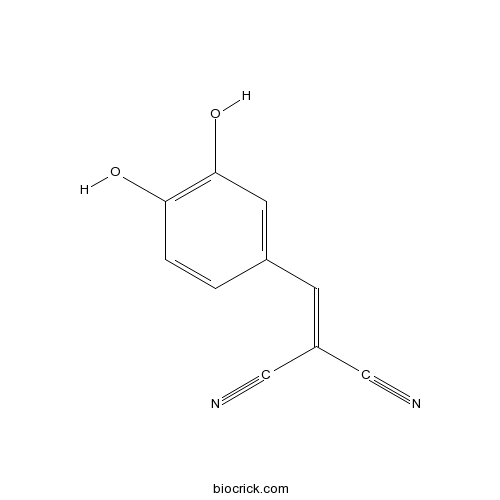
AG-18EGFR/PDGFR inhibitor CAS# 118409-57-7 |

Quality Control & MSDS
3D structure
Package In Stock
Number of papers citing our products

| Cas No. | 118409-57-7 | SDF | Download SDF |
| PubChem ID | 2052 | Appearance | Powder |
| Formula | C10H6N2O2 | M.Wt | 186.17 |
| Type of Compound | N/A | Storage | Desiccate at -20°C |
| Synonyms | RG-50810, Tyrphostin A23 | ||
| Solubility | DMSO : 100 mg/mL (537.14 mM; Need ultrasonic) | ||
| Chemical Name | 2-[(3,4-dihydroxyphenyl)methylidene]propanedinitrile | ||
| SMILES | C1=CC(=C(C=C1C=C(C#N)C#N)O)O | ||
| Standard InChIKey | VTJXFTPMFYAJJU-UHFFFAOYSA-N | ||
| Standard InChI | InChI=1S/C10H6N2O2/c11-5-8(6-12)3-7-1-2-9(13)10(14)4-7/h1-4,13-14H | ||
| General tips | For obtaining a higher solubility , please warm the tube at 37 ℃ and shake it in the ultrasonic bath for a while.Stock solution can be stored below -20℃ for several months. We recommend that you prepare and use the solution on the same day. However, if the test schedule requires, the stock solutions can be prepared in advance, and the stock solution must be sealed and stored below -20℃. In general, the stock solution can be kept for several months. Before use, we recommend that you leave the vial at room temperature for at least an hour before opening it. |
||
| About Packaging | 1. The packaging of the product may be reversed during transportation, cause the high purity compounds to adhere to the neck or cap of the vial.Take the vail out of its packaging and shake gently until the compounds fall to the bottom of the vial. 2. For liquid products, please centrifuge at 500xg to gather the liquid to the bottom of the vial. 3. Try to avoid loss or contamination during the experiment. |
||
| Shipping Condition | Packaging according to customer requirements(5mg, 10mg, 20mg and more). Ship via FedEx, DHL, UPS, EMS or other couriers with RT, or blue ice upon request. | ||
| Description | Inhibitor of epidermal growth factor receptor (EGFR) and platelet-derived growth factor receptor (PDGFR) kinase (IC50 values are 35 and 25 μM respectively). Inhibits EGF-stimulated cell proliferation. Also acts as a mitochondrial uncoupler that alters phosphorylation-dependent cell signaling. |

AG-18 Dilution Calculator

AG-18 Molarity Calculator
| 1 mg | 5 mg | 10 mg | 20 mg | 25 mg | |
| 1 mM | 5.3714 mL | 26.8572 mL | 53.7143 mL | 107.4287 mL | 134.2859 mL |
| 5 mM | 1.0743 mL | 5.3714 mL | 10.7429 mL | 21.4857 mL | 26.8572 mL |
| 10 mM | 0.5371 mL | 2.6857 mL | 5.3714 mL | 10.7429 mL | 13.4286 mL |
| 50 mM | 0.1074 mL | 0.5371 mL | 1.0743 mL | 2.1486 mL | 2.6857 mL |
| 100 mM | 0.0537 mL | 0.2686 mL | 0.5371 mL | 1.0743 mL | 1.3429 mL |
| * Note: If you are in the process of experiment, it's necessary to make the dilution ratios of the samples. The dilution data above is only for reference. Normally, it's can get a better solubility within lower of Concentrations. | |||||

Calcutta University

University of Minnesota

University of Maryland School of Medicine

University of Illinois at Chicago

The Ohio State University

University of Zurich

Harvard University

Colorado State University

Auburn University

Yale University

Worcester Polytechnic Institute

Washington State University

Stanford University

University of Leipzig

Universidade da Beira Interior

The Institute of Cancer Research

Heidelberg University

University of Amsterdam

University of Auckland

TsingHua University

The University of Michigan

Miami University

DRURY University

Jilin University

Fudan University

Wuhan University

Sun Yat-sen University

Universite de Paris

Deemed University

Auckland University

The University of Tokyo

Korea University
AG18 is an inhibitor of EGFR kinase with IC50 values of 35μM. [3]
AG18 acts as general tyrosine kinase inhibitor to block tyrosine phosphorylation and events downstream of tyrosine phosphorylation. [1]
AG18 and AG10 reduced cellular ATP by 90% and increased the rate of oxygen consumption in the absence of the muscarinic agonist carbachol, indicating that these tyrphostins uncouple mitochondria. AG18 and AG10 blocked parotid phosphorylation events only at concentrations that reduced ATP content. AG18 and AG10 also activated AMPK and/or uncoupled mitochondria inHEK293, PC12, and HeLa cells. [1]
In granulosa-lutein cells, tyrphostin AG18 reversiblely arrested the FSH-induced accumulation of P450scc mRNA with IC50 value of 15 mu M. However, AG18 did not accelerate the mRNA degradation process. Moreover, even the extremely high levels of P450scc mRNA in granulosa-lutein cells, were not affected by the addition of AG18 in culture. Aromatase cytochrome P450 and 3 beta-hydroxysteroid dehydrogenase-I, were inhibited by AG18 at their mRNA levels. [2]
References:
[1]. Soltoff SP. Evidence that tyrphostins AG10 and AG18 are mitochondrial uncouplers that alter phosphorylation-dependent cell signaling. J Biol Chem. 2004 Mar 19;279(12):10910-8. Epub 2003 Dec 19.
[2]. Orly J, Rei Z, Greenberg NM et al. Tyrosine kinase inhibitor AG18 arrests follicle-stimulating hormone-induced granulosa cell differentiation: use of reverse transcriptase-polymerase chain reaction assay for multiple messenger ribonucleic acids. Endocrinology. 1994 Jun;134(6):2336-46.
[3]. Gazit A, Yaish P, Gilon C et al. Tyrphostins I: synthesis and biological activity of protein tyrosine kinase inhibitors. J Med Chem. 1989 Oct;32(10):2344-52.
- Trimethylamine oxide
Catalog No.:BCN1819
CAS No.:1184-78-7
- Australine
Catalog No.:BCN2053
CAS No.:118396-02-4
- Licoricesaponin A3
Catalog No.:BCN7905
CAS No.:118325-22-7
- Tazarotene
Catalog No.:BCC2540
CAS No.:118292-40-3
- 6-O-Acetylscandoside
Catalog No.:BCN8320
CAS No.:118292-15-2
- AF-DX 384
Catalog No.:BCC7024
CAS No.:118290-26-9
- Lafutidine
Catalog No.:BCC4544
CAS No.:118288-08-7
- Isodorsmanin A
Catalog No.:BCN6460
CAS No.:118266-99-2
- 1,4-Dicaffeoylquinic acid
Catalog No.:BCN5912
CAS No.:1182-34-9
- Soyasaponin Ab
Catalog No.:BCN2896
CAS No.:118194-13-1
- EMD638683
Catalog No.:BCC1551
CAS No.:1181770-72-8
- 2-Picenecarboxylic acid
Catalog No.:BCN3063
CAS No.:118172-80-8
- UNC 926 hydrochloride
Catalog No.:BCC2445
CAS No.:1184136-10-4
- nTZDpa
Catalog No.:BCC7268
CAS No.:118414-59-8
- MK 886
Catalog No.:BCC7017
CAS No.:118414-82-7
- Licoricesaponin G2
Catalog No.:BCN7897
CAS No.:118441-84-2
- Arcyriaflavin A
Catalog No.:BCC7370
CAS No.:118458-54-1
- Cyclo(L-Phe-trans-4-hydroxy-L-Pro)
Catalog No.:BCN3989
CAS No.:118477-06-8
- Fmoc-D-Tyr(tBu)-OH
Catalog No.:BCC3569
CAS No.:118488-18-9
- 5,7-Dichlorokynurenic acid sodium salt
Catalog No.:BCC7758
CAS No.:1184986-70-6
- TRIS hydrochloride
Catalog No.:BCC7589
CAS No.:1185-53-1
- Myelin Basic Protein (87-99)
Catalog No.:BCC1028
CAS No.:118506-26-6
- LP 12 hydrochloride
Catalog No.:BCC7517
CAS No.:1185136-22-4
- DPPE fumarate
Catalog No.:BCC5669
CAS No.:1185241-83-1
Reactions of a cyclodimethylsiloxane (Me2SiO)6 with silver salts of weakly coordinating anions; crystal structures of [Ag(Me2SiO)6][Al] ([Al] = [FAl{OC(CF3)3}3], [Al{OC(CF3)3}4]) and their comparison with [Ag(18-crown-6)]2[SbF6]2.[Pubmed:23445274]
Inorg Chem. 2013 Mar 18;52(6):3113-26.
Two silver-cyclodimethylsiloxane cation salts [AgD6][Al] ([Al] = [Al(ORF)4](1) or [FAl(OR(F))3](2), R(F) = C(CF3)3, D = Me2SiO) were prepared by the reactions of Ag[Al] with D6 in SO2(l). For a comparison the [Ag(18-crown-6)]2[SbF6]2(3) salt was prepared by the reaction of Ag[SbF6] and 18-crown-6 in SO2(l). The compounds were characterized by IR, multinuclear NMR, and single crystal X-ray crystallography. The structures of 1 and 2 show that D6 acts as a pseudo crown ether toward Ag(+). The stabilities and bonding of [MDn](+) and [M(18-crown-6)](+) (M = Ag, Li, n = 4-8) complexes were studied with theoretical calculations. The calculations predicted that D6 adopts a puckered C(i) symmetric structure in the gas phase in contrast to previous reports. 18-Crown-6 was calculated to bind more strongly to Li(+) and Ag(+) than D6. (29)Si[(1)H] NMR results in solution, and calculations in the gas phase established that a hard Lewis acid Li(+) binds more strongly to D6 than Ag(+). A comparison of the [MD(n)](+) complex stabilities showed D7 to form the most stable metal complexes in the gas phase and the solid state and explained why [AgD7][SbF6] was isolated in a previous reaction where ring transformations resulted in an equilibrium of [AgD(n)](+) complexes. In contrast, the isolations of 1 and 2 were possible because the corresponding equilibrium of [AgD(n)](+) complexes was not observed with [Al](-) anions. The formation of the dinuclear complex salt 3 instead of the corresponding mononuclear complex salt was shown to be driven by the gain in lattice enthalpy in the solid state. The bonding to Li(+) in D6 and 18-crown-6 metal complexes was described by a quantum theory of atoms in molecules (QTAIM) analysis to be mostly electrostatic while the bonding to Ag(+) also had a significant charge transfer component. The charge transfer from both D6 and 18-crown-6 to Ag(+) and Li(+) metal ions was depicted by the QTAIM analysis to be of similar strength, and the difference in the stabilities of the complexes was attributed mostly to more attractive electrostatic interactions between 18-crown-6 and the metal ions despite the more negative oxygen atomic charges calculated for D6.
Electronic structure of the mononuclear Ag(ii) complex [Ag([18]aneS(4)O(2))](2+) ([18]aneS(4)O(2) = 1,10-dioxa-4,7,13,16-tetrathiacyclooctadecane).[Pubmed:18389115]
Chem Commun (Camb). 2008 Mar 21;(11):1305-7.
The structure of [Ag([18]aneS(4)O(2))](PF(6))(2).CH(2)Cl(2) shows a highly unusual and unexpected boat conformation for the macrocycle with square-planar S(4)-coordination at the formal Ag(ii) centre and the two ether O-centres lying on the same side of the S(4) plane; the SOMO in [Ag([18]aneS(4)O(2))](2+) possesses 22.7% Ag 4d(xy) character, as determined by multi-frequency EPR spectroscopy and supported by DFT calculations.
Evidence that tyrphostins AG10 and AG18 are mitochondrial uncouplers that alter phosphorylation-dependent cell signaling.[Pubmed:14688271]
J Biol Chem. 2004 Mar 19;279(12):10910-8.
Receptor agonists that initiate fluid secretion in salivary gland epithelial cells also increase protein phosphorylation. To assess contributions of tyrosine phosphorylation to secretion, changes in muscarinic receptor-initiated secretion (estimated from sodium pump-dependent increases in oxygen consumption) were measured in parotid acinar cells exposed to tyrosine kinase inhibitors. However, like the mitochondrial uncoupler carbonyl cyanide p-trifluoromethoxyphenyl hydrazone, tyrphostins AG10 and AG18 increased the rate of oxygen consumption and reduced cellular ATP by approximately 90% in the absence of the muscarinic agonist carbachol, indicating that these tyrphostins uncouple mitochondria. Exposure of isolated mitochondria to five structurally related tyrphostins demonstrated that their relative potencies as uncouplers differed from their in vitro kinase-inhibitory potencies due to different molecular requirements for the two effects. AG10 and AG18 blocked parotid phosphorylation events only at concentrations that reduced ATP content. The tyrosine kinase inhibitor genistein reduced ATP content by 15-20% and weakly uncoupled isolated mitochondria, but its inhibition of carbachol-mediated protein kinase Cdelta tyrosine phosphorylation and ERK1/2 activation appeared attributable to blocking tyrosine kinases directly. Carbachol itself rapidly reduced ATP content by 15-20%. Carbachol, 3'-O-(4-benzoyl)benzoyl adenosine 5'-triphosphate (P2X(7) receptor agonist), AG10, AG18, and carbonyl cyanide p-trifluoromethoxyphenyl hydrazone rapidly activated the fuel sensor AMP-activated protein kinase (AMPK); however, only AMPK activation by carbachol and BzATP was due to sodium pump stimulation. AG10 and AG18 also activated AMPK and/or uncoupled mitochondria in PC12, HeLa, and HEK293 cells. These studies demonstrate that some tyrosine kinase inhibitors produce cellular effects that are mechanistically different from their primary in vitro characterizations and, as do salivary secretory stimuli, promote rapid metabolic alterations that initiate secondary signaling events.
Tyrosine kinase inhibition: an approach to drug development.[Pubmed:7892601]
Science. 1995 Mar 24;267(5205):1782-8.
Protein tyrosine kinases (PTKs) regulate cell proliferation, cell differentiation, and signaling processes in the cells of the immune system. Uncontrolled signaling from receptor tyrosine kinases and intracellular tyrosine kinases can lead to inflammatory responses and to diseases such as cancer, atherosclerosis, and psoriasis. Thus, inhibitors that block the activity of tyrosine kinases and the signaling pathways they activate may provide a useful basis for drug development. This article summarizes recent progress in the development of PTK inhibitors and demonstrates their potential use in the treatment of disease.
Tyrphostins inhibit epidermal growth factor (EGF)-receptor tyrosine kinase activity in living cells and EGF-stimulated cell proliferation.[Pubmed:2788167]
J Biol Chem. 1989 Aug 25;264(24):14503-9.
Synthetic compounds called tyrphostins were examined for their effects on cells which are mitogenically responsive to epidermal growth factor (EGF). We studied in detail the effects of two tyrphostins on EGF binding, tyrosine phosphorylation in intact cells, EGF-receptor internalization, and mitogenesis. These compounds inhibited EGF-stimulated [3H]thymidine incorporation in a specific manner and the degree of selectivity varied. Both compounds inhibited EGF-stimulated receptor autophosphorylation and tyrosine phosphorylation of endogenous substrates in intact cells at doses that correlated with the IC50 for [3H] thymidine incorporation. These results are consistent with the notion that tyrosine phosphorylation is a crucial signal in transduction of the mitogenic message delivered by EGF. The compound RG50864 demonstrated specificity at inhibiting EGF-stimulated cell growth compared with stimulation with either platelet-derived growth factor or serum. For both compounds RG50864 and RG50810, long term exposure (16 h) of cells to tyrphostins was required for optimal inhibition because of the instability and slow action of these compounds. Tyrphostins did not alter cell surface display of EGF-receptor, EGF binding or EGF-induced internalization, degradation, and down-regulation of EGF receptors. These novel synthetic inhibitors, specific for EGF-receptor kinase, offer a new method to inhibit EGF-stimulated cell proliferation which may be useful in treating specific pathological conditions involving cellular proliferation, including different types of cancers.
Tyrphostins I: synthesis and biological activity of protein tyrosine kinase inhibitors.[Pubmed:2552117]
J Med Chem. 1989 Oct;32(10):2344-52.
A novel class of low molecular weight protein tyrosine kinase inhibitors is described. These compounds constitute a systematic series of molecules with a progressive increase in affinity toward the substrate site of the EGF receptor kinase domain. These competitive inhibitors also effectively block the EGF-dependent autophosphorylation of the receptor. The potent EGF receptor kinase blockers examined were found to competitively inhibit the homologous insulin receptor kinase at 10(2)-10(3) higher inhibitor concentrations in spite of the significant homology between these protein tyrosine kinases. These results demonstrate the ability to synthesize selective tyrosine kinase inhibitors. The most potent EGF receptor kinase inhibitors also inhibit the EGF-dependent proliferation of A431/clone 15 cells with little or no effect on EGF independent cell growth. These results demonstrate the potential use of protein tyrosine kinase inhibitors as selective antiproliferative agents for proliferative diseases caused by the hyperactivity of protein tyrosine kinases. We have suggested the name "tyrphostins" for this class of antiproliferative compounds which act as protein tyrosine kinase blockers.
Blocking of EGF-dependent cell proliferation by EGF receptor kinase inhibitors.[Pubmed:3263702]
Science. 1988 Nov 11;242(4880):933-5.
A systematic series of low molecular weight protein tyrosine kinase inhibitors were synthesized; they had progressively increasing affinity over a 2500-fold range toward the substrate site of epidermal growth factor (EGF) receptor kinase domain. These compounds inhibited EGF receptor kinase activity up to three orders of magnitude more than they inhibited insulin receptor kinase, and they also effectively inhibited the EGF-dependent autophosphorylation of the receptor. The most potent compounds effectively inhibited the EGF-dependent proliferation of A431/clone 15 cells with little or no effect on the EGF-independent proliferation of these cells. The potential use of tyrosine protein kinase inhibitors as antiproliferative agents is demonstrated.


