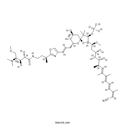
Calyculin AProtein phosphatase inhibitor CAS# 101932-71-2 |

- Fumonisin B1
Catalog No.:BCC2461
CAS No.:116355-83-0
- Calcineurin Autoinhibitory Peptide
Catalog No.:BCC2456
CAS No.:148067-21-4
- DL-AP3
Catalog No.:BCC2459
CAS No.:20263-06-3
- Ceramide
Catalog No.:BCC2458
CAS No.:3102-57-6
- Fostriecin sodium salt
Catalog No.:BCC2460
CAS No.:87860-39-7
Quality Control & MSDS
3D structure
Package In Stock
Number of papers citing our products

| Cas No. | 101932-71-2 | SDF | Download SDF |
| PubChem ID | 5280780 | Appearance | Powder |
| Formula | C50H81N4O15P | M.Wt | 1009.18 |
| Type of Compound | N/A | Storage | Desiccate at -20°C |
| Synonyms | (-)-Calyculin A | ||
| Solubility | Soluble to 50 mM in DMSO | ||
| Chemical Name | [(2R,3R,5S,7S,8R,9R)-2-[(1S,3S,4R,5R,6R,7E,9E,11E,13Z)-14-cyano-3,5-dihydroxy-1-methoxy-4,6,8,9,13-pentamethyltetradeca-7,9,11,13-tetraenyl]-9-[(E)-3-[2-[(2S)-4-[[(2S,3S,4S)-4-(dimethylamino)-2,3-dihydroxy-5-methoxypentanoyl]amino]butan-2-yl]-1,3-oxazol-4-yl]prop-2-enyl]-7-hydroxy-4,4,8-trimethyl-1,10-dioxaspiro[4.5]decan-3-yl] dihydrogen phosphate | ||
| SMILES | CC1C(CC2(C(C(C(O2)C(CC(C(C)C(C(C)C=C(C)C(=CC=CC(=CC#N)C)C)O)O)OC)OP(=O)(O)O)(C)C)OC1CC=CC3=COC(=N3)C(C)CCNC(=O)C(C(C(COC)N(C)C)O)O)O | ||
| Standard InChIKey | FKAWLXNLHHIHLA-QJLNTFFJSA-N | ||
| Standard InChI | InChI=1S/C50H81N4O15P/c1-29(20-22-51)16-14-17-30(2)32(4)24-33(5)42(57)35(7)38(55)25-41(65-13)45-46(69-70(61,62)63)49(8,9)50(68-45)26-39(56)34(6)40(67-50)19-15-18-36-27-66-48(53-36)31(3)21-23-52-47(60)44(59)43(58)37(28-64-12)54(10)11/h14-18,20,24,27,31,33-35,37-46,55-59H,19,21,23,25-26,28H2,1-13H3,(H,52,60)(H2,61,62,63)/b16-14+,18-15+,29-20-,30-17+,32-24+/t31-,33+,34+,35+,37-,38-,39-,40+,41-,42+,43-,44-,45+,46-,50-/m0/s1 | ||
| General tips | For obtaining a higher solubility , please warm the tube at 37 ℃ and shake it in the ultrasonic bath for a while.Stock solution can be stored below -20℃ for several months. We recommend that you prepare and use the solution on the same day. However, if the test schedule requires, the stock solutions can be prepared in advance, and the stock solution must be sealed and stored below -20℃. In general, the stock solution can be kept for several months. Before use, we recommend that you leave the vial at room temperature for at least an hour before opening it. |
||
| About Packaging | 1. The packaging of the product may be reversed during transportation, cause the high purity compounds to adhere to the neck or cap of the vial.Take the vail out of its packaging and shake gently until the compounds fall to the bottom of the vial. 2. For liquid products, please centrifuge at 500xg to gather the liquid to the bottom of the vial. 3. Try to avoid loss or contamination during the experiment. |
||
| Shipping Condition | Packaging according to customer requirements(5mg, 10mg, 20mg and more). Ship via FedEx, DHL, UPS, EMS or other couriers with RT, or blue ice upon request. | ||
| Description | Potent and selective cell-permeable inhibitor of protein phosphatase 1 (IC50 = 0.3 - 0.7 nM) and protein phosphatase 2A (IC50 = 0.5 - 1 nM). Displays > 10,000,000-fold selectivity over PP2B and PP2C. |

Calyculin A Dilution Calculator

Calyculin A Molarity Calculator
| 1 mg | 5 mg | 10 mg | 20 mg | 25 mg | |
| 1 mM | 0.9909 mL | 4.9545 mL | 9.909 mL | 19.8181 mL | 24.7726 mL |
| 5 mM | 0.1982 mL | 0.9909 mL | 1.9818 mL | 3.9636 mL | 4.9545 mL |
| 10 mM | 0.0991 mL | 0.4955 mL | 0.9909 mL | 1.9818 mL | 2.4773 mL |
| 50 mM | 0.0198 mL | 0.0991 mL | 0.1982 mL | 0.3964 mL | 0.4955 mL |
| 100 mM | 0.0099 mL | 0.0495 mL | 0.0991 mL | 0.1982 mL | 0.2477 mL |
| * Note: If you are in the process of experiment, it's necessary to make the dilution ratios of the samples. The dilution data above is only for reference. Normally, it's can get a better solubility within lower of Concentrations. | |||||

Calcutta University

University of Minnesota

University of Maryland School of Medicine

University of Illinois at Chicago

The Ohio State University

University of Zurich

Harvard University

Colorado State University

Auburn University

Yale University

Worcester Polytechnic Institute

Washington State University

Stanford University

University of Leipzig

Universidade da Beira Interior

The Institute of Cancer Research

Heidelberg University

University of Amsterdam

University of Auckland

TsingHua University

The University of Michigan

Miami University

DRURY University

Jilin University

Fudan University

Wuhan University

Sun Yat-sen University

Universite de Paris

Deemed University

Auckland University

The University of Tokyo

Korea University
Calyculin A is a natural, cell-permeable inhibitor of the serine-threonine protein phosphatases (PP) PP1 and PP2A (IC50s = 0.3-0.7 and 0.5-1 nM, respectively). It is without significant effect against PP2B, PP2C, and PP4.Through its effects on PP1 and PP2A, calyculin A has been shown to either promote or inhibit cancer cell growth in tumor cell lines and animal models.
- Regorafenib monohydrate
Catalog No.:BCC1884
CAS No.:1019206-88-2
- Dabigatran etexilate benzenesulfonate
Catalog No.:BCC8925
CAS No.:1019206-65-5
- sodium 4-pentynoate
Catalog No.:BCC1958
CAS No.:101917-30-0
- LX-4211
Catalog No.:BCC1714
CAS No.:1018899-04-1
- 7-Z-Trifostigmanoside I
Catalog No.:BCN7869
CAS No.:1018898-17-3
- Diclazuril
Catalog No.:BCC8937
CAS No.:101831-37-2
- Butenafine HCl
Catalog No.:BCC4768
CAS No.:101827-46-7
- TG 100801 Hydrochloride
Catalog No.:BCC1997
CAS No.:1018069-81-2
- Desacetylmatricarin
Catalog No.:BCN7258
CAS No.:10180-88-8
- 7-O-Demethyl-3-isomangostin hydrate
Catalog No.:BCN7882
CAS No.:
- Elliotinol
Catalog No.:BCN5833
CAS No.:10178-31-1
- Boc-N-Me-Ser-OH
Catalog No.:BCC2613
CAS No.:101772-29-6
- S0859
Catalog No.:BCC1914
CAS No.:1019331-10-2
- Octacosyl (E)-ferulate
Catalog No.:BCN5834
CAS No.:101959-37-9
- Zardaverine
Catalog No.:BCC2069
CAS No.:101975-10-4
- GW791343 dihydrochloride
Catalog No.:BCC1613
CAS No.:1019779-04-4
- Acetoacetanilide
Catalog No.:BCC8803
CAS No.:102-01-2
- 3,4-Dihydroxyphenylacetic Acid
Catalog No.:BCC8281
CAS No.:102-32-9
- Phenyethyl 3-methylcaffeate
Catalog No.:BCN8457
CAS No.:71835-85-3
- Sulfaclozine
Catalog No.:BCC9155
CAS No.:102-65-8
- 20,24-Epoxy-24-methoxy-23(24-25)abeo-dammaran-3-one
Catalog No.:BCN1639
CAS No.:1020074-97-8
- SGI-1027
Catalog No.:BCC4588
CAS No.:1020149-73-8
- DCC-2036 (Rebastinib)
Catalog No.:BCC4390
CAS No.:1020172-07-9
- (R)-4-Benzyl-2-oxazolidinone
Catalog No.:BCC8395
CAS No.:102029-44-7
Dose response of multiple parameters for calyculin A-induced premature chromosome condensation in human peripheral blood lymphocytes exposed to high doses of cobalt-60 gamma-rays.[Pubmed:27542714]
Mutat Res Genet Toxicol Environ Mutagen. 2016 Sep 1;807:47-54.
Many studies have investigated exposure biomarkers for high dose radiation. However, no systematic study on which biomarkers can be used in dose estimation through premature chromosome condensation (PCC) analysis has been conducted. The present study aims to screen the high-dose radiation exposure indicator in Calyculin A-induced PCC. The dose response of multiple biological endpoints, including G2/A-PCC (G2/M and M/A-PCC) index, PCC ring (PCC-R), ratio of the longest/shortest length (L/L ratio), and length and width ratio of the longest chromosome (L/B ratio), were investigated in Calyculin A-induced G2/A-PCC spreads in human peripheral blood lymphocytes exposed to 0-20Gy (dose-rate of 1Gy/min) cobalt-60 gamma-rays. The G2/A-PCC index was decreased with enhanced absorbed doses of 4-20Gy gamma-rays. The G2/A PCC-R at 0-12Gy gamma-rays conformed to Poisson distribution. Three types of PCC-R were scored according to their shape and their solidity or hollowness. The frequencies of hollow PCC-R and PCC-R including or excluding solid ring in G2/A-PCC spreads were enhanced with increased doses. The length and width of the longest chromosome, as well as the length of the shortest chromosome in each G2/M-PCC or M/A-PCC spread, were measured. All L/L or L/B ratios in G2/M-PCC or M/A-PCC spread increased with enhanced doses. A blind test with two new irradiated doses was conducted to validate which biomarker could be used in dose estimation. Results showed that hollow PCC-R and PCC-R including solid ring can be utilized for accurate dose estimation, and that hollow PCC-R was optimal for practical application.
Translocation of Tektin 3 to the equatorial segment of heads in bull spermatozoa exposed to dibutyryl cAMP and calyculin A.[Pubmed:27883267]
Mol Reprod Dev. 2017 Jan;84(1):30-43.
Tektins (TEKTs) are filamentous proteins associated with microtubules in cilia, flagella, basal bodies, and centrioles. Five TEKTs (TEKT1, -2, -3, -4, and -5) have been identified as components of mammalian sperm flagella. We previously reported that TKET1 and -3 are also present in the heads of rodent spermatozoa. The present study clearly demonstrates that TEKT2 is present at the acrosome cap whereas TEKT3 resides just beneath the plasma membrane of the post-acrosomal region of sperm heads in unactivated bull spermatozoa, and builds on the distributional differences of TEKT1, -2, and -3 on sperm heads. We also discovered that hyperactivation of bull spermatozoa by cell-permeable cAMP and Calyculin A, a protein phosphatase inhibitor, promoted translocation of TEKT3 from the post-acrosomal region to the equatorial segment in sperm heads, and that TEKT3 accumulated at the equatorial segment is lost upon acrosome reaction. Thus, translocation of TEKT3 to the equatorial segment may be a capacitation- or hyperactivation-associated phenomenon in bull spermatozoa. Mol. Reprod. Dev. 84: 30-43, 2017. (c) 2016 Wiley Periodicals, Inc.
Stimulation of Suicidal Erythrocyte Death by Phosphatase Inhibitor Calyculin A.[Pubmed:27855413]
Cell Physiol Biochem. 2016;40(1-2):163-171.
BACKGROUND/AIMS: The serine/threonine protein phosphatase 1 and 2a inhibitor Calyculin A may trigger suicidal death or apoptosis of tumor cells. Similar to apoptosis of nucleated cells, erythrocytes may enter eryptosis, the suicidal erythrocyte death characterized by cell shrinkage and cell membrane scrambling with phosphatidylserine translocation to the erythrocyte surface. Triggers of eryptosis include increase of cytosolic Ca2+ activity ([Ca2+] i). Eryptosis is fostered by activation of staurosporine sensitive protein kinase C, SB203580 sensitive p38 kinase, and D4476 sensitive casein kinase. Eryptosis may further involve zVAD sensitive caspases. The present study explored, whether Calyculin A induces eryptosis and, if so, whether its effect requires Ca2+ entry, kinases and/or caspases Methods: Phosphatidylserine exposure at the cell surface was estimated from annexin-V-binding, cell volume from forward scatter, and [Ca2+] i from Fluo-3 fluorescence, as determined by flow cytometry. RESULTS: A 48 hours exposure of human erythrocytes to Calyculin A (>/= 2.5 nM) significantly increased the percentage of annexin-V-binding cells, significantly decreased forward scatter and significantly increased Fluo-3 fluorescence. The effect of Calyculin A on annexin-V-binding was significantly blunted by removal of extracellular Ca2+, by staurosorine (1 microM), SB203580 (2 microM), D4476 (10 microM), and zVAD (10 microM). CONCLUSIONS: Calyculin A triggers cell shrinkage and phospholipid scrambling of the erythrocyte cell membrane, an effect at least in part requiring Ca2+ entry, kinase activity and caspase activation.
Differential inhibition and posttranslational modification of protein phosphatase 1 and 2A in MCF7 cells treated with calyculin-A, okadaic acid, and tautomycin.[Pubmed:9153244]
J Biol Chem. 1997 May 23;272(21):13856-63.
Calyculin-A (CA), okadaic acid (OA), and tautomycin (TAU) are potent inhibitors of protein phosphatases 1 (PP1) and 2A (PP2A) and are widely used on cells in culture. Despite their well characterized selectivity in vitro, their exact intracellular effects on PP1 and PP2A cannot be directly deduced from their extracellular concentration because their cell permeation properties are not known. Here we demonstrate that, due to the tight binding of the inhibitors to PP1 and/or PP2A, their cell penetration could be monitored by measuring PP1 and PP2A activities in cell-free extracts. Treatment of MCF7 cells with 10 nM CA for 2 h simultaneously inhibited PP1 and PP2A activities by more than 50%. A concentration of 1 microM OA was required to obtain a similar time course of PP2A inhibition in MCF7 cells to that observed with 10 nM CA, whereas PP1 activity was unaffected. PP1 was predominantly inhibited in MCF7 cells treated with TAU but even at 10 microM TAU PP1 inhibition was much slower than that observed with 10 nM CA. Furthermore, binding of inhibitors to PP2Ac and/or PP1c in MCF7 cells led to differential posttranslational modifications of the carboxyl termini of the proteins as demonstrated by Western blotting. OA and CA, in contrast to TAU, induced demethylation of the carboxyl-terminal Leu309 residue of PP2Ac. On the other hand, CA and TAU, in contrast to OA, elicited a marked decrease in immunoreactivity of the carboxyl terminus of the alpha-isoform of PP1c, probably reflecting proteolysis of the protein. These results suggest that in MCF7 cells OA selectively inhibits PP2A and TAU predominantly affects PP1, a conclusion supported by their differential effects on cytokeratins in this cell line.
The binding mode of calyculin A to protein phosphatase-1. A novel spiroketal vector model.[Pubmed:9287341]
J Biol Chem. 1997 Sep 12;272(37):23312-6.
The catalytic subunits of serine/threonine protein phosphatases 1 and 2A are subject to inhibition by various toxins such as the microcystins, the nodularins, okadaic acid, tautomycin, and the calyculins. A recent paper (Bagu, J. R., Sykes, B. D, Craig, M. M., and Holmes, C. F. B. (1997) J. Biol. Chem. 272, 5087-5097) reported the successful docking of the crystal structure of Calyculin A to the crystal structure of protein phosphatase-1. Unfortunately, the model presented there is based on the structure of the unnatural enantiomer of Calyculin A and must therefore be incorrect. We have developed a spiroketal vector model which appears to account for the spatial orientation of the hydrophobic and basic chains extending from the spiroketal-phosphate core of Calyculin A. The model also clearly demonstrates why the unnatural enantiomer of Calyculin A does not fit properly into the pocket of the active site. Based on our model, we present a possible open binding mode for Calyculin A in the enzyme. This open structure is conceptually similar to the predicted binding mode of the peptide inhibitor DARPP-32 to the enzyme; the hydrophobic, metal-binding, and electrostatic interactions are all retained in this model.
Calyculin A and okadaic acid: inhibitors of protein phosphatase activity.[Pubmed:2539153]
Biochem Biophys Res Commun. 1989 Mar 31;159(3):871-7.
Calyculin A and okadaic acid induce contraction in smooth muscle fibers. Okadaic acid is an inhibitor of phosphatase activity and the aims of this study were to determine if Calyculin A also inhibits phosphatase and to screen effects of both compounds on various phosphatases. Neither compound inhibited acid or alkaline phosphatases, nor the phosphotyrosine protein phosphatase. Both compounds were potent inhibitors of the catalytic subunit of type-2A phosphatase, with IC50 values of 0.5 to 1 nM. With the catalytic subunit of protein phosphatase type-1, Calyculin A was a more effective inhibitor than okadaic acid, IC50 values for Calyculin A were about 2 nM and for okadaic acid between 60 and 500 nM. The endogenous phosphatase of smooth muscle myosin B was inhibited by both compounds with IC50 values of 0.3 to 0.7 nM and 15 to 70 nM, for Calyculin A and okadaic acid, respectively. The partially purified catalytic subunit from myosin B had IC50 values of 0.7 and 200 nM for Calyculin A and okadaic acid, respectively. The pattern of inhibition for the phosphatase in myosin B therefore is similar to that of the type-1 enzyme.


