
DesmosterolCAS# 313-04-2 |
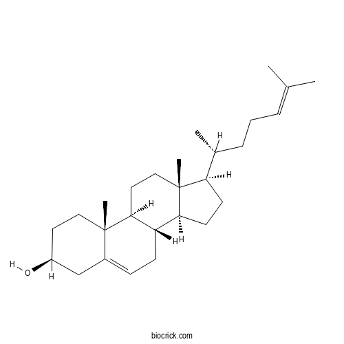
| Cas No. | 313-04-2 | SDF | File under preparation. |
| PubChem ID | 439577.0 | Appearance | Powder |
| Formula | C27H44O | M.Wt | 384.6 |
| Type of Compound | N/A | Storage | Desiccate at -20°C |
| Solubility | Soluble in Chloroform,Dichloromethane,Ethyl Acetate,DMSO,Acetone,etc. | ||
| Chemical Name | (3S,8S,9S,10R,13R,14S,17R)-10,13-dimethyl-17-[(2R)-6-methylhept-5-en-2-yl]-2,3,4,7,8,9,11,12,14,15,16,17-dodecahydro-1H-cyclopenta[a]phenanthren-3-ol | ||
| SMILES | CC(CCC=C(C)C)C1CCC2C1(CCC3C2CC=C4C3(CCC(C4)O)C)C | ||
| Standard InChIKey | AVSXSVCZWQODGV-DPAQBDIFSA-N | ||
| Standard InChI | InChI=1S/C27H44O/c1-18(2)7-6-8-19(3)23-11-12-24-22-10-9-20-17-21(28)13-15-26(20,4)25(22)14-16-27(23,24)5/h7,9,19,21-25,28H,6,8,10-17H2,1-5H3/t19-,21+,22+,23-,24+,25+,26+,27-/m1/s1 | ||
| General tips | For obtaining a higher solubility , please warm the tube at 37 ℃ and shake it in the ultrasonic bath for a while.Stock solution can be stored below -20℃ for several months. We recommend that you prepare and use the solution on the same day. However, if the test schedule requires, the stock solutions can be prepared in advance, and the stock solution must be sealed and stored below -20℃. In general, the stock solution can be kept for several months. Before use, we recommend that you leave the vial at room temperature for at least an hour before opening it. |
||
| About Packaging | 1. The packaging of the product may be reversed during transportation, cause the high purity compounds to adhere to the neck or cap of the vial.Take the vail out of its packaging and shake gently until the compounds fall to the bottom of the vial. 2. For liquid products, please centrifuge at 500xg to gather the liquid to the bottom of the vial. 3. Try to avoid loss or contamination during the experiment. |
||
| Shipping Condition | Packaging according to customer requirements(5mg, 10mg, 20mg and more). Ship via FedEx, DHL, UPS, EMS or other couriers with RT, or blue ice upon request. | ||
Quality Control & MSDS
3D structure
Package In Stock
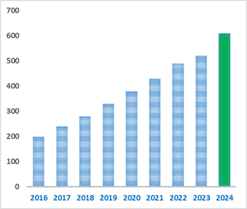
Number of papers citing our products

Desmosterol Dilution Calculator
Desmosterol Molarity Calculator
-
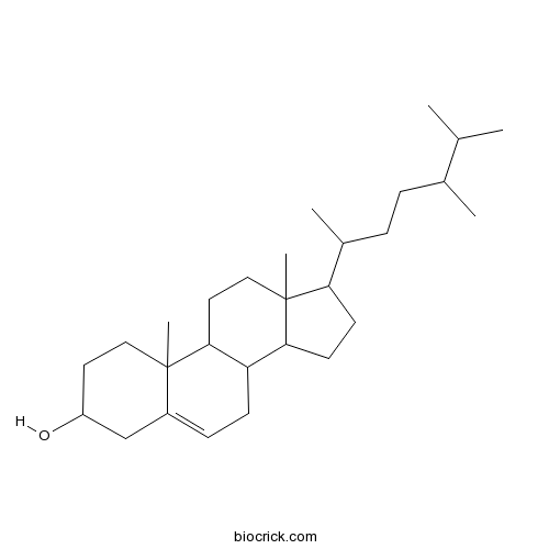 CampesterolCatalog No.: BCN3181CAS No.: 474-62-4
-
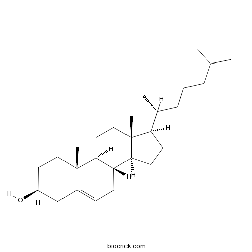 CholesterolCatalog No.: BCN2199CAS No.: 57-88-5
-
 Delta 5-avenasterolCatalog No.: BCN3211CAS No.: 18472-36-1
-
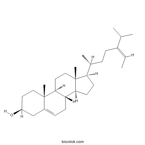 FucosterolCatalog No.: BCN6427CAS No.: 17605-67-3
-
 ClerosterolCatalog No.: BCN2905CAS No.: 2364-23-0
-
 27-HydroxycholesterolCatalog No.: BCN2750CAS No.: 20380-11-4
-
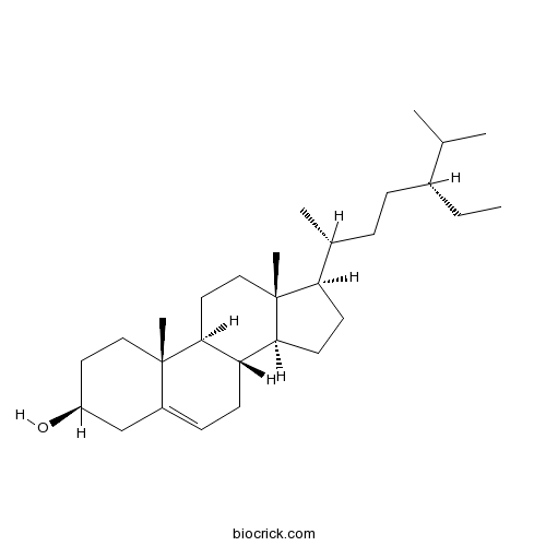 Beta-SitosterolCatalog No.: BCN1015CAS No.: 83-46-5
-
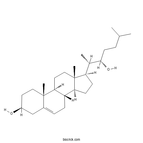 22(S)-HydroxycholesterolCatalog No.: BCX2366CAS No.: 22348-64-7
-
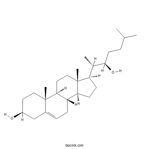 (22R)-HydroxycholesterolCatalog No.: BCX2463CAS No.: 17954-98-2
-
 BrassicasterolCatalog No.: BCN2613CAS No.: 474-67-9
-
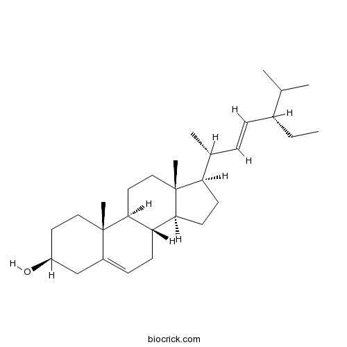 StigmasterolCatalog No.: BCN4376CAS No.: 83-48-7
-
 22-DehydroclerosterolCatalog No.: BCN5141CAS No.: 26315-07-1
-
 AndrostenediolCatalog No.: BCC8828CAS No.: 521-17-5
| 1 mg | 5 mg | 10 mg | 20 mg | 25 mg | |
| 1 mM | 2.6001 mL | 13.0005 mL | 26.001 mL | 52.0021 mL | 65.0026 mL |
| 5 mM | 0.52 mL | 2.6001 mL | 5.2002 mL | 10.4004 mL | 13.0005 mL |
| 10 mM | 0.26 mL | 1.3001 mL | 2.6001 mL | 5.2002 mL | 6.5003 mL |
| 50 mM | 0.052 mL | 0.26 mL | 0.52 mL | 1.04 mL | 1.3001 mL |
| 100 mM | 0.026 mL | 0.13 mL | 0.26 mL | 0.52 mL | 0.65 mL |
| * Note: If you are in the process of experiment, it's necessary to make the dilution ratios of the samples. The dilution data above is only for reference. Normally, it's can get a better solubility within lower of Concentrations. | |||||
- 2,2-Dimethyl-2,3-dihydroquinazolin-4(1H )-one
Catalog No.:BCX2360
CAS No.:77726-78-4
- Uridine 5'-diphosphogalactose disodium
Catalog No.:BCX2359
CAS No.:137868-52-1
- Uridine 5'-diphosphoxylose disodium
Catalog No.:BCX2358
CAS No.:108320-89-4
- Adenosine triphosphate
Catalog No.:BCX2357
CAS No.:56-65-5
- Cisplatin
Catalog No.:BCX2356
CAS No.:15663-27-1
- Uridine 5'-diphosphogaluronic acid trisodium
Catalog No.:BCX2355
CAS No.:148407-07-2
- Phenylephrine
Catalog No.:BCX2354
CAS No.:59-42-7
- Dotriacontane
Catalog No.:BCX2353
CAS No.:544-85-4
- 5-Pentadecylresorcinol
Catalog No.:BCX2352
CAS No.:3158-56-3
- 2-tert-Butyl-4-methoxyphenol
Catalog No.:BCX2351
CAS No.:121-00-6
- Pipemidic acid
Catalog No.:BCX2350
CAS No.:51940-44-4
- Methylaervine
Catalog No.:BCX2349
CAS No.:86293-40-5
- 7-Dehydrocholestero
Catalog No.:BCX2362
CAS No.:434-16-2
- Acetyl CoA
Catalog No.:BCX2363
CAS No.:102029-73-2
- CoA
Catalog No.:BCX2364
CAS No.:85-61-0
- Caffeoyl CoA
Catalog No.:BCX2365
CAS No.:53034-79-0
- 22(S)-Hydroxycholesterol
Catalog No.:BCX2366
CAS No.:22348-64-7
- Feruloyl CoA
Catalog No.:BCX2367
CAS No.:30802-02-9
- Palmitoyl CoA
Catalog No.:BCX2368
CAS No.:188174-64-3
- Benzoyl CoA
Catalog No.:BCX2369
CAS No.:102185-37-5
- Cinnamoyl CoA
Catalog No.:BCX2370
CAS No.:76109-04-1
- Sinapoyl CoA
Catalog No.:BCX2371
CAS No.:54429-80-0
- NADPH tetrasodium salt
Catalog No.:BCX2372
CAS No.:2646-71-1
- p-Coumaroyl CoA
Catalog No.:BCX2373
CAS No.:119785-99-8
Multi-Omics-Guided Discovery of Holothuria scabra-Derived Drug Candidates Targeting Ferroptosis and the Bone Tumor Microenvironment in Osteosarcoma.[Pubmed:42505966]
Mar Drugs. 2026 Jun 28;24(7):226.
Osteosarcoma remains the most common primary malignant bone tumor in adolescents and is characterized by aggressive metastasis, resistance to therapy, and extensive bone microenvironment remodeling. Therefore, the identification of novel multi-target therapeutic agents capable of simultaneously inducing ferroptosis and disrupting tumor-supportive signaling is urgently needed. This study employed a multi-omics-guided approach to investigate the anti-osteosarcoma potential of metabolites derived from the sea cucumber Holothuria scabra. LC-MS/MS profiling identified major bioactive constituents, including holothurins, scabrasides, fucosterol, Desmosterol, and 24-methylenecholesterol. Integrated transcriptomic analysis of the GSE42352 dataset revealed key ferroptosis- and bone microenvironment-associated targets, including CXCR4, CTSK, RUNX2, VEGFA, and TFRC. In silico pharmacological prediction and molecular docking demonstrated favorable anticancer properties and strong binding affinities of several metabolites toward these targets, with fucosterol and holothurin A exhibiting the most promising interactions. Functional validation in MG-63 osteosarcoma cells showed concentration-dependent reductions in cell viability and migration following H. scabra treatment. Furthermore, treatment decreased GPX4, NRF2, and GSH levels while increasing TFRC and MDA, indicating activation of ferroptotic cell death. In a MG-63/RAW264.7 co-culture model, H. scabra suppressed RANKL, VEGFA, MMP9, and TRAP-positive osteoclast formation, suggesting inhibition of osteoclastogenesis, angiogenesis, and metastatic potential. Collectively, these findings identify H. scabra as a promising marine source of multi-target compounds for osteosarcoma management through coordinated induction of ferroptosis and remodeling of the bone tumor microenvironment.
DHCR24 inhibition upregulates desmosterol without promoting regression of atherosclerotic plaques in APOE*3-Leiden.CETP mice.[Pubmed:42457066]
Steroids. 2026 Jul 15;235-236:109830.
BACKGROUND AND AIMS: Inhibition of Delta24-dehydrocholesterol reductase (DHCR24) leads to accumulation of the cholesterol biosynthesis intermediate Desmosterol, which is the endogenous ligand for liver X receptors (LXRs). Activation of LXRs is considered to be atheroprotective as it suppresses inflammation and stimulates cholesterol efflux in macrophages via upregulation of ATP-binding cassette transporters A1 and G1. In the current study we aimed to investigate the effect of the selective DHCR24 inhibitor SH42 on regression of atherosclerotic plaques in APOE*3-Leiden.CETP mice, a well-established model for human-like lipid metabolism. METHODS: Mice were fed a Western-type diet (16% fat, 0.15% cholesterol) for 20 weeks to promote plaque development. Subsequently, treatment was started with vehicle or SH42 for 8 weeks while the animals were kept on a regular chow diet. RESULTS: Treatment with SH42 significantly upregulated Desmosterol levels in liver and plasma, and led to a small reduction in plasma triglyceride, nonHDL-cholesterol and HDL-cholesterol levels on top of the lipid-lowering effect caused by the dietary switch. While SH42 treatment did not affect immune cell numbers in blood, bone marrow, or spleen, SH42 strongly reduced the number of macrophages found in the peritoneum after thioglycolate injection. CONCLUSIONS: While the dietary switch halted atherosclerotic lesion progression, SH42 treatment did not accelerate atherosclerotic lesion regression further.
Endothelial cytochrome P450 -derived cholesterol limits angiogenesis.[Pubmed:42435651]
Redox Biol. 2026 Sep;95:104289.
The cytochrome P450 redox system is composed of a cytochrome P450 reductase (POR) and multiple CYP450 enzymes (CYP450). Of the CYP450 isoenzymes, CYP51A1 is essential for endogenous cholesterol biosynthesis. Elevated circulating cholesterol is a well-established risk factor for cardiovascular disease, however, the role of intracellular cholesterol synthesis in normal endothelial function remains unclear. To investigate this, we generated CRISPR/Cas9 knockouts of the cytochrome P450 reductase in primary human endothelial cells (EC) and studied an endothelial-specific, tamoxifen-inducible POR knockout mouse (ecPOR(-/-)). Deletion of POR led to the accumulation of lanosterol, the substrate of POR/CYP51A1, and a reduction in Desmosterol. Functionally, POR deficiency promoted basal and VEGF-induced angiogenesis in spheroids and mouse aortic segments. Retinal angiogenesis was increased in ecPOR(-/-) mice in vivo. Mechanistically, POR deletion activated the Sterol Regulatory Element Binding Transcription Factor (SREBP2) regulatory pathway, as shown by increased nuclear translocation of cleaved SREBP2 in EC and in en face-stained mouse aortae. Overexpression of nuclear SREBP2 in endothelial cells mimicked the angiogenic phenotype observed upon POR deletion. Conversely, double deletion of POR and SREBP2 normalized angiogenesis to levels of control cells. RNAseq of POR-deficient EC revealed an upregulation of PI3K-related signaling pathways and genes involved in cholesterol homeostasis, including enhanced expression of pro-angiogenic factors. In line with these findings, knockout of POR increased cellular PIP3 levels, AKT phosphorylation, and activation of downstream targets such as p70 S6 kinase. These findings demonstrate that inhibition of the endothelial POR/CYP51A1 axis impairs endogenous cholesterol synthesis, activates SREBP2, and enhances angiogenesis via PI3K/AKT/mTOR signaling, highlighting a critical and novel link between intracellular cholesterol metabolism and vascular growth.
Peroxidation rate constants and mechanisms of isoprenoid-derived lipids and their roles in ferroptosis.[Pubmed:42273072]
Redox Biochem Chem. 2025 Sep;13:100058.
Ferroptosis is a type of regulated cell death that is dependent on iron and driven by lipid peroxidation. Polyunsaturated fatty acids (PUFAs) sensitize cells to ferroptosis as they are prone to lipid peroxidation while monounsaturated fatty acids confer resistance to ferroptosis when incorporated into the lipid membrane as they are much less reactive toward lipid peroxidation. Recently, in addition to fatty acid-derived lipids, isoprenoid-derived lipids have been found to regulate ferroptosis. Specifically, ferroptosis suppressor protein 1 (FSP1) was found to be anti-ferroptotic as it reduces the oxidized forms of coenzyme Q(10) and vitamin K to their reduced quinol forms, which are phenolic radical-trapping antioxidants. Vitamins D(3) and A have also been found to inhibit ferroptosis in cancer cells. Furthermore, it has been shown that metabolites along the cholesterol synthesis pathway, including squalene, cholesterol, Desmosterol, and 7-dehydrocholesterol (7-DHC), can protect cells against ferroptosis in vitro. Despite large variations in the reactivities of these lipids toward lipid peroxidation, they generally exhibit anti-ferroptotic properties. In this review, we will discuss the peroxidation rate constants and mechanisms of these isoprenoid-derived lipids and how they might contribute to their roles in ferroptosis.
Arginase 2 regulates cholesterol biosynthesis in endothelial cells.[Pubmed:41997283]
Exp Cell Res. 2026 Jun 15;459(2):115029.
BACKGROUND: Arginase 2 (Arg 2) is a mitochondrial enzyme that hydrolyzes L-arginine to L-ornithine and urea, influencing endothelial nitric oxide (NO) bioavailability and vascular function. Although Arg 2 is implicated in endothelial dysfunction under hypercholesterolemic and oxidative stress conditions, its potential role in endothelial cholesterol metabolism remains unknown. METHODS: Arg 2 was deleted in immortalized human umbilical vein endothelial cells (HUVECs) using CRISPR/Cas9, followed by transcriptomic analyses. Differential gene expression was validated by quantitative RT-PCR and immunoblotting. Overexpression of wild-type Arg 2 and the catalytically inactive Arg 2 (H160F) mutant was achieved using recombinant lentiviral transduction. Arginase activity was quantified by measuring urea production using a colorimetric assay. Cholesterol intermediates were quantified by LC-MS. RESULTS: RNA sequencing revealed that Arg 2 deletion markedly downregulated genes involved in the mevalonate and steroid biosynthesis pathways, including HMGCS1, FDFT1, FDPS, SQLE, and DHCR7. These transcriptional changes were accompanied by reduced protein levels of key cholesterol biosynthetic enzymes and decreased cellular concentrations of sterols, lanosterol, Desmosterol, and cholesterol. Conversely, either overexpression of wild-type Arg 2 or the catalytically inactive Arg 2 (H160F) mutant enhanced the expression of these enzymes. CONCLUSIONS: These findings identify a previously unrecognized role of Arg 2 in promoting endothelial cholesterol biosynthesis. Beyond competing with endothelial NO synthase for L-arginine, Arg 2 may regulate vascular homeostasis through modulation the mevalonate pathway, independent of its enzymatic activity. This dual function may link amino acid and lipid metabolism in the endothelium and suggests new mechanisms by which Arg 2 contributes to endothelial dysfunction and atherosclerotic progression.
Physiological Functions of Side-Chain-Retaining Sterols in the Brain and Their Roles in Neurodegenerative Diseases.[Pubmed:41893338]
Metabolites. 2026 Mar 11;16(3):189.
Although the brain comprises only 2% of total body weight, it contains approximately 23% of the total cholesterol of the body. In the brain, cholesterol plays a critical role as a structural component of cell membranes and myelin sheaths. However, the blood-brain barrier restricts cholesterol influx from the systemic circulation into the brain. As a result, the brain synthesizes cholesterol de novo and regulates its metabolism independently. Desmosterol, a cholesterol precursor produced during cholesterol biosynthesis, and cholesterol metabolites, 24S-hydroxycholesterol and chenodeoxycholic acid, are sterols with structurally retained side chains. These side-chain-retaining sterols have traditionally been regarded as intermediates in the cholesterol synthesis process or as metabolites for cholesterol excretion, but accumulating evidence indicates that they also function as physiologically active signaling molecules that influence brain function via nuclear receptors, such as liver X receptors, and membrane receptors, such as NMDA receptors. Through nuclear receptors, these side-chain-retaining sterols regulate the transcription of genes involved in lipid transport, inflammation control, and amyloid clearance, while their membrane receptor action enables rapid synaptic effects. These side-chain-retaining sterols mediate metabolic crosstalk between neurons and glial cells and contribute to maintaining cholesterol balance in the developing brain. Furthermore, these side-chain-retaining sterols have been shown to affect amyloid-beta clearance, alpha-synuclein aggregation, neuroinflammation, mitochondrial function, and remyelination. Dysregulation of these side-chain-retaining sterols is associated with neurodegenerative diseases such as Alzheimer's disease and Parkinson's disease. Overall, side-chain-retaining sterols are important regulators of brain physiology. This review focuses on the current knowledge regarding the physiological functions of side-chain-retaining sterols in the brain and their roles in neurodegenerative diseases.
Differential lipid dynamics regulation by sterols in ceramide and sphingomyelin containing membranes.[Pubmed:41864600]
Biochim Biophys Acta Biomembr. 2026 Jul;1868(3):184523.
The role of cholesterol in membrane lipid organization and ordering has been evidenced over the past twenty years. The participation of cholesterol precursors and other cholesterol derivatives in the modulation of cell membrane physicochemical properties is less well documented. Herein, an investigation into the effects of cholesterol, two cholesterol precursors (Desmosterol and lanosterol) and two hydroxylated metabolites (24S-hydroxycholesterol and 25-hydroxycholesterol) on membrane dynamics was performed. The differences between sphingomyelin- and ceramide-containing membranes were investigated using membrane models that mimic the plasma membrane. Two fluorescent probes were used to study the ordering power and molecular mobility: Laurdan, a sensor of membrane polarity and lipid order near the lipid head groups, and cholesterol-pyrene, a sensor of order near the center of the membrane. The results showed that the double bond in the sterol tail of Desmosterol modified the molecular dynamics near the bilayer center resulting in less order in ceramide membranes than in sphingomyelin membranes. The methyl groups on the sterol rings of lanosterol induced increased polarity and decreased order near the lipid head groups, and the hydroxyl groups in the 24 and 25 carbons of the sterol tail changed membrane properties across the membrane axis in a complex manner. These results suggest that the differences in the intra- and intermolecular hydrogen bonding capacities of ceramide and sphingomyelin with specific sterols are in part responsible for specific physicochemical modifications of membranes properties.
Dysregulated lipid metabolism and hypomyelination in postnatal peroxisome-deficient Pex2 knockout Zellweger mice.[Pubmed:41815956]
Front Mol Neurosci. 2026 Feb 24;19:1636268.
Peroxisomes are dynamic organelles that play a crucial role in cellular metabolism, particularly in fatty acid degradation, cholesterol homeostasis and reactive oxygen species metabolism. Their dysfunction is associated with severe neurological disorders, including Zellweger spectrum disorders (ZSD) and X-linked adrenoleukodystrophy (X-ALD). In this study, we investigated the relationship between cholesterol homeostasis and myelination in postnatal peroxisome-deficient Pex2 knockout mice. We dissected the central nervous system (CNS) of 10-day-old (P10) control and Pex2 (-/-) mice into five regions: spinal cord, brainstem, cerebellum, diencephalon and cerebral cortex. Catalase activity, a marker enzyme of peroxisomes, was significantly increased in CNS regions of Pex2 (-/-) mice, indicating an oxidative imbalance. Proteomic analysis revealed significant alterations in peroxisomal proteins and pathways related to neurodegenerative diseases, cholesterol and fatty acid metabolism and mRNA processing. Cholesterol biosynthesis was particularly dysregulated: enzyme activities, mRNA, and protein levels were reduced in white matter regions but increased in the cerebral cortex. The elevated Desmosterol levels in the brain of Pex2 (-/-) mice indicate impaired cholesterol synthesis. Sphingolipid metabolism was also altered in the peroxisome-deficient CNS, as the protein levels of enzymes dihydroceramide desaturase 1, ceramide synthase 2, fatty acid 2-hydroxylase, and UDP-glycosyltransferase 8 were significantly decreased. Myelination was significantly reduced throughout the CNS, as evidenced by decreased activities of the myelin marker 2',3'-cyclic nucleotide 3'-phosphodiesterase (CNP) and decreased mRNA and protein levels of myelin-associated proteins. The consistent decrease in ribosomal protein S6 phosphorylation in the CNS of Pex2 (-/-) mice suggests that decreased mechanistic target of rapamycin complex 1 (mTORC1) activity contributes to hypomyelination. Gene expression analysis revealed an upregulation of pro-inflammatory cytokines and altered expression of some homeostatic and disease-associated microglial (DAM) genes. However, full DAM activation was not yet observed in Pex2 (-/-) mice at P10. In conclusion, this study shows that systemic peroxisome deficiency leads to severe hypomyelination and dysregulation of cholesterol and fatty acid metabolism in the CNS, providing new insights into the pathophysiology of peroxisomal disorders.
The sterols-LXR axis alleviates IL-17-mediated acute inflammatory response in Apostichopus japonicus.[Pubmed:41783566]
Mar Life Sci Technol. 2025 Oct 13;8(1):116-128.
Inflammation plays a crucial role in defending the host against pathogens and other harmful stimuli. However, uncontrolled acute inflammation has been implicated in a multitude of diseases, including those that affect aquatic animals. While the processes that initiate the inflammatory response are relatively well understood, the roles of lipids, their derivatives, and the mechanisms by which they contribute to alleviating acute inflammation remain less well characterized. In the present study, lipidomic analyses were performed to identify changes in lipid metabolism in the coelomocytes of the sea cucumber Apostichopus japonicus after infection with Vibrio splendidus. The omics data indicated significant increases in specific sterols, including Desmosterol, 25-hydroxycholesterol, and 24,25-epoxycholesterol, during the late phase of the inflammatory response. The accumulation of these sterols was found to be responsible for the suppression of A. japonicus interleukin-17 (AjIL-17)-mediated inflammatory response. A subsequent biolayer interferometry assay revealed that A. japonicus liver X receptor (AjLXR) served as the downstream receptor of these sterols. The knockdown of AjLXR reversed the Desmosterol-dependent suppression of AjIL-17 expression. Conversely, the activation of AjLXR by the synthetic agonist GW3965 inhibited the expression of AjIL-17 induced by infection with V. splendidus or stimulation with LPS. Moreover, AjLXR repressed AjIL-17 transcription by directly binding to the AjIL-17 promoter in a SUMOylation-independent manner. In conclusion, our findings highlight the crucial role of the sterols-LXR axis in mitigating IL-17-mediated acute inflammation, suggesting that targeting this axis could be a promising strategy for preventing and treating chronic inflammation-associated diseases. SUPPLEMENTARY INFORMATION: The online version contains supplementary material available at 10.1007/s42995-025-00322-9.
Sex-Specific Metabolic Footprint of Ketogenic Diet in C57BL/6J Mice.[Pubmed:41751361]
Biomedicines. 2026 Feb 19;14(2):462.
Background/Objectives: The ketogenic diet (KD) induces profound metabolic shifts, yet the sex-specific long-term effects on skeletal muscle metabolism and sterol homeostasis across tissues remain insufficiently characterized. This study tested the hypothesis that a prolonged KD would elicit distinct, sex-dependent metabolic and sterol adaptations in mice. Methods: We examined how a 12-week KD, compared with a standard diet, affected body mass, the skeletal muscle metabolome, hepatic lipid and collagen content, and sterol profiles in the skeletal muscle, liver, spleen, and serum in male and female C57BL/6J mice. Three-month-old mice of both sexes were randomized to a KD or standard diet and evaluated using the histological quantification of hepatic steatosis and collagen deposition, matrix-assisted laser desorption/ionization time-of-flight imaging mass spectrometry (MALDI-TOF IMS) of skeletal muscle, and LC-MS/MS-based sterol profiling. Results: The KD induced rapid body mass gain in males and delayed weight gain in females, promoted hepatic steatosis in both sexes, and generated clearly segregated, sex- and diet-specific skeletal muscle metabolomic signatures. These signatures included reduced tricarboxylic acid cycle precursors and a marked decrease in S-adenosylmethionine in KD-fed females. Across tissues, the KD consistently suppressed precursor sterols, including 7-dehydrocholesterol and Desmosterol in the skeletal muscle, liver, and spleen, while elevating serum cholesterol and Desmosterol (male-biased), with changes generally more pronounced in males. Conclusions: Collectively, these findings demonstrate that a long-term KD drives sex- and organ-specific metabolic remodeling, with evidence of greater metabolic flexibility but a shared risk of hepatic steatosis in females. These results underscore the importance of personalized, sex-stratified approaches when considering long-term ketogenic interventions.
Clinical and genetic analysis of an early-onset sitosterolemia family caused by a novel compound heterozygous ABCG5 mutation.[Pubmed:41691973]
J Chromatogr B Analyt Technol Biomed Life Sci. 2026 Apr 1;1273:124963.
BACKGROUND: Sitosterolemia (STSL) is a rare autosomal recessive disorder caused by biallelic mutations in ABCG5 or ABCG8, which encode sterolin efflux transporters. Defective transport leads to intestinal hyperabsorption and reduced biliary excretion of plant sterols and cholesterol, causing their accumulation and heterogeneous phenotypes often misdiagnosed as familial hypercholesterolemia. METHODS: Whole-exome sequencing was performed in a 3-year-old child with early-onset hypercholesterolemia. Candidate variants were validated by Sanger sequencing of family members, followed by genotype-phenotype analysis. Plasma plant sterols were quantified by gas chromatography-mass spectrometry, and structural modeling was used to predict the impact of variants. RESULTS: The proband developed multiple xanthomas over the knees, elbows, ankles, finger joints and skin folds at 2 years of age. Her father and grandfather had obesity and hypercholesterolemia with fatty liver but no xanthomas. Genetic analysis identified a novel compound heterozygous ABCG5 mutation: c.1337G > A (p.Arg446Gln), inherited from the father and classified as likely pathogenic, and c.1396G > C (p.Ala466Pro), inherited from the mother and classified as a variant of uncertain significance according to ACMG guidelines. Segregation supported the genotype-phenotype correlation. Plasma sterol profiling showed elevated cholestanol, Desmosterol, campesterol, stigmasterol and sitosterol with normal squalene and lathosterol. Ezetimibe plus a low-plant-sterol diet improved xanthomas and biochemical parameters. Structural modeling demonstrated conformational changes and reduced hydrogen bonding for both variants, suggesting impaired protein stability. CONCLUSION: The missense variants c.1337G > A (p.Arg446Gln) and c.1396G > C (p.Ala466Pro) in exon 10 of ABCG5 likely represent the molecular genetic basis of STSL in this family.
A genome-wide association meta-analysis of cholesterol synthesis intermediates identifies three associations for lanosterol.[Pubmed:41619350]
EBioMedicine. 2026 Feb;124:106144.
BACKGROUND: Cholesterol is a main contributor to coronary artery disease (CAD). Although the genetic basis of blood cholesterol concentration is well studied, there is currently a lack of studies investigating the genetics of its precursors from de novo biosynthesis. METHODS: We conducted a genome-wide association meta-analysis, combining data from KORA, LIFE-Heart, LIFE-Adult, LURIC, the Sorbs study, and YFS, resulting in up to 10,519 individuals. We investigated 14 traits related to serum concentrations of lanosterol, Desmosterol, and cholesterol. Direct and indirect effects of lanosterol on CAD were investigated with a Mendelian randomisation mediation analysis. FINDINGS: Our analysis revealed four genome-wide significant (p < 5 x 10(-8)) associations not previously reported in the GWAS catalogue. These include two loci without prior connection to cholesterol, associated with lanosterol (7q21.2, CYP51A1) and free cholesterol (11q14.1), and two associations with lanosterol at loci previously reported for cholesterol (5q13.3, HMGCR; 11q23.3, APO cluster). We also replicated eight loci previously reported for associations with cholesterol-related traits. Lanosterol exhibited significant total and indirect effects on CAD, but its direct effect was not significant. INTERPRETATION: We demonstrate that the investigation of intermediate phenotypes can help to functionally fine map previously reported associations for cholesterol, improving our understanding of genetic regulation of cholesterol concentrations. Further, the effect of lanosterol on CAD is probably fully mediated by total cholesterol. FUNDING: This investigation was primarily funded by the ministry for science and health of the Rhineland-Palatinate through the CoAGE graduate programme. A complete list of funding organisations is provided in the acknowledgements.
DHCR24-related desmosterolosis in the first reported Turkish patient: Expanding the genotypic and phenotypic spectrum.[Pubmed:41500862]
J Clin Lipidol. 2026 Feb;20(2):451-456.
BACKGROUND: Desmosterolosis is a ultra-rare autosomal recessive disorder caused by biallelic variants in the DHCR24 gene, which encodes 3-beta-hydroxysterol delta-24-reductase-an enzyme involved in the final step of cholesterol biosynthesis. Here, we report a 3.5-year-old female with previously unreported compound heterozygous DHCR24 variants: c.1412A>G (p.Tyr471Cys), and c.275C>T (p.Thr92Met). CASE PRESENTATION: The patient presented with agenesis of the corpus callosum, hypotonia, developmental delay, and dysmorphic facial features. METHOD AND RESULTS: Trio-clinical exome sequencing confirmed the trans configuration of the variants. Plasma Desmosterol levels were elevated >50-fold (134 ng/L; reference Desmosterolosis and expands the variant spectrum, emphasizing the importance of integrating clinical, biochemical, and computational approaches in diagnosing rare metabolic disorders.

Calcutta University

University of Minnesota

University of Maryland School of Medicine

University of Illinois at Chicago

The Ohio State University

University of Zurich

Harvard University

Colorado State University

Auburn University

Yale University

Worcester Polytechnic Institute

Washington State University

Stanford University

University of Leipzig

Universidade da Beira Interior

The Institute of Cancer Research

Heidelberg University

University of Amsterdam

University of Auckland

TsingHua University

The University of Michigan

Miami University

DRURY University

Jilin University

Fudan University

Wuhan University

Sun Yat-sen University

Universite de Paris

Deemed University

Auckland University

The University of Tokyo

Korea University