
Adenosine triphosphateCAS# 56-65-5 |
| Cas No. | 56-65-5 | SDF | File under preparation. |
| PubChem ID | 5957.0 | Appearance | Powder |
| Formula | C10H16N5O13P3 | M.Wt | 507.2 |
| Type of Compound | N/A | Storage | Desiccate at -20°C |
| Solubility | Soluble in Chloroform,Dichloromethane,Ethyl Acetate,DMSO,Acetone,etc. | ||
| Chemical Name | [[(2R,3S,4R,5R)-5-(6-aminopurin-9-yl)-3,4-dihydroxyoxolan-2-yl]methoxy-hydroxyphosphoryl] phosphono hydrogen phosphate | ||
| SMILES | C1=NC(=C2C(=N1)N(C=N2)C3C(C(C(O3)COP(=O)(O)OP(=O)(O)OP(=O)(O)O)O)O)N | ||
| Standard InChIKey | ZKHQWZAMYRWXGA-KQYNXXCUSA-N | ||
| Standard InChI | InChI=1S/C10H16N5O13P3/c11-8-5-9(13-2-12-8)15(3-14-5)10-7(17)6(16)4(26-10)1-25-30(21,22)28-31(23,24)27-29(18,19)20/h2-4,6-7,10,16-17H,1H2,(H,21,22)(H,23,24)(H2,11,12,13)(H2,18,19,20)/t4-,6-,7-,10-/m1/s1 | ||
| General tips | For obtaining a higher solubility , please warm the tube at 37 ℃ and shake it in the ultrasonic bath for a while.Stock solution can be stored below -20℃ for several months. We recommend that you prepare and use the solution on the same day. However, if the test schedule requires, the stock solutions can be prepared in advance, and the stock solution must be sealed and stored below -20℃. In general, the stock solution can be kept for several months. Before use, we recommend that you leave the vial at room temperature for at least an hour before opening it. |
||
| About Packaging | 1. The packaging of the product may be reversed during transportation, cause the high purity compounds to adhere to the neck or cap of the vial.Take the vail out of its packaging and shake gently until the compounds fall to the bottom of the vial. 2. For liquid products, please centrifuge at 500xg to gather the liquid to the bottom of the vial. 3. Try to avoid loss or contamination during the experiment. |
||
| Shipping Condition | Packaging according to customer requirements(5mg, 10mg, 20mg and more). Ship via FedEx, DHL, UPS, EMS or other couriers with RT, or blue ice upon request. | ||
Quality Control & MSDS
3D structure
Package In Stock
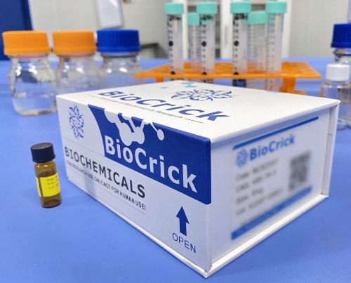
Number of papers citing our products

Adenosine triphosphate Dilution Calculator
Adenosine triphosphate Molarity Calculator
-
 Adenosine 5'-monophosphateCatalog No.: BCC8809CAS No.: 61-19-8
-
 α,β-Methyleneadenosine 5'-triphosphate trisodium saltCatalog No.: BCC3591CAS No.: 7292-42-4
-
 ATP disodium saltCatalog No.: BCC5160CAS No.: 987-65-5
-
 alpha,beta-Methyleneadenosine 5'-triphosphate trisodium saltCatalog No.: BCC7603CAS No.: 1343364-54-4
-
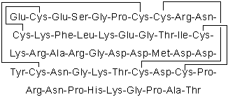 Echistatin, α1 isoformCatalog No.: BCC5988CAS No.: 154303-05-6
-
 ATPγS tetralithium saltCatalog No.: BCC7855CAS No.: 93839-89-5
-
 Mirificin-4'-O-glucosideCatalog No.: BCN9051CAS No.: 168035-01-6
-
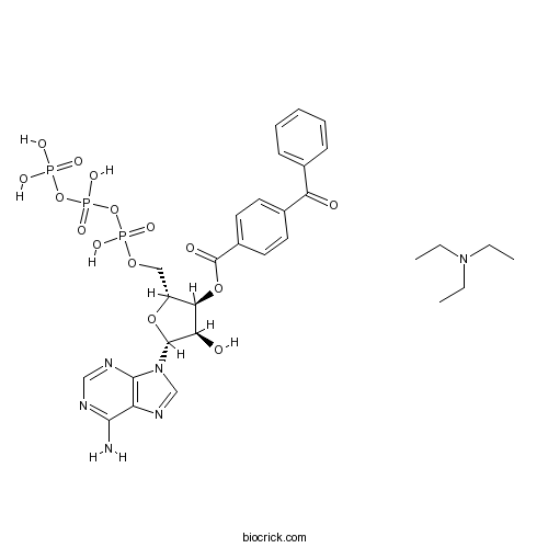 BzATP triethylammonium saltCatalog No.: BCC7643CAS No.: 112898-15-4
-
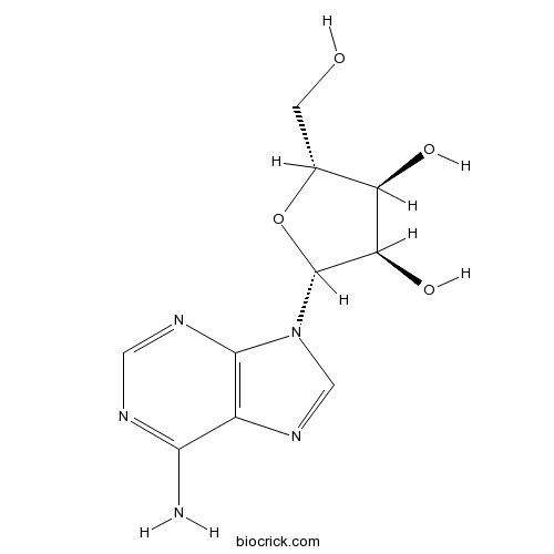 AdenosineCatalog No.: BCN5796CAS No.: 58-61-7
-
 VidarabineCatalog No.: BCC4877CAS No.: 5536-17-4
-
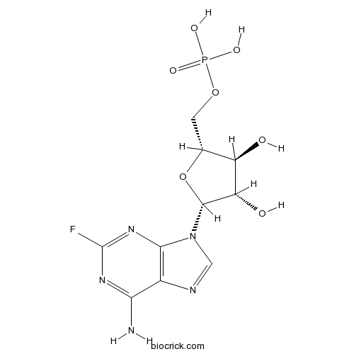 Fludarabine Phosphate (Fludara)Catalog No.: BCC3681CAS No.: 75607-67-9
| 1 mg | 5 mg | 10 mg | 20 mg | 25 mg | |
| 1 mM | 1.9716 mL | 9.858 mL | 19.7161 mL | 39.4322 mL | 49.2902 mL |
| 5 mM | 0.3943 mL | 1.9716 mL | 3.9432 mL | 7.8864 mL | 9.858 mL |
| 10 mM | 0.1972 mL | 0.9858 mL | 1.9716 mL | 3.9432 mL | 4.929 mL |
| 50 mM | 0.0394 mL | 0.1972 mL | 0.3943 mL | 0.7886 mL | 0.9858 mL |
| 100 mM | 0.0197 mL | 0.0986 mL | 0.1972 mL | 0.3943 mL | 0.4929 mL |
| * Note: If you are in the process of experiment, it's necessary to make the dilution ratios of the samples. The dilution data above is only for reference. Normally, it's can get a better solubility within lower of Concentrations. | |||||
- Cisplatin
Catalog No.:BCX2356
CAS No.:15663-27-1
- Uridine 5'-diphosphogaluronic acid trisodium
Catalog No.:BCX2355
CAS No.:148407-07-2
- Phenylephrine
Catalog No.:BCX2354
CAS No.:59-42-7
- Dotriacontane
Catalog No.:BCX2353
CAS No.:544-85-4
- 5-Pentadecylresorcinol
Catalog No.:BCX2352
CAS No.:3158-56-3
- 2-tert-Butyl-4-methoxyphenol
Catalog No.:BCX2351
CAS No.:121-00-6
- Pipemidic acid
Catalog No.:BCX2350
CAS No.:51940-44-4
- Methylaervine
Catalog No.:BCX2349
CAS No.:86293-40-5
- Pyridoxal phosphate
Catalog No.:BCX2348
CAS No.:54-47-7
- (+)-Licarin B
Catalog No.:BCX2347
CAS No.:42016-15-9
- Caffealdehyde
Catalog No.:BCX2346
CAS No.:141632-15-7
- Acridone
Catalog No.:BCX2345
CAS No.:578-95-0
- Uridine 5'-diphosphoxylose disodium
Catalog No.:BCX2358
CAS No.:108320-89-4
- Uridine 5'-diphosphogalactose disodium
Catalog No.:BCX2359
CAS No.:137868-52-1
- 2,2-Dimethyl-2,3-dihydroquinazolin-4(1H )-one
Catalog No.:BCX2360
CAS No.:77726-78-4
- Desmosterol
Catalog No.:BCX2361
CAS No.:313-04-2
- 7-Dehydrocholestero
Catalog No.:BCX2362
CAS No.:434-16-2
- Acetyl CoA
Catalog No.:BCX2363
CAS No.:102029-73-2
- CoA
Catalog No.:BCX2364
CAS No.:85-61-0
- Caffeoyl CoA
Catalog No.:BCX2365
CAS No.:53034-79-0
- 22(S)-Hydroxycholesterol
Catalog No.:BCX2366
CAS No.:22348-64-7
- Feruloyl CoA
Catalog No.:BCX2367
CAS No.:30802-02-9
- Palmitoyl CoA
Catalog No.:BCX2368
CAS No.:188174-64-3
- Benzoyl CoA
Catalog No.:BCX2369
CAS No.:102185-37-5
Biallelic ABCG8 Variants or Biochemical Evidence of Sitosterolemia: A Case Report.[Pubmed:42828365]
Cureus. 2026 Sep 2;18(9):e115644.
Sitosterolemia is a rare lipid disorder characterized by xanthomas, premature atherosclerotic cardiovascular disease (ASCVD), and abnormally elevated circulating low-density lipoprotein cholesterol (LDL-c) measurements. Pathogenic variants in Adenosine triphosphate (ATP)-binding cassette transporters lead to the accumulation of phytosterols. A child presented to the pediatric cardiology clinic with an incidentally noted presumed pathogenic variant and a variant of unknown significance in a sitosterolemia gene but had normal fasting lipids and no accumulation of circulating phytosterols. This case underscores the importance of phenotype-driven management and universal lipid screening, while highlighting the challenges of a gene-first testing/screening approach.
SIRT7 upregulation protects against TKI-induced cardiotoxicity by inhibiting MCU-mediated mitochondrial calcium overload.[Pubmed:42827607]
Front Pharmacol. 2026 Sep 18;17:1929231.
BACKGROUND: Tyrosine kinase inhibitors (TKIs) are potent anticancer agents; however, their clinical application is often restricted because of cardiotoxic effects. The mechanisms underlying cardiotoxicity that are shared across different TKIs remain inadequately understood. PURPOSE: This study aimed to investigate the role of sirtuin 7 (SIRT7) in regulating mitochondrial calcium homeostasis during TKI-induced cardiomyocyte injury and determine whether the SIRT7-mitochondrial calcium uniporter (MCU) axis could serve as a common protective target. METHODS: AC16 cells were treated for 72 h with three TKIs: sunitinib (4.11 microM, IC(30)), trametinib (12.21 microM, IC(30)), and imatinib (21.84 microM, IC(30)). Cell viability, cardiomyocyte injury markers (N-terminal pro-brain natriuretic peptide and cardiac troponin I), cytosolic and mitochondrial Ca(2+) levels, mitochondrial membrane potential, Adenosine triphosphate (ATP) content, and endoplasmic reticulum-mitochondria contacts assessed using transmission electron microscopy were evaluated. Proteomic analysis was performed to identify differentially expressed proteins shared across treatments. In human induced pluripotent stem cell-derived cardiomyocytes (hiPSC-CMs), contraction amplitude was measured using an impedance-based system, and mitochondrial Ca(2+) overload was evaluated. RESULTS: All three TKIs induced concentration-dependent injury in AC16 cells, accompanied by mitochondrial Ca(2+) overload, ATP depletion exceeding 50%, and increased mitochondria-endoplasmic reticulum (ER) contacts. Proteomic analysis identified consistent SIRT7 downregulation across the three TKI treatments. SIRT7 overexpression mitigated TKI-induced cellular damage, reversed MCU upregulation and functional activation, restored mitochondrial function, and reduced excessive mitochondria-ER contacts. In hiPSC-CMs, SIRT7 overexpression reduced mitochondrial Ca(2+) overload and prevented TKI-induced contraction inhibition. CONCLUSION: SIRT7 upregulation protects against TKI-induced cardiotoxicity by inhibiting MCU-mediated mitochondrial calcium overload.
Creatine supplementation in soccer players: a systematic review.[Pubmed:42826085]
J Int Soc Sports Nutr. 2026 Dec 31;23(sup1):2735569.
AIM: Soccer places repeated demands on short-duration, high-intensity performance, including sprints, accelerations, jumps, and technical actions performed over the course of a match. Because these efforts depend heavily on rapid Adenosine triphosphate (ATP) resynthesis, creatine is a plausible ergogenic aid for soccer players. However, findings in soccer-specific studies have been inconsistent, and previous reviews have addressed narrower questions using smaller evidence sets. This systematic review therefore examined the effects of creatine supplementation on performance, body composition, and recovery/fatigue-related outcomes in soccer players. METHODS: PubMed, Scopus, and Web of Science were searched from inception to 15 May 2025 in accordance with the Preferred Reporting Items for Systematic Reviews and Meta-Analyses (PRISMA) 2020 statement. We included controlled clinical trials of creatine supplementation in male or female soccer players of any age or competitive level when an eligible placebo, control, or comparison condition was available. Outcomes were grouped into performance, body composition, and recovery/fatigue domains. Domains from the revised Cochrane risk-of-bias tool for randomised trials (RoB 2) and the Risk Of Bias In Non-randomised Studies - of Interventions (ROBINS-I) tool were used to produce study-level methodological summaries rather than formal result-specific risk-of-bias assessments. Because the studies differed substantially in design, supplementation protocols, populations, and outcomes, findings were synthesised qualitatively rather than pooled in a meta-analysis. RESULTS: Source-level verification left 45 eligible reports representing 41 unique studies: 28 randomised parallel-group studies, 2 randomised crossover/repeated-condition studies, and 11 non-randomised or allocation-unverifiable controlled studies. Some trials reported favourable findings for repeated-sprint performance, anaerobic power, and muscular strength, but the comparative evidence was inconsistent and the certainty of evidence was very low. Findings for vertical jump performance and technical skills were mixed, while change-of-direction and endurance outcomes showed no consistent benefit. Recovery- and fatigue-related findings were also largely null or inconsistent. Body mass often increased, particularly after loading protocols, whereas evidence for gains in lean mass was limited and inconsistent. CONCLUSION: The soccer-specific evidence for creatine supplementation remains suggestive rather than conclusive. Favourable findings have been reported for some repeated-sprint, anaerobic-power, and strength outcomes, but the comparative evidence is heterogeneous, often limited by unclear or nonsignificant between-group effects, and of very low certainty. Firm conclusions about efficacy are therefore not possible. Body mass commonly increased, especially after loading protocols, while effects on lean mass, endurance, change-of-direction ability, and recovery/fatigue-related outcomes remain uncertain.
Scutellarein promotes diabetic wound repair and preserves fibroblast calcium-mitochondrial homeostasis.[Pubmed:42824297]
Front Pharmacol. 2026 Sep 17;17:1925629.
BACKGROUND: Diabetic wounds are characterized by delayed repair associated with persistent inflammation, oxidative stress, fibroblast dysfunction, mitochondrial injury, and impaired angiogenesis. Scutellarein (SCU), a structurally defined plant-derived flavonoid metabolite, has documented antioxidant and anti-inflammatory activities; however, its role and the mechanistically relevant pathways engaged in diabetic wound repair, particularly those involving calcium-mitochondrial homeostasis and purinergic signaling, remain incompletely defined. METHODS: We administered local SCU in a streptozotocin-induced diabetic mouse cutaneous wound model and, in H2O2 -injured L929 fibroblasts, assessed scratch closure, survival, redox balance, mitochondrial morphology and function, and calcium overload. We then used RNA sequencing (RNA-seq) and Adenosine triphosphate (ATP) challenge/reversal experiments to examine purinergic calcium signaling. These analyses identified P2rx1-the gene encoding P2RX1 (P2X purinoceptor 1), an ATP-gated cation channel that mediates rapid calcium entry-as a candidate purinergic signal rather than a validated target. RESULTS: SCU accelerated wound closure and increased re-epithelialization, Masson-positive collagen deposition, proliferative alpha -SMA-positive wound cells, and angiogenic indices in diabetic wounds. In injured fibroblasts, SCU increased scratch closure, reduced cell death and reactive oxygen species (ROS) accumulation, restored the superoxide dismutase (SOD)/malondialdehyde (MDA) balance, preserved mitochondrial morphology and membrane potential, and increased cellular ATP content. RNA-seq showed P2rx1 downregulation after SCU treatment and enrichment of calcium transport, mitochondrial, inflammatory, and wound-healing pathways. ATP addition attenuated the SCU-associated reductions in calcium overload, mitochondrial depolarization, and inflammatory gene expression, consistent with a contribution from broad purinergic calcium signaling. CONCLUSION: In experimental models, SCU promoted diabetic wound repair and protected fibroblasts from oxidative injury. The data support preservation of calcium-mitochondrial homeostasis as a mechanistically relevant axis, and broad purinergic signaling, possibly involving P2RX1, may contribute to these effects; direct receptor-perturbation studies are warranted.
Exposure to 4-hydroxy-4'-isopropoxydiphenylsulfone causes retinal structural damage and visual impairment in zebrafish.[Pubmed:42822976]
J Environ Sci (China). 2026 Nov;169:856-866.
4-Hydroxy-4'-isopropoxydiphenylsulfone (BPSIP), an emerging substitute for bisphenol A (BPA), has been increasingly detected in the environment, yet its toxicological profile remains poorly understood. In this study, zebrafish larvae were exposed to BPSIP at various concentrations, including an environmentally relevant level (50 mug/L), for 5 days. Transcriptomic analysis indicated that BPSIP predominantly disrupted biological processes associated with optic nerve development, including oxidative stress and energy metabolism. Corresponding biochemical assays confirmed metabolic disturbance, marked by altered levels of Adenosine triphosphate (ATP), pyruvate, and lactate. Oxidative stress was further demonstrated by elevated superoxide dismutase (SOD) activity, coupled with reduced catalase (CAT) activity, total antioxidant capacity (T-AOC), and glutathione (GSH) content. Multi-level toxicological endpoints confirmed optic nerve dysfunction: locomotor behavior assays revealed impaired photoresponse, which was consistent with systemic downregulation of key phototransduction-related genes and histopathological damage in the retina and lens. Additionally, increased thyroid hormone levels and altered expression of hypothalamic-pituitary-thyroid (HPT) axis-related genes indicated thyroid disruption. Together, these results demonstrate that BPSIP exposure disrupts visual function in zebrafish larvae, likely mediated by interference in phototransduction, oxidative homeostasis, energy metabolism, and thyroid signaling. This study provides new insights into the toxicity of BPSIP and underscores its potential ecological and health risks.
Comparative glycolipid metabolic toxicities and mechanisms of the rubber-derived contaminants 6PPD and its derivative 6PPD-Q in zebrafish.[Pubmed:42822933]
J Environ Sci (China). 2026 Nov;169:379-392.
Emerging pollutants 6PPD and its oxidation product 6PPD-Q enter the environment via rubber product abrasion, with scarce toxicological data for risk assessment. This study compared their toxicity and mechanisms on zebrafish glucolipid metabolism at environmentally relevant and sublethal concentrations. Both induced developmental malformations dose-dependently, with 120hpf larvae LC(50) of 991.6 mug/L (6PPD) and 165.2 mug/L (6PPD-Q), showing 6PPD-Q has higher teratogenicity and lethality. RNA-seq and bioinformatics revealed shared differentially expressed genes (DEGs) enrichment in ether lipid, glycerophospholipid, glycerolipid metabolism, and insulin signaling pathways, indicating common disruption of glucose-lipid homeostasis. Oil red o(ORO) staining and assays confirmed hepatic lipid accumulation (elevated total cholesterol (TC), triglycerides (TG), low-density lipoprotein cholesterol (LDL-C), reduced high-density lipoprotein cholesterol (HDL-C)) and glucose metabolism disorders (hyperglycemia, decreased insulin, lactate accumulation, etc.), suggesting insulin resistance and suppressed tricarboxylic acid cycle (TCA) cycle. Energy metabolism was dysfunctional (reduced Adenosine triphosphate (ATP), impaired mitochondrial membrane potential (DeltaPsim)), inhibiting swimming vitality, with more severe disturbances in 6PPD-Q group. PPI network analysis and RT-qPCR showed 6-PPD disrupts glucolipid metabolism via phospholipase A2(PLA2)-mediated pathways, while 6PPD-Q acts through PLA2, phospholipase D1(PLD1), and lysophosphatidylcholine acyltransferases (LPCATs). This study confirms their induction of glucolipid metabolic disorders with distinct mechanisms, providing a basis for environmental health risk assessment and related strategies.
Soil functional microbial pathways related to GHGs emissions of coastal degraded mangroves-rice ecologies in Sundarban, India: A soil metagenomic approach.[Pubmed:42822929]
J Environ Sci (China). 2026 Nov;169:344-357.
Mangroves and their adjacent rice ecologies play a critical role in regulating greenhouse gas (GHGs) emissions, particularly in tropical coastal wetlands. This study assessed the impact of mangrove degradation on methane (CH(4)) and nitrous oxide (N(2)O) emissions by comparing two distinct ecosystems; degraded mangroves, and adjacent rice system in the Sundarban, India. Annual CH(4) and N(2)O emissions were quantified and linked to soil microbial energy metabolism pathways; methanogenesis, methanotrophy, sulphur (S), and nitrogen (N) metabolism by using whole-genome metagenomic analysis. The Kyoto Encyclopedia of Genes and Genomes (KEGG) orthologs was used for functional microbial pathway analysis. Acetoclastic pathway was found to be the dominant CH(4) production pathway in rice, where relatively low soil salinity favored CO(2) to CH(4) conversion. Methane oxidation was higher in degraded mangroves, driven by the serine and xylulose monophosphate pathways, reflecting microbial adaptation to saline and alkaline conditions. The assimilatory pathway (AMP) dominated S metabolism, with adenosine phosphosulfate kinase more abundant in degraded mangroves and Adenosine triphosphate sulfurylase in rice ecology. Denitrification was the primary N pathway in degraded mangroves and adjacent rice systems, while dissimilatory nitrate reduction to ammonium (DNRA) prevailed in rice. A higher abundance of enzymes involved in acetate to methane production pathway was observed in rice systems, indicating greater CH(4) production potential, whereas a higher abundance of phosphoserine aminotransferase in serine pathways in degraded mangroves suggested relatively higher potential for CH(4) oxidation. These results demonstrate that targeted microbial management in mangrove-rice ecologies could serve as a promising strategy for climate change mitigation in coastal ecology.
Astrocytic NOX2 contributes to ROS-associated mitochondrial dysfunction in cerebral ischemia-reperfusion injury: modulation by electroacupuncture.[Pubmed:42822758]
Neuroscience. 2026 Oct 1:S0306-4522(26)00660-3.
BACKGROUND: Cerebral ischemia-reperfusion injury (CIRI) causes severe neuronal damage, in which oxidative stress and mitochondrial dysfunction are major pathological events. Astrocytes are important regulators of redox homeostasis in the injured brain, yet whether astrocytic NADPH oxidase 2 (NOX2) contributes to mitochondrial impairment after ischemia remains poorly understood. Electroacupuncture (EA) has been reported to protect against ischemic brain injury, but the cellular mechanisms involved remain unclear. METHODS: In this study, a mouse middle cerebral artery occlusion/reperfusion (MCAO/R) model was combined with astrocyte-specific NOX2 knockdown using stereotactic viral delivery. Neurological deficits, infarct size, cerebral blood flow, reactive oxygen species (ROS) accumulation, Adenosine triphosphate (ATP) levels, mitochondrial morphology, and mitochondrial dynamics-associated proteins were examined. RESULTS: MCAO/R resulted in increased astrocytic NOX2 expression, excessive ROS generation, mitochondrial fragmentation, impaired energy metabolism, and neurological dysfunction. EA treatment alleviated these changes by reducing NOX2 activation and ROS accumulation, improving mitochondrial morphology and ATP production, and restoring the balance between mitochondrial fission and fusion. These effects were reflected by decreased Drp1 and Fis1 expression and increased Mfn2 and Opa1 expression. Notably, astrocytic NOX2 knockdown produced similar protective effects and further strengthened the benefits of EA. CONCLUSION: Astrocytic NOX2 contributes to ROS accumulation, mitochondrial dysfunction, and ischemic brain injury. EA treatment is accompanied by reduced astrocytic NOX2 expression, attenuated oxidative stress, and restoration of mitochondrial homeostasis, suggesting that regulation of astrocytic NOX2 may contribute to the protective effects of EA. Targeting astrocytic NOX2 may represent a potential therapeutic strategy for ischemic stroke.
The key mechanisms and intervention strategies of cell-type-specific mitochondrial metabolism in ophthalmic diseases.[Pubmed:42818878]
Front Cell Dev Biol. 2026 Sep 16;14:1961856.
The eye is one of the most metabolically active organs, and mitochondria serve as the central hub for cellular energy metabolism by providing Adenosine triphosphate through oxidative phosphorylation. Disruption of mitochondrial metabolism-including insufficient Adenosine triphosphate synthesis, excessive production of reactive oxygen species, and dysregulated quality control-can trigger various ocular diseases. Common blinding diseases such as myopia, diabetic retinopathy, and glaucoma are closely associated with mitochondrial dysfunction in specific cell types. This review delineates the mechanisms by which metabolic adaptations in scleral fibroblasts induce myopia and the synergistic action of lipofuscin, the high-glucose-triggered mitochondrial reactive oxygen species bursts and metabolic memory in diabetic retinopathy, and the roles of mitochondrial dynamics imbalance and energy exhaustion in trabecular meshwork cells in glaucoma. Based on these mechanisms, the article proposes intervention strategies targeting mitochondrial antioxidant defense, enhancing mitochondrial biogenesis, regulating mitochondrial dynamics, and restoring mitophagy, aiming for precise prevention and treatment of ocular diseases at the mitochondrial level.

Calcutta University

University of Minnesota

University of Maryland School of Medicine

University of Illinois at Chicago

The Ohio State University

University of Zurich

Harvard University

Colorado State University

Auburn University

Yale University

Worcester Polytechnic Institute

Washington State University

Stanford University

University of Leipzig

Universidade da Beira Interior

The Institute of Cancer Research

Heidelberg University

University of Amsterdam

University of Auckland

TsingHua University

The University of Michigan

Miami University

DRURY University

Jilin University

Fudan University

Wuhan University

Sun Yat-sen University

Universite de Paris

Deemed University

Auckland University

The University of Tokyo

Korea University