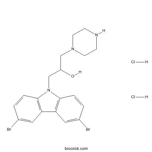
Bax channel blockerInhibitor of Bax-mediated mitochondrial cytochrome c release CAS# 335165-68-9 |

- Bavisant dihydrochloride hydrate
Catalog No.:BCC1404
CAS No.:1103522-80-0
- Lidocaine
Catalog No.:BCC1084
CAS No.:137-58-6
- Mianserin HCl
Catalog No.:BCC1114
CAS No.:21535-47-7
- Loratadine
Catalog No.:BCC1262
CAS No.:79794-75-5
- Bavisant dihydrochloride
Catalog No.:BCC1403
CAS No.:929622-09-3
Quality Control & MSDS
3D structure
Package In Stock
Number of papers citing our products

| Cas No. | 335165-68-9 | SDF | Download SDF |
| PubChem ID | 2729026 | Appearance | Powder |
| Formula | C19H23Br2Cl2N3O | M.Wt | 540.12 |
| Type of Compound | N/A | Storage | Desiccate at -20°C |
| Solubility | DMSO : 18.67 mg/mL (39.96 mM; Need ultrasonic) H2O : < 0.1 mg/mL (insoluble) | ||
| Chemical Name | 1-(3,6-dibromocarbazol-9-yl)-3-piperazin-1-ylpropan-2-ol;dihydrochloride | ||
| SMILES | C1CN(CCN1)CC(CN2C3=C(C=C(C=C3)Br)C4=C2C=CC(=C4)Br)O.Cl.Cl | ||
| Standard InChIKey | HWFKCAFKXZFOQT-UHFFFAOYSA-N | ||
| Standard InChI | InChI=1S/C19H21Br2N3O.2ClH/c20-13-1-3-18-16(9-13)17-10-14(21)2-4-19(17)24(18)12-15(25)11-23-7-5-22-6-8-23;;/h1-4,9-10,15,22,25H,5-8,11-12H2;2*1H | ||
| General tips | For obtaining a higher solubility , please warm the tube at 37 ℃ and shake it in the ultrasonic bath for a while.Stock solution can be stored below -20℃ for several months. We recommend that you prepare and use the solution on the same day. However, if the test schedule requires, the stock solutions can be prepared in advance, and the stock solution must be sealed and stored below -20℃. In general, the stock solution can be kept for several months. Before use, we recommend that you leave the vial at room temperature for at least an hour before opening it. |
||
| About Packaging | 1. The packaging of the product may be reversed during transportation, cause the high purity compounds to adhere to the neck or cap of the vial.Take the vail out of its packaging and shake gently until the compounds fall to the bottom of the vial. 2. For liquid products, please centrifuge at 500xg to gather the liquid to the bottom of the vial. 3. Try to avoid loss or contamination during the experiment. |
||
| Shipping Condition | Packaging according to customer requirements(5mg, 10mg, 20mg and more). Ship via FedEx, DHL, UPS, EMS or other couriers with RT, or blue ice upon request. | ||

Bax channel blocker Dilution Calculator

Bax channel blocker Molarity Calculator
| 1 mg | 5 mg | 10 mg | 20 mg | 25 mg | |
| 1 mM | 1.8514 mL | 9.2572 mL | 18.5144 mL | 37.0288 mL | 46.286 mL |
| 5 mM | 0.3703 mL | 1.8514 mL | 3.7029 mL | 7.4058 mL | 9.2572 mL |
| 10 mM | 0.1851 mL | 0.9257 mL | 1.8514 mL | 3.7029 mL | 4.6286 mL |
| 50 mM | 0.037 mL | 0.1851 mL | 0.3703 mL | 0.7406 mL | 0.9257 mL |
| 100 mM | 0.0185 mL | 0.0926 mL | 0.1851 mL | 0.3703 mL | 0.4629 mL |
| * Note: If you are in the process of experiment, it's necessary to make the dilution ratios of the samples. The dilution data above is only for reference. Normally, it's can get a better solubility within lower of Concentrations. | |||||

Calcutta University

University of Minnesota

University of Maryland School of Medicine

University of Illinois at Chicago

The Ohio State University

University of Zurich

Harvard University

Colorado State University

Auburn University

Yale University

Worcester Polytechnic Institute

Washington State University

Stanford University

University of Leipzig

Universidade da Beira Interior

The Institute of Cancer Research

Heidelberg University

University of Amsterdam

University of Auckland

TsingHua University

The University of Michigan

Miami University

DRURY University

Jilin University

Fudan University

Wuhan University

Sun Yat-sen University

Universite de Paris

Deemed University

Auckland University

The University of Tokyo

Korea University
IC50: 0.52 μM in Bax assay
Bax channel blocker is an inhibitor of Bax-mediated mitochondrial cytochrome c release.
In the cytosol, cytochrome c is found to form a complex with dATP, Apaf-1, and procaspase-9, which results in caspase 9 activation followed by downstream activation of other caspases, such as caspase 8, ultimately leading to the cell death. After caspase 8 cleavage, the 15.5 kDa C-terminal fragment of Bid interacts with Bak and Bax.
In vitro: Bax channel blocker, a 3,6-dibromocarbazole derivative, was observed to inhibit cytochrome c releasing from mitochondria by Bax channel modulation. The monohydroxy analogue Bax channel blocker remained the unprecedented inhibition of Bax-induced cytochrome c release at 10 μM. The IC50 value of Bax channel blocker was determined to be 0.52 μM, indicating that Bax channel blocker was a Bax channel inhibitor as hypothesized. Moreover, in the liposome assay, Bax channel blocker showing significant inhibition (>65%) of cytochrome c release at 10 μM also demonstrated sub-micromolar IC50 value [1].
In vivo: So far, there is no animal in vivo study conducted for Bax channel blocker.
Clinical trial: N/A
Reference:
[1] Bombrun A,Gerber P,Casi G,Terradillos O,Antonsson B,Halazy S. 3,6-dibromocarbazole piperazine derivatives of 2-propanol as first inhibitors of cytochrome c release via Bax channel modulation. J Med Chem.2003 Oct 9;46(21):4365-8.
- 6'-Iodoresiniferatoxin
Catalog No.:BCC7114
CAS No.:335151-55-8
- Luteinizing Hormone Releasing Hormone (LHRH)
Catalog No.:BCC1049
CAS No.:33515-09-2
- Fucoxanthin
Catalog No.:BCN2948
CAS No.:3351-86-8
- Substance P
Catalog No.:BCC6957
CAS No.:33507-63-0
- MEK inhibitor
Catalog No.:BCC1738
CAS No.:334951-92-7
- (S)-HexylHIBO
Catalog No.:BCC7167
CAS No.:334887-48-8
- HexylHIBO
Catalog No.:BCC7166
CAS No.:334887-43-3
- Gisadenafil besylate
Catalog No.:BCC7871
CAS No.:334827-98-4
- Laburnine
Catalog No.:BCN1992
CAS No.:3348-73-0
- Tracheloside
Catalog No.:BCN2738
CAS No.:33464-71-0
- 3-Aminopiperidine dihydrochloride
Catalog No.:BCC8619
CAS No.:334618-23-4
- AH 6809
Catalog No.:BCC1332
CAS No.:33458-93-4
- iMAC2
Catalog No.:BCC2396
CAS No.:335166-36-4
- Raddeanoside 20
Catalog No.:BCN2796
CAS No.:335354-79-5
- Cyclo(Phe-Leu)
Catalog No.:BCN2418
CAS No.:3354-31-2
- (-)-Bilobalide
Catalog No.:BCN1279
CAS No.:33570-04-6
- Polpunonic acid
Catalog No.:BCN7136
CAS No.:33600-93-0
- Myricanol
Catalog No.:BCN5258
CAS No.:33606-81-4
- Ispinesib (SB-715992)
Catalog No.:BCC2509
CAS No.:336113-53-2
- Britannilactone
Catalog No.:BCN3509
CAS No.:33620-72-3
- Desoxyrhapontigenin
Catalog No.:BCN6479
CAS No.:33626-08-3
- Britannin
Catalog No.:BCN2366
CAS No.:33627-28-0
- 1-O-Acetylbritannilactone
Catalog No.:BCN7715
CAS No.:33627-41-7
- (S)-(+)-Ketamine hydrochloride
Catalog No.:BCC7930
CAS No.:33643-47-9
Azoxystrobin Induces Apoptosis of Human Esophageal Squamous Cell Carcinoma KYSE-150 Cells through Triggering of the Mitochondrial Pathway.[Pubmed:28567017]
Front Pharmacol. 2017 May 17;8:277.
Recent studies indicate that mitochondrial pathways of apoptosis are potential chemotherapeutic target for the treatment of esophageal cancer. Azoxystrobin (AZOX), a methoxyacrylate derived from the naturally occurring strobilurins, is a known fungicide acting as a ubiquinol oxidation (Qo) inhibitor of mitochondrial respiratory complex III. In this study, the effects of AZOX on human esophageal squamous cell carcinoma KYSE-150 cells were examined and the underlying mechanisms were investigated. AZOX exhibited inhibitory effects on the proliferation of KYSE-150 cells with inhibitory concentration 50% (IC50) of 2.42 mug/ml by 48 h treatment. Flow cytometry assessment revealed that the inhibitory effect of AZOX on KYSE-150 cell proliferation occurred with cell cycle arrest at S phase and increased cell apoptosis in time-dependent and dose-dependent manners. Cleaved poly ADP ribose polymerase (PARP), caspase-3 and caspase-9 were increased significantly by AZOX. It is worth noted that the Bcl-2/Bax ratios were decreased because of the down-regulated Bcl-2 and up-regulated Bax expression level. Meanwhile, the cytochrome c release was increased by AZOX in KYSE-150 cells. AZOX-induced cytochrome c expression and caspase-3 activation was significantly blocked by Bax channel blocker. Intragastric administration of AZOX effectively decreased the tumor size generated by subcutaneous inoculation of KYSE-150 cells in nude mice. Consistently, decreased Bcl-2 expression, increased cytochrome c and PARP level, and activated caspase-3 and caspase-9 were observed in the tumor samples. These results indicate that AZOX can effectively induce esophageal cancer cell apoptosis through the mitochondrial pathways of apoptosis, suggesting AZOX or its derivatives may be developed as potential chemotherapeutic agents for the treatment of esophageal cancer.
Fibroblast growth factor 2 protects against renal ischaemia/reperfusion injury by attenuating mitochondrial damage and proinflammatory signalling.[Pubmed:28544332]
J Cell Mol Med. 2017 Nov;21(11):2909-2925.
Ischaemia-reperfusion injury (I/RI) is a common cause of acute kidney injury (AKI). The molecular basis underlying I/RI-induced renal pathogenesis and measures to prevent or reverse this pathologic process remains to be resolved. Basic fibroblast growth factor (FGF2) is reported to have protective roles of myocardial infarction as well as in several other I/R related disorders. Herein we present evidence that FGF2 exhibits robust protective effect against renal histological and functional damages in a rat I/RI model. FGF2 treatment greatly alleviated I/R-induced acute renal dysfunction and largely blunted I/R-induced elevation in serum creatinine and blood urea nitrogen, and also the number of TUNEL-positive tubular cells in the kidney. Mechanistically, FGF2 substantially ameliorated renal I/RI by mitigating several mitochondria damaging parameters including pro-apoptotic alteration of Bcl2/Bax expression, caspase-3 activation, loss of mitochondrial membrane potential and KATP channel integrity. Of note, the protective effect of FGF2 was significantly compromised by the KATP channel blocker 5-HD. Interestingly, I/RI alone resulted in mild activation of FGFR, whereas FGF2 treatment led to more robust receptor activation. More significantly, post-I/RI administration of FGF2 also exhibited robust protection against I/RI by reducing cell apoptosis, inhibiting the release of damage-associated molecular pattern molecule HMBG1 and activation of its downstream inflammatory cytokines such as IL-1alpha, IL-6 and TNF alpha. Taken together, our data suggest that FGF2 offers effective protection against I/RI and improves animal survival by attenuating mitochondrial damage and HMGB1-mediated inflammatory response. Therefore, FGF2 has the potential to be used for the prevention and treatment of I/RI-induced AKI.
Cysteine-rich buccal gland protein suppressed the proliferation, migration and invasion of hela cells through akt pathway.[Pubmed:28945311]
IUBMB Life. 2017 Nov;69(11):856-866.
Cysteine-rich buccal gland protein (CRBGP) as a member of cysteine-rich secretory proteins (CRISPs) superfamily was isolated from the buccal glands of Lampetra japonica, the blood suckers in the marine. Previous studies showed CRBGP could suppress angiogenesis probably due to its ion channel blocking activity. Whether CRBGP could also affect the activity of tumor cells has not been reported yet. In this study, CRBGP suppressed the proliferation of Hela cells with an IC50 of 6.7 muM by inducing apoptosis. Both microscopic observation and Western blot indicated that CRBGP was able to induce the nuclei shrinking, downregulate the protein level of BCL2 and caspase 3 as well as upregulate the level of BAX in Hela cells, suggested that CRBGP might induce apoptosis of Hela cells in a mitochondrial-dependent pathway. Furthermore, CRBGP could disturb F-actin organization, which would finally cause the Hela cells to lose their shape and to lessen their abilities on adhesion, migration and invasion. Finally, CRBGP was shown to reduce the phosphorylation level of Akt, which indicated that CRBGP might inhibit the proliferation and metastasis of Hela cells through Akt pathway. CRBGP, as a voltage-gated sodium channel blocker, also possesses the anti-tumor abilities which provided information on the effects and action manner of the other CRISPs. (c) 2017 IUBMB Life, 69(11):856-866, 2017.
Sarcolemmal ATP-sensitive potassium channel protects cardiac myocytes against lipopolysaccharide-induced apoptosis.[Pubmed:27430376]
Int J Mol Med. 2016 Sep;38(3):758-66.
The sarcolemmal ATP-sensitive K+ (sarcKATP) channel plays a cardioprotective role during stress. However, the role of the sarcKATP channel in the apoptosis of cardiomyocytes and association with mitochondrial calcium remains unclear. For this purpose, we developed a model of LPS-induced sepsis in neonatal rat cardiomyocytes (NRCs). The TUNEL assay was performed in order to detect the apoptosis of cardiac myocytes and the MTT assay was performed to determine cellular viability. Exposure to LPS significantly decreased the viability of the NRCs as well as the expression of Bcl-2, whereas it enhanced the activity and expression of the apoptosis-related proteins caspase-3 and Bax, respectively. The sarcKATP channel blocker, HMR-1098, increased the apoptosis of NRCs, whereas the specific sarcKATP channel opener, P-1075, reduced the apoptosis of NRCs. The mitochondrial calcium uniporter inhibitor ruthenium red (RR) partially inhibited the pro-apoptotic effect of HMR-1098. In order to confirm the role of the sarcKATP channel, we constructed a recombinant adenovirus vector carrying the sarcKATP channel mutant subunit Kir6.2AAA to inhibit the channel activity. Kir6.2AAA adenovirus infection in NRCs significantly aggravated the apoptosis of myocytes induced by LPS. Elucidating the regulatory mechanisms of the sarcKATP channel in apoptosis may facilitate the development of novel therapeutic targets and strategies for the management of sepsis and cardiac dysfunction.
Acid-sensing ion channel 1a regulates the survival of nucleus pulposus cells in the acidic environment of degenerated intervertebral discs.[Pubmed:27746861]
Iran J Basic Med Sci. 2016 Aug;19(8):812-820.
OBJECTIVES: Activation of acid-sensing ion channel 1a (ASIC1a) is responsible for tissue injury caused by acidosis in nervous systems. But its physiological and pathological roles in nucleus pulposus cells (NPCs) are unclear. The aim of this study is to investigate whether ASIC1a regulates the survival of NPCs in the acidic environment of degenerated discs. MATERIALS AND METHODS: NPCs were isolated and cultured followed by immunofluorescent staining and Western-blot analysis for ASIC1a. Intracellular calcium ([Ca(2+)]i) was determined by Ca(2+)-imaging using Fura-2-AM. Cell necrosis, apoptosis, and senescence following acid exposure were determined using lactate dehydrogenase (LDH) release assay, annexin V-fluorescein isothiocyanate/propidium iodide dual-staining and cell cycle analysis, respectively, followed by Western-blot analysis for apoptosis-related proteins (Bax, Bcl-2, and caspase-3) and senescence-related proteins (p53, p21, and p16). Effects of treatment with psalmotoxin-1 (PcTX1, blocker of ASIC1a) on [Ca(2+)]i and cell survival were investigated. RESULTS: ASIC1a was detected in healthy NPCs, and its expression was significantly higher in degenerated cells. When NPCs were treated with PcTX1, acid-induced increases in [Ca(2+)]i were significantly inhibited. PcTX1 treatment also resulted in decreased LDH release, cell apoptosis and cell cycle arrest in acid condition. Acid exposure decreased the expression of Bcl-2 and increased the expression of Bax, cleaved caspase-3 and senescence-related proteins (p53, p21, and p16), which was inhibited by PcTX1. CONCLUSION: The present findings suggest that further understanding of ASIC1a functionality may provide not only a novel insight into intervertebral disc biology but also a novel therapeutic target for intervertebral disc degeneration.


