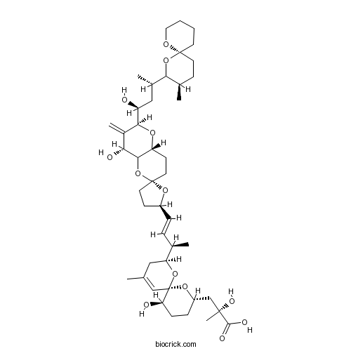
Okadaic acidProtein phosphatase 1 inhibitor CAS# 78111-17-8 |

- Calyculin A
Catalog No.:BCC2457
CAS No.:101932-71-2
- Calcineurin Autoinhibitory Peptide
Catalog No.:BCC2456
CAS No.:148067-21-4
- DL-AP3
Catalog No.:BCC2459
CAS No.:20263-06-3
- Ceramide
Catalog No.:BCC2458
CAS No.:3102-57-6
- Fostriecin sodium salt
Catalog No.:BCC2460
CAS No.:87860-39-7
Quality Control & MSDS
3D structure
Package In Stock
Number of papers citing our products

| Cas No. | 78111-17-8 | SDF | Download SDF |
| PubChem ID | 11953808 | Appearance | Powder |
| Formula | C44H68O13 | M.Wt | 805.01 |
| Type of Compound | N/A | Storage | Desiccate at -20°C |
| Solubility | Soluble to 100 mM in DMSO | ||
| Chemical Name | (2R)-3-[(2S,6R,8S,11R)-2-[(E,2R)-4-[(2S,2'R,4R,6R,8aR)-4-hydroxy-2-[(1S,3S)-1-hydroxy-3-[(3R,6S)-3-methyl-1,7-dioxaspiro[5.5]undecan-2-yl]butyl]-3-methylidenespiro[4a,7,8,8a-tetrahydro-4H-pyrano[3,2-b]pyran-6,5'-oxolane]-2'-yl]but-3-en-2-yl]-11-hydroxy-4-methyl-1,7-dioxaspiro[5.5]undec-4-en-8-yl]-2-hydroxy-2-methylpropanoic acid | ||
| SMILES | CC1CCC2(CCCCO2)OC1C(C)CC(C3C(=C)C(C4C(O3)CCC5(O4)CCC(O5)C=CC(C)C6CC(=CC7(O6)C(CCC(O7)CC(C)(C(=O)O)O)O)C)O)O | ||
| Standard InChIKey | QNDVLZJODHBUFM-AAWJMCDUSA-N | ||
| Standard InChI | InChI=1S/C44H68O13/c1-25-21-34(55-44(23-25)35(46)12-11-31(54-44)24-41(6,50)40(48)49)26(2)9-10-30-14-18-43(53-30)19-15-33-39(57-43)36(47)29(5)38(52-33)32(45)22-28(4)37-27(3)13-17-42(56-37)16-7-8-20-51-42/h9-10,23,26-28,30-39,45-47,50H,5,7-8,11-22,24H2,1-4,6H3,(H,48,49)/b10-9+/t26-,27-,28+,30+,31+,32+,33-,34+,35-,36-,37?,38+,39?,41-,42+,43-,44-/m1/s1 | ||
| General tips | For obtaining a higher solubility , please warm the tube at 37 ℃ and shake it in the ultrasonic bath for a while.Stock solution can be stored below -20℃ for several months. We recommend that you prepare and use the solution on the same day. However, if the test schedule requires, the stock solutions can be prepared in advance, and the stock solution must be sealed and stored below -20℃. In general, the stock solution can be kept for several months. Before use, we recommend that you leave the vial at room temperature for at least an hour before opening it. |
||
| About Packaging | 1. The packaging of the product may be reversed during transportation, cause the high purity compounds to adhere to the neck or cap of the vial.Take the vail out of its packaging and shake gently until the compounds fall to the bottom of the vial. 2. For liquid products, please centrifuge at 500xg to gather the liquid to the bottom of the vial. 3. Try to avoid loss or contamination during the experiment. |
||
| Shipping Condition | Packaging according to customer requirements(5mg, 10mg, 20mg and more). Ship via FedEx, DHL, UPS, EMS or other couriers with RT, or blue ice upon request. | ||
| Description | Potent inhibitor of protein phosphatase 1 (IC50 = 3 nM) and protein phosphatase 2A (IC50 = 0.2-1 nM). Displays > 100,000,000-fold selectivity over PP2B and PP2C. Tumor promoter. Shown to activate atypical protein kinase C in adipocytes. |

Okadaic acid Dilution Calculator

Okadaic acid Molarity Calculator
| 1 mg | 5 mg | 10 mg | 20 mg | 25 mg | |
| 1 mM | 1.2422 mL | 6.2111 mL | 12.4222 mL | 24.8444 mL | 31.0555 mL |
| 5 mM | 0.2484 mL | 1.2422 mL | 2.4844 mL | 4.9689 mL | 6.2111 mL |
| 10 mM | 0.1242 mL | 0.6211 mL | 1.2422 mL | 2.4844 mL | 3.1056 mL |
| 50 mM | 0.0248 mL | 0.1242 mL | 0.2484 mL | 0.4969 mL | 0.6211 mL |
| 100 mM | 0.0124 mL | 0.0621 mL | 0.1242 mL | 0.2484 mL | 0.3106 mL |
| * Note: If you are in the process of experiment, it's necessary to make the dilution ratios of the samples. The dilution data above is only for reference. Normally, it's can get a better solubility within lower of Concentrations. | |||||

Calcutta University

University of Minnesota

University of Maryland School of Medicine

University of Illinois at Chicago

The Ohio State University

University of Zurich

Harvard University

Colorado State University

Auburn University

Yale University

Worcester Polytechnic Institute

Washington State University

Stanford University

University of Leipzig

Universidade da Beira Interior

The Institute of Cancer Research

Heidelberg University

University of Amsterdam

University of Auckland

TsingHua University

The University of Michigan

Miami University

DRURY University

Jilin University

Fudan University

Wuhan University

Sun Yat-sen University

Universite de Paris

Deemed University

Auckland University

The University of Tokyo

Korea University
Okadaic acid is a potent inhibitor of protein phosphatase 1/2A with IC50 values of 19 nM and 0.2 nM for protein phosphatase 1 and protein phosphatase 2A, respectively [1].
Protein phosphatase 1 (PP1) and protein phosphatase 2A (PP2A) are two major mammalian serine/threonine protein phosphatases and are activated in response to Ca2+ cascades as well as increased protein kinase A activity [2].
In confluent rabbit lens epithelial cells (RLECs), okadaic acid (100 nM) significantly induced cell apoptosis. Also, okadaic acid induced the expression of p53 and bax, which are necessary for the apoptotic programs. In N/N1003A cells, okadaic acid (10 nM) decreased total phosphatase activity by 20% and mainly inhibited PP-2A activity, while okadaic acid (100 nM) reduced 81% total phosphatase activity and inhibited PP-1 and PP-2A activity [3].
In the rat striatum, okadaic acid (0.005, 0.05 and 0.5 nM) increased Elk-1 and CREB phosphorylation and c-Fos immunoreactivity in a dose-dependant way. Also, okadaic acid (0.05 and 0.5 nM) increased c-fos mRNA expression in a dose-dependent way [2].
References:
[1]. Holmes CF. Liquid chromatography-linked protein phosphatase bioassay; a highly sensitive marine bioscreen for okadaic acid and related diarrhetic shellfish toxins. Toxicon, 1991, 29(4-5): 469-477.
[2]. Choe ES, Parelkar NK, Kim JY, et al. The protein phosphatase 1/2A inhibitor okadaic acid increases CREB and Elk-1 phosphorylation and c-fos expression in the rat striatum in vivo. J Neurochem, 2004, 89(2): 383-390.
[3]. Li DW, Fass U, Huizar I, et al. Okadaic acid-induced lens epithelial cell apoptosis requires inhibition of phosphatase-1 and is associated with induction of gene expression including p53 and bax. Eur J Biochem, 1998, 257(2): 351-361.
- Aztreonam
Catalog No.:BCC2557
CAS No.:78110-38-0
- 2-Acetylfluorene
Catalog No.:BCC8516
CAS No.:781-73-7
- (R)-(+)-8-Hydroxy-DPAT hydrobromide
Catalog No.:BCC6929
CAS No.:78095-19-9
- Fmoc-Lys(Fmoc)-OH
Catalog No.:BCC3521
CAS No.:78081-87-5
- MMPX
Catalog No.:BCC6692
CAS No.:78033-08-6
- 1,3-Diacetylvilasinin
Catalog No.:BCN4580
CAS No.:78012-28-9
- Zeylasterone
Catalog No.:BCN8057
CAS No.:78012-25-6
- Linalool
Catalog No.:BCN6339
CAS No.:78-70-6
- Isophorone
Catalog No.:BCN8329
CAS No.:78-59-1
- Mulberrofuran C
Catalog No.:BCN4032
CAS No.:77996-04-4
- Secologanin dimethyl acetal
Catalog No.:BCN4581
CAS No.:77988-07-9
- Z-Glycinol
Catalog No.:BCC3095
CAS No.:77987-49-6
- DAMGO
Catalog No.:BCC6958
CAS No.:78123-71-4
- MK-0974
Catalog No.:BCC1756
CAS No.:781649-09-0
- CDPPB
Catalog No.:BCC7610
CAS No.:781652-57-1
- 4-Hydroxyisoleucine
Catalog No.:BCN1211
CAS No.:781658-23-9
- YM155
Catalog No.:BCC2251
CAS No.:781661-94-7
- Nirtetralin
Catalog No.:BCN3755
CAS No.:78185-63-4
- 20(S)-Ginsenoside Rh2
Catalog No.:BCN1070
CAS No.:78214-33-2
- Paroxetine HCl
Catalog No.:BCC5054
CAS No.:78246-49-8
- [Orn5]-URP
Catalog No.:BCC5985
CAS No.:782485-03-4
- Hydroxysafflor yellow A
Catalog No.:BCN1049
CAS No.:78281-02-4
- Nepafenac
Catalog No.:BCC1258
CAS No.:78281-72-8
- Ecliptasaponin A
Catalog No.:BCN3843
CAS No.:78285-90-2
ACA, an inhibitor phospholipases A2 and transient receptor potential melastatin-2 channels, attenuates okadaic acid induced neurodegeneration in rats.[Pubmed:28363841]
Life Sci. 2017 May 1;176:10-20.
AIM: In recent studies, it has been shown that the Transient Receptor Potential Melastatin-2 Channels (TRPM2) and Phospholipases A2 (PLA2) inhibitors may have a protective effect on neurons. This study was aimed to investigate the protective effect of TRPM2 and PLA2 inhibitor N-(p-amylcinnamoyl) Anthranilic Acid (ACA) in a neurodegenerative model induced by Okadaic acid (OKA). MAIN METHODS: OKA (200ng/10mul) was administered bilateral intracerebroventricularly as a single injection. KEY FINDINGS: OKA-treated rats showed significant impairments of spatial memory in Morris Water Maze Test. OKA-induced memory-impaired rats showed increased numbers of degenerated neurons and Caspase-3, tau phosphorylated ser396, beta-amyloid positive cells in the hippocampus and cerebral cortex. Furthermore, OKA-treated rats exhibited significantly increased MDA, TNF-alpha levels, and decreased SOD, GSH-PX enzyme activates and GSH levels of the tissues. ACA administration ameliorated OKA-induced memory impairment in rats. The ACA treatment also increased SOD and GSH-PX enzyme activation and GSH levels, and conversely decreased the levels of MDA, TNF-alpha. It was found that the numbers of the degenerated neurons and Caspase-3 positive cells of cortex and hippocampus regions were significantly reduced. SIGNIFICANCE: ACA administration attenuates the oxidative stress and neuroinflammation of OKA-induced neurodegeneration; and ameliorates the cognitive decline and neurodegeneration.
Sulfated diesters of okadaic acid and DTX-1: Self-protective precursors of diarrhetic shellfish poisoning (DSP) toxins.[Pubmed:28366404]
Harmful Algae. 2017 Mar;63:85-93.
Many toxic secondary metabolites used for defense are also toxic to the producing organism. One important way to circumvent toxicity is to store the toxin as an inactive precursor. Several sulfated diesters of the diarrhetic shellfish poisoning (DSP) toxin Okadaic acid have been reported from cultures of various dinoflagellate species belonging to the genus Prorocentrum. It has been proposed that these sulfated diesters are a means of toxin storage within the dinoflagellate cell, and that a putative enzyme mediated two-step hydrolysis of sulfated diesters such as DTX-4 and DTX-5 initially leads to the formation of diol esters and ultimately to the release of free Okadaic acid. However, only one diol ester and no sulfated diesters of DTX-1, a closely related DSP toxin, have been isolated leading some to speculate that this toxin is not stored as a sulfated diester and is processed by some other means. DSP components in organic extracts of two large scale Prorocentrum lima laboratory cultures have been investigated. In addition to the usual suite of Okadaic acid esters, as well as the free acids Okadaic acid and DTX-1, a group of corresponding diol- and sulfated diesters of both Okadaic acid and DTX-1 have now been isolated and structurally characterized, confirming that both Okadaic acid and DTX-1 are initially formed in the dinoflagellate cell as the non-toxic sulfated diesters.
Effects of algal toxin okadaic acid on the non-specific immune and antioxidant response of bay scallop (Argopecten irradians).[Pubmed:28323217]
Fish Shellfish Immunol. 2017 Jun;65:111-117.
Okadaic acid (OA) is produced by dinoflagellates during harmful algal blooms and is a diarrhetic shellfish-poisoning (DSP) toxin. This toxin is particularly problematic for bivalves that are cultured for human consumption. This study aimed to reveal the effects of exposure to OA on the non-specific immune responses of bay scallop, Argopecten irradians. Various immunological parameters (superoxide dismutase (SOD), acid phosphatase (ACP), alkaline phosphatase (ALP), lysozyme activities, and total protein level) were assessed in the hemolymph of bay scallops at 3, 6, 12, 24, and 48 h post-exposure (hpe) to different concentrations (50, 100, and 500 nM) of OA. Moreover, the expression of immune system-related genes (MnSOD, PrxV, PGRP, and BD) was also measured. Results showed that SOD and ACP activities were decreased between 12 and 48 hpe. The ALP, lysozyme activities, and total protein levels were also modulated after exposure to different concentrations of OA. The expression of immune-system-related genes was also assessed at different time points during the exposure period. Overall, our results suggest that the exposure to OA had negative effects on the antioxidant and non-specific immune responses, and even disrupted the metabolism of bay scallops, making them more vulnerable to environmental stress-inducing agents; they provide a better understanding of the response status of bivalves against DSP toxins.
SLM, a novel carbazole-based fluorophore attenuates okadaic acid-induced tau hyperphosphorylation via down-regulating GSK-3beta activity in SH-SY5Y cells.[Pubmed:28359686]
Eur J Pharm Sci. 2017 Dec 15;110:101-108.
Phosphorylated tau dissociates from microtubules and aggregates to form neurofibrillary tangles resulting in neuronal toxicity and cognitive deficits. Attenuating tau hyperphosphorylation is considered as an effective therapeutic approach for Alzheimer's disease (AD). From our previous study, SLM, a carbazole-based fluorophore prevents Abeta aggregation, reduced glycogen synthase kinase-3beta (GSK-3beta) activity and tau hyperphosphorylation in triple transgenic mouse model of AD. However, the mechanism by which SLM attenuates tau hyperphosphorylation warrants further investigation. In the current study, we intend to evaluate the effects of SLM against Okadaic acid (OA)-induced tau hyperphosphorylation and microtubules instability in human neuroblastoma (SH-SY5Y) cells. The results showed that, SLM reduced the OA-induced cell neurotoxicity and tau hyperphosphorylation in SH-SY5Y cells. SLM treatment down-regulated GSK-3beta activity. However, in the presence of GSK-3beta inhibitor (SB216763, 10muM), SLM treatment could not reduce GSK-3beta activity and tau hyperphosphorylation as compared with SB216763 treatment alone. Furthermore, SLM treatment also ameliorated OA-induced microtubules instability and cytoskeleton damage. Collectively, SLM attenuated OA-induced tau hyperphosphorylation via down-regulating GSK-3beta activity in SH-SY5Y cells. Therefore, this study supports SLM as a potential compound for AD and other tau pathology-related neurodegenerative disorders.
Okadaic acid activates atypical protein kinase C (zeta/lambda) in rat and 3T3/L1 adipocytes. An apparent requirement for activation of Glut4 translocation and glucose transport.[Pubmed:10318822]
J Biol Chem. 1999 May 14;274(20):14074-8.
Okadaic acid, an inhibitor of protein phosphatases 1 and 2A, is known to provoke insulin-like effects on GLUT4 translocation and glucose transport, but the underlying mechanism is obscure. Presently, we found in both rat adipocytes and 3T3/L1 adipocytes that Okadaic acid provoked partial insulin-like increases in glucose transport, which were inhibited by phosphatidylinositol (PI) 3-kinase inhibitors, wortmannin and LY294002, and inhibitors of atypical protein kinase C (PKC) isoforms, zeta and lambda. Moreover, in both cell types, Okadaic acid provoked increases in the activity of immunoprecipitable PKC-zeta/lambda by a PI 3-kinase-dependent mechanism. In keeping with apparent PI 3-kinase dependence of stimulatory effects of Okadaic acid on glucose transport and PKC-zeta/lambda activity, Okadaic acid provoked insulin-like increases in membrane PI 3-kinase activity in rat adipocytes; the mechanism for PI 3-kinase activation was uncertain, however, because it was not apparent in phosphotyrosine immunoprecipitates. Of further note, Okadaic acid provoked partial insulin-like increases in the translocation of hemagglutinin antigen-tagged GLUT4 to the plasma membrane in transiently transfected rat adipocytes, and these stimulatory effects on hemagglutinin antigen-tagged GLUT4 translocation were inhibited by co-expression of kinase-inactive forms of PKC-zeta and PKC-lambda but not by a double mutant (T308A, S473A), activation-resistant form of protein kinase B. Our findings suggest that, as with insulin, PI 3-kinase-dependent atypical PKCs, zeta and lambda, are required for Okadaic acid-induced increases in GLUT4 translocation and glucose transport in rat adipocytes and 3T3/L1 adipocytes.
Okadaic acid-induced apoptosis in neuronal cells: evidence for an abortive mitotic attempt.[Pubmed:9489733]
J Neurochem. 1998 Mar;70(3):1124-33.
There is increasing evidence that apoptosis in postmitotic neurons is associated with a frustrated attempt to reenter the mitotic cycle. Okadaic acid, a specific protein phosphatase inhibitor, is currently used in models of Alzheimer's research to increase the degree of phosphorylation of various proteins, such as the microtubule-associated protein tau. Okadaic acid induces programmed cell death in the human neuroblastoma cell lines TR14 and NT2-N, as evidenced by fragmentation of DNA and attenuation of this process by protein synthesis inhibitors. In differentiated TR14 cells, Okadaic acid increases the fraction of cells in the S phase, induces the appearance of cyclin B1 and cyclin D1 markers of the cell cycle, and triggers a time-dependent increase in DNA fragmentation after release of a thymidine block. Fully differentiated NT2-N cells are forced to enter the mitotic cycle as shown by DNA staining. Chromatin condensation and chromosome formation are initiated, but the cells fail to complete their mitotic cycle. These data suggest that Okadaic acid forces differentiated neuronal cells into the mitotic cycle. This pattern of cyclin up-regulation and cell cycle shift is compared with apoptosis induced by neurotrophic factor deprivation in differentiated rat pheochromocytoma PC12 cells.
Okadaic acid: a new probe for the study of cellular regulation.[Pubmed:2158158]
Trends Biochem Sci. 1990 Mar;15(3):98-102.
The tumour promoter Okadaic acid is a potent and specific inhibitor of protein phosphatases 1 and 2A. Here we review recent studies which demonstrate that this toxin is extremely useful for identifying biological processes that are controlled through the reversible phosphorylation of proteins.
Effects of the tumour promoter okadaic acid on intracellular protein phosphorylation and metabolism.[Pubmed:2562908]
Nature. 1989 Jan 5;337(6202):78-81.
Okadaic acid is a polyether derivative of 38-carbon fatty acid, and is implicated as the causative agent of diarrhetic shellfish poisoning. It is a potent tumour promoter that is not an activator of protein kinase C, but is a powerful inhibitor of protein phosphatases-1 and -2A (PP1 and PP2A) in vitro. We report here that Okadaic acid rapidly stimulates protein phosphorylation in intact cells, and behaves like a specific protein phosphatase inhibitor in a variety of metabolic processes. Our results indicate that PP1 and PP2A are the dominant protein phosphatases acting on a wide range of phosphoproteins in vivo. We also find that Okadaic acid mimics the effect of insulin on glucose transport in adipocytes, which suggests that this process is stimulated by a serine/threonine phosphorylation event.


