
TryptamineCAS# 61-54-1 |
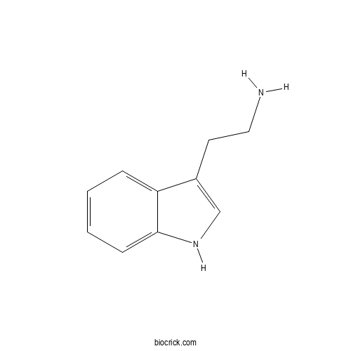
| Cas No. | 61-54-1 | SDF | File under preparation. |
| PubChem ID | 1150.0 | Appearance | Powder |
| Formula | C10H12N2 | M.Wt | 160.2 |
| Type of Compound | N/A | Storage | Desiccate at -20°C |
| Solubility | Soluble in Chloroform,Dichloromethane,Ethyl Acetate,DMSO,Acetone,etc. | ||
| Chemical Name | 2-(1H-indol-3-yl)ethanamine | ||
| SMILES | C1=CC=C2C(=C1)C(=CN2)CCN | ||
| Standard InChIKey | APJYDQYYACXCRM-UHFFFAOYSA-N | ||
| Standard InChI | InChI=1S/C10H12N2/c11-6-5-8-7-12-10-4-2-1-3-9(8)10/h1-4,7,12H,5-6,11H2 | ||
| General tips | For obtaining a higher solubility , please warm the tube at 37 ℃ and shake it in the ultrasonic bath for a while.Stock solution can be stored below -20℃ for several months. We recommend that you prepare and use the solution on the same day. However, if the test schedule requires, the stock solutions can be prepared in advance, and the stock solution must be sealed and stored below -20℃. In general, the stock solution can be kept for several months. Before use, we recommend that you leave the vial at room temperature for at least an hour before opening it. |
||
| About Packaging | 1. The packaging of the product may be reversed during transportation, cause the high purity compounds to adhere to the neck or cap of the vial.Take the vail out of its packaging and shake gently until the compounds fall to the bottom of the vial. 2. For liquid products, please centrifuge at 500xg to gather the liquid to the bottom of the vial. 3. Try to avoid loss or contamination during the experiment. |
||
| Shipping Condition | Packaging according to customer requirements(5mg, 10mg, 20mg and more). Ship via FedEx, DHL, UPS, EMS or other couriers with RT, or blue ice upon request. | ||
Quality Control & MSDS
3D structure
Package In Stock
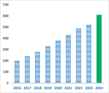
Number of papers citing our products

Tryptamine Dilution Calculator
Tryptamine Molarity Calculator


| 1 mg | 5 mg | 10 mg | 20 mg | 25 mg | |
| 1 mM | 6.2422 mL | 31.211 mL | 62.422 mL | 124.8439 mL | 156.0549 mL |
| 5 mM | 1.2484 mL | 6.2422 mL | 12.4844 mL | 24.9688 mL | 31.211 mL |
| 10 mM | 0.6242 mL | 3.1211 mL | 6.2422 mL | 12.4844 mL | 15.6055 mL |
| 50 mM | 0.1248 mL | 0.6242 mL | 1.2484 mL | 2.4969 mL | 3.1211 mL |
| 100 mM | 0.0624 mL | 0.3121 mL | 0.6242 mL | 1.2484 mL | 1.5605 mL |
| * Note: If you are in the process of experiment, it's necessary to make the dilution ratios of the samples. The dilution data above is only for reference. Normally, it's can get a better solubility within lower of Concentrations. | |||||
- Coniferyl acetate
Catalog No.:BCX2255
CAS No.:94930-81-1
- Scinamine C
Catalog No.:BCX2254
CAS No.:2184979-75-5
- 3''-Hydroxypyrrolezanthine
Catalog No.:BCX2253
CAS No.:2647391-04-4
- Lyxose
Catalog No.:BCX2252
CAS No.:65-42-9
- 3'-O-Methylhesperidin
Catalog No.:BCX2251
CAS No.:28034-80-2
- Benzoic acid,2-[(5,10-dihydro-3,7,8-trihydroxy-5,10-dioxo...
Catalog No.:BCX2910
CAS No.:60202-70-2
- Osmanthuside B
Catalog No.:BCX2909
CAS No.:94492-23-6
- Podoverine A
Catalog No.:BCX2908
CAS No.:107882-43-9
- Benzoic acid,2-(5,10-dihydro-2,3,7,8-tetrahydroxy...
Catalog No.:BCX2907
CAS No.:103744-88-3
- Bacogenin A4 (Ebelinlacton)
Catalog No.:BCX2906
CAS No.:3649-76-1
- Feruloylputrescine
Catalog No.:BCX2905
CAS No.:501-13-3
- Cistanoside D
Catalog No.:BCX2904
CAS No.:94492-21-4
- Benzil
Catalog No.:BCX2257
CAS No.:134-81-6
- Lobetyolin
Catalog No.:BCX2258
CAS No.:129277-38-9
- 4-(2-(Carboxymethyl)-3,5-dihydroxyphenoxy)benzoic acid
Catalog No.:BCX2259
CAS No.:2193189-82-9
- 4,5-Di-O-caffeoylquinic acid
Catalog No.:BCX2260
CAS No.:89886-30-6
- Malic acid
Catalog No.:BCX2261
CAS No.:636-61-3
- Picropodophyllin
Catalog No.:BCX2262
CAS No.:477-47-4
- α-Santalene
Catalog No.:BCX2263
CAS No.:512-61-8
- Valerianol
Catalog No.:BCX2264
CAS No.:20489-45-6
- 7-epi-Teucrenone
Catalog No.:BCX2265
CAS No.:165898-48-6
- Isosyringinoside
Catalog No.:BCX2266
CAS No.:152686-85-6
- Taccalonolide Q
Catalog No.:BCX2267
CAS No.:488791-19-1
- 7-O-Galloyltricetiflavan
Catalog No.:BCX2268
CAS No.:889447-89-6
The therapeutic potential of melatonin: Focus on the SIRT1 signaling pathway.[Pubmed:42821192]
Mol Biol Rep. 2026 Oct 1;53(1):1667.
Melatonin (N-acetyl-5-methoxyTryptamine) is a pleiotropic indoleamine involved in diverse physiological and cellular processes and has consequently been investigated in numerous disease models. This molecule is primarily produced in the pineal gland and is well-known for its diverse functions including antitumor, immunomodulatory, antioxidant, antihypertensive, and sedative effects. It is also involved in the regulation of body weight, gastrointestinal function, reproduction, thermoregulation, and cardioprotection. Melatonin exerts its protective effects by modulating various molecular pathways. Among the signaling pathways implicated in these effects, the sirtuin-1 (SIRT1) pathway has received considerable attention. SIRT1 is a well-studied member of the mammalian sirtuin family that functions as a nicotinamide adenine dinucleotide (NAD+)-dependent deacetylase. SIRT1 shuttles between the cytoplasm and the nucleus, where it deacetylates histones and other proteins. This activity impacts various cellular functions such as cell survival, metabolism, growth, aging, and stress resistance. Available evidence suggests that melatonin may modulate SIRT1 activity and thereby influence processes involved in inflammation, oxidative stress, apoptosis, and autophagy in various pathological conditions. However, evidence regarding the role of SIRT1 in the effects of melatonin remains limited In this review, we examine the pharmacological effects of melatonin across several disease models, with particular emphasis on the potential involvement of SIRT1 signaling.
Peripheral neurotransmitter metabolic signatures in schizophrenia: a targeted metabolomics study.[Pubmed:42818814]
Front Psychiatry. 2026 Sep 16;17:1935417.
OBJECTIVE: To investigate peripheral metabolic alterations in patients with schizophrenia using a two-stage metabolomic workflow of untargeted analysis and targeted re-analysis,explore the associated metabolic pathways, identify potential peripheral metabolic biomarkers, and elucidate the underlying metabolic dysregulation mechanisms. METHODS: Plasma samples were collected from 24 schizophrenia patients and 10 healthy controls for non-derivatized targeted metabolomics analysis. Differential metabolites were identified and visualized using box plots. Hierarchical clustering analysis was performed to evaluate expression pattern differences across samples, and KEGG pathway enrichment analysis was conducted to map the involved biological pathways. False discovery rate (FDR) correction was applied for multiple comparisons. RESULTS: Among the 21 quantified metabolites, four exhibited significant differences after FDR correction (FDR < 0.05). alpha-Ketoglutarate was significantly elevated in the patient group, whereas succinic acid, Tryptamine, and amino butyric acid were significantly reduced. The opposite variations of alpha-ketoglutarate and succinic acid suggest a peripheral TCA cycle metabolic bottleneck. Hierarchical clustering based on these four metabolites effectively distinguished patients from healthy controls. KEGG enrichment demonstrated that these metabolites converged predominantly on amino acid metabolism, energy metabolism, and most notably the GABAergic synapse pathway, offering peripheral evidence supporting the core position of GABAergic dysfunction in schizophrenia. CONCLUSION: Schizophrenia patients display significant peripheral metabolic disturbances characterized by dysregulated amino acid metabolism, synaptic transmission abnormalities, and energy metabolism imbalance. The four-metabolite panel (alpha-ketoglutarate, succinic acid, Tryptamine, and amino butyric acid) may provide experimental basis for further mechanistic research and therapeutic development.
Functional characterization of AtAAS as a 5-hydroxytryptophan decarboxylase linking serotonin formation to melatonin metabolism in Arabidopsis thaliana.[Pubmed:42812410]
Front Plant Sci. 2026 Sep 15;17:1961230.
INTRODUCTION: Serotonin and melatonin are plant signaling molecules involved in growth, stress responses, and redox balance. While animals produce serotonin via 5‑hydroxytryptophan (5‑HTP), plants are thought to use the Tryptamine pathway. Although 5‑HTP exists in plants, its conversion to serotonin lacks a genetically defined enzyme. METHODS: We performed phylogenetic and structural analyses of Arabidopsis aromatic aldehyde synthase (AtAAS), expressed recombinant AtAAS, and examined its 5-HTP decarboxylase activity in vitro. Enzyme kinetic parameters were determined, and endogenous 5-HTP levels in Arabidopsis were quantified. A T-DNA insertion mutant of AtAAS was further used to assess serotonin accumulation following 5-HTP feeding and to determine endogenous levels of serotonin, N-acetylserotonin, and melatonin under normal growth conditions. RESULTS: AtAAS showed phylogenetic and structural similarities to animal aromatic amino acid decarboxylases and efficiently converted 5-HTP to serotonin in vitro (optimal pH 7.5, 55 degrees C; Km = 1.806 mM, Vmax = 3.089 nmol/min/mg). Endogenous 5-HTP was detectable in Arabidopsis. Compared with wild-type plants, the AtAAS T-DNA insertion mutant exhibited reduced serotonin accumulation following 5-HTP feeding and decreased endogenous levels of downstream N-acetylserotonin and melatonin under normal growth conditions. DISCUSSION: his discovery unveils enzymatic plasticity in plant serotonin biosynthesis and provides evidence for a previously unrecognized 5-HTP-dependent route in Arabidopsis.
Rivastigmine-centered molecular hybridization strategies for multi-targeted intervention in Alzheimer's disease: Advances (2020-2026).[Pubmed:42810176]
Bioorg Med Chem. 2026 Sep 25;143:118811.
Alzheimer's disease (AD) is a progressive and multifactorial neurodegenerative disorder characterized by cholinergic dysfunction, amyloid-beta (Abeta) aggregation, tau hyperphosphorylation, oxidative stress, mitochondrial impairment, metal dyshomeostasis, and chronic neuroinflammation, collectively leading to irreversible cognitive decline and neuronal degeneration. Despite decades of intensive research, currently approved therapeutic agents, including cholinesterase inhibitors (ChEI) and NMDA receptor antagonists, provide only symptomatic relief and do not substantially alter disease progression. Among the clinically available ChEI, rivastigmine (RIV) has attracted considerable attention owing to its dual inhibitory activity against acetylcholinesterase (AChE) and butyrylcholinesterase (BChE), pseudo-irreversible mode of action, and established clinical efficacy in AD management. In recent years, RIV has emerged as a highly promising pharmacophoric scaffold for the development of multifunctional hybrid molecules designed according to the multi-target-directed ligand (MTDL) strategy to modulate multiple pathological pathways involved in AD simultaneously. This review comprehensively summarizes and critically analyzes advances reported from 2020 to 2026 in the design, synthesis, biological evaluation, and therapeutic potential of RIV-based hybrid molecules and scaffold modifications for AD treatment. The review places particular emphasis on the rational hybridization of RIV with diverse bioactive pharmacophores, including Tryptamine, coumarin, flavonoids, melatonin, tacrine, ferulic acid, cannabidiol, benzimidazole, piperidine, aryloxacetic acid, and antioxidant motifs, to generate multifunctional agents with enhanced pharmacological profiles. The reviewed hybrids demonstrated remarkable multifunctional properties, including potent inhibition of AChE and BChE, suppression of Abeta aggregation, antioxidant and metal-chelating activities, anti-inflammatory effects, modulation of monoamine oxidases, neuroprotection, mitochondrial stabilization, and improved blood-brain barrier permeability. Several derivatives exhibited significantly greater potency and broader neuroprotective profiles than RIV itself, highlighting the therapeutic value of molecular hybridization approaches in AD drug discovery.
Altered tryptophan metabolism and AhR-associated epithelial barrier dysfunction in diet-induced obese mice with DSS-induced colitis.[Pubmed:42805069]
Int Immunopharmacol. 2026 Sep 28;190:117468.
Obesity may modify intestinal inflammation through altered microbial metabolism and epithelial barrier regulation. We combined acute and chronic dextran sulffate sodium (DSS) colitis model in high-fat diet (HFD)-induced obese mice with fecal microbiota sequencing, fecal and colonic metabolomic profiling, pharmacological intervention and Caco-2 barrier assays with aryl hydrocarbon receptor (AhR) knockdown. Obese mice developed more severe colitis accompanied by alterations in gut microbiota composition and tryptophan metabolism and reduced colonic AhR, CYP1A1, and ZO-1 levels. Fecal Tryptamine levels were positively correlated with Ligilactobacillus abundance, while colonic tryptophan metabolite profiles were also altered. In Caco-2 cells, palmitic acid (300 muM) and oleic acid (150 muM), combined with tumor necrosis factor-alpha and interferon-gamma, impaired barrier function. Tryptamine (100 muM) and 6-formylindolo[3,2-b]carbazole (FICZ; 50 nM) increased nuclear AhR abundance and CYP1A1 protein levels. Both treatments partially ameliorated barrier dysfunction, increasing transepithelial electrical resistance, reducing FITC-dextran permeability, and enhancing ZO-1 staining, without restoring claudin-3. Following AhR knockdown, Tryptamine treatment was associated with a lower mean basolateral FITC-dextran concentration but no significant restoration of CYP1A1 or ZO-1; FICZ did not significantly improve permeability. FICZ also attenuated colonic mucosal injury in both acute and chronic colitis models. These findings extend the evidence for AhR-mediated barrier regulation to obesity-associated colitis and provide a basis for further investigation of potential causal links between altered microbial metabolism and epithelial barrier dysfunction.

Calcutta University

University of Minnesota

University of Maryland School of Medicine

University of Illinois at Chicago

The Ohio State University

University of Zurich

Harvard University

Colorado State University

Auburn University

Yale University

Worcester Polytechnic Institute

Washington State University

Stanford University

University of Leipzig

Universidade da Beira Interior

The Institute of Cancer Research

Heidelberg University

University of Amsterdam

University of Auckland

TsingHua University

The University of Michigan

Miami University

DRURY University

Jilin University

Fudan University

Wuhan University

Sun Yat-sen University

Universite de Paris

Deemed University

Auckland University

The University of Tokyo

Korea University