
D-Glucuronic acidCAS# 6556-12-3 |
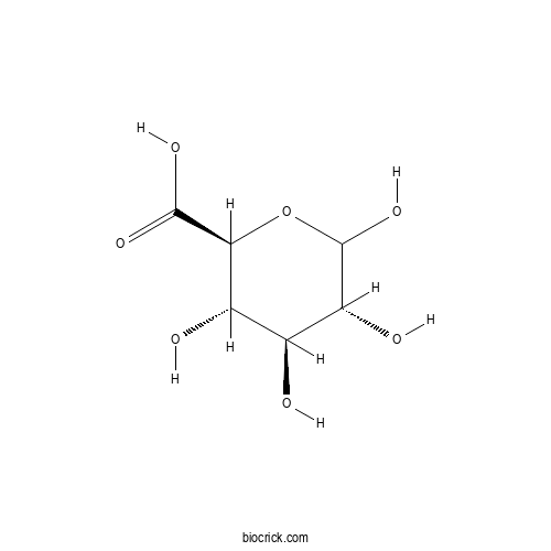
| Cas No. | 6556-12-3 | SDF | File under preparation. |
| PubChem ID | 94715.0 | Appearance | Powder |
| Formula | C6H10O7 | M.Wt | 194.1 |
| Type of Compound | N/A | Storage | Desiccate at -20°C |
| Solubility | Soluble in Chloroform,Dichloromethane,Ethyl Acetate,DMSO,Acetone,etc. | ||
| Chemical Name | (2S,3S,4S,5R)-3,4,5,6-tetrahydroxyoxane-2-carboxylic acid | ||
| SMILES | C1(C(C(OC(C1O)O)C(=O)O)O)O | ||
| Standard InChIKey | AEMOLEFTQBMNLQ-AQKNRBDQSA-N | ||
| Standard InChI | InChI=1S/C6H10O7/c7-1-2(8)4(5(10)11)13-6(12)3(1)9/h1-4,6-9,12H,(H,10,11)/t1-,2-,3+,4-,6?/m0/s1 | ||
| General tips | For obtaining a higher solubility , please warm the tube at 37 ℃ and shake it in the ultrasonic bath for a while.Stock solution can be stored below -20℃ for several months. We recommend that you prepare and use the solution on the same day. However, if the test schedule requires, the stock solutions can be prepared in advance, and the stock solution must be sealed and stored below -20℃. In general, the stock solution can be kept for several months. Before use, we recommend that you leave the vial at room temperature for at least an hour before opening it. |
||
| About Packaging | 1. The packaging of the product may be reversed during transportation, cause the high purity compounds to adhere to the neck or cap of the vial.Take the vail out of its packaging and shake gently until the compounds fall to the bottom of the vial. 2. For liquid products, please centrifuge at 500xg to gather the liquid to the bottom of the vial. 3. Try to avoid loss or contamination during the experiment. |
||
| Shipping Condition | Packaging according to customer requirements(5mg, 10mg, 20mg and more). Ship via FedEx, DHL, UPS, EMS or other couriers with RT, or blue ice upon request. | ||
Quality Control & MSDS
3D structure
Package In Stock
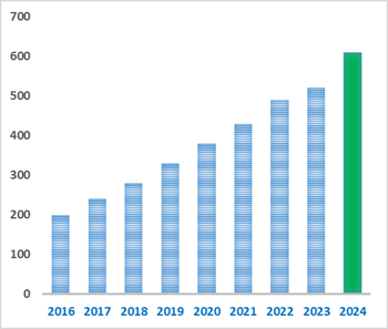
Number of papers citing our products

D-Glucuronic acid Dilution Calculator
D-Glucuronic acid Molarity Calculator
-
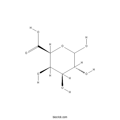 D-Galacturonic acid hydrateCatalog No.: BCX2315CAS No.: 685-73-4
-
 L-octaguluronic acid octasodium saltCatalog No.: BCX1424CAS No.: 862694-88-0
-
 L-heptaguluronic acid heptasodium saltCatalog No.: BCX1425CAS No.: 862694-87-9
-
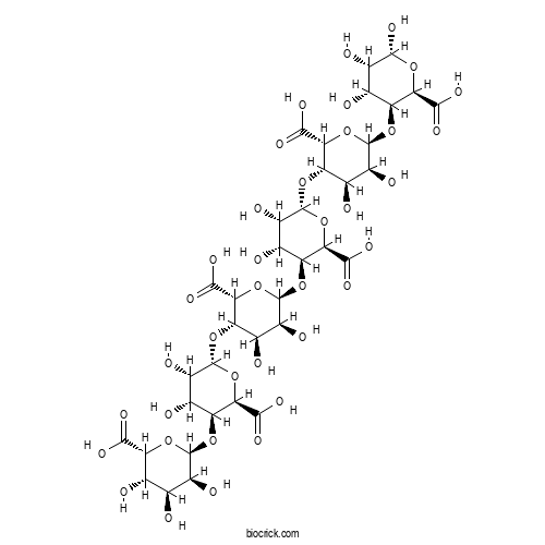 L-hexaguluronic acid hexasodium saltCatalog No.: BCX1426CAS No.: 183668-74-8
-
 L-pentaguluronic acid pentasodium saltCatalog No.: BCX1427CAS No.: 183668-72-6
-
 L-triguluronic acid trisodium saltCatalog No.: BCX1429CAS No.: 66754-14-1
-
 L-diguluronic acid disodium saltCatalog No.: BCX1430CAS No.: 34044-54-7
-
 D-octamannuronic acid octasodium saltCatalog No.: BCX1432CAS No.: 862694-98-2
-
 D-heptamannuronic acid heptasodium saltCatalog No.: BCX1433CAS No.: 862694-97-1
-
 D-hexamannuronic acid hexasodium saltCatalog No.: BCX1434CAS No.: 183668-52-2
-
 D-pentamannuronic acid pentasodium saltCatalog No.: BCX1435CAS No.: 183668-50-0
-
 D-trimannuronic acid trisodium saltCatalog No.: BCX1437CAS No.: 66754-13-0
-
 D-dimannuronic acid disodium saltCatalog No.: BCX1438CAS No.: 34044-53-6
| 1 mg | 5 mg | 10 mg | 20 mg | 25 mg | |
| 1 mM | 5.152 mL | 25.7599 mL | 51.5198 mL | 103.0397 mL | 128.7996 mL |
| 5 mM | 1.0304 mL | 5.152 mL | 10.304 mL | 20.6079 mL | 25.7599 mL |
| 10 mM | 0.5152 mL | 2.576 mL | 5.152 mL | 10.304 mL | 12.88 mL |
| 50 mM | 0.103 mL | 0.5152 mL | 1.0304 mL | 2.0608 mL | 2.576 mL |
| 100 mM | 0.0515 mL | 0.2576 mL | 0.5152 mL | 1.0304 mL | 1.288 mL |
| * Note: If you are in the process of experiment, it's necessary to make the dilution ratios of the samples. The dilution data above is only for reference. Normally, it's can get a better solubility within lower of Concentrations. | |||||
- 6-Hydroxyflavanone
Catalog No.:BCX2341
CAS No.:190831-08-4
- Guanidinoacetic acid
Catalog No.:BCX2340
CAS No.:352-97-6
- 3-Amino-3-(3,4-dimethoxyphenyl)propionic acid
Catalog No.:BCX2339
CAS No.:54160-63-3
- 3-Methyl-4-isopropylphenol
Catalog No.:BCX2338
CAS No.:3228-02-2
- Turanose
Catalog No.:BCX2337
CAS No.:547-25-1
- Doxofylline
Catalog No.:BCX2336
CAS No.:69975-86-6
- 5-Methoxytryptamine
Catalog No.:BCX2335
CAS No.:608-07-1
- L-Mannose
Catalog No.:BCX2334
CAS No.:10030-80-5
- L-Galactose
Catalog No.:BCX2333
CAS No.:15572-79-9
- L-Arabinose
Catalog No.:BCX2332
CAS No.:5328-37-0
- Norcantharidin
Catalog No.:BCX2331
CAS No.:29745-04-8
- Dopamine
Catalog No.:BCX2330
CAS No.:51-61-6
- Bryodulcosigenin
Catalog No.:BCX2343
CAS No.:88930-16-9
- Lactose
Catalog No.:BCX2344
CAS No.:63-42-3
- Acridone
Catalog No.:BCX2345
CAS No.:578-95-0
- Caffealdehyde
Catalog No.:BCX2346
CAS No.:141632-15-7
- (+)-Licarin B
Catalog No.:BCX2347
CAS No.:42016-15-9
- Pyridoxal phosphate
Catalog No.:BCX2348
CAS No.:54-47-7
- Methylaervine
Catalog No.:BCX2349
CAS No.:86293-40-5
- Pipemidic acid
Catalog No.:BCX2350
CAS No.:51940-44-4
- 2-tert-Butyl-4-methoxyphenol
Catalog No.:BCX2351
CAS No.:121-00-6
- 5-Pentadecylresorcinol
Catalog No.:BCX2352
CAS No.:3158-56-3
- Dotriacontane
Catalog No.:BCX2353
CAS No.:544-85-4
- Phenylephrine
Catalog No.:BCX2354
CAS No.:59-42-7
Utahmycin C, a new glycosylated derivative of a rare azaanthraquinone produced by Streptomyces sp. YD248.[Pubmed:42819103]
Front Microbiol. 2026 Sep 16;17:1921234.
Chemical investigation of Streptomyces sp. YD248 led to the isolation of utahmycin C (1), a new glycosylated member of the rare bacterial 1,3-dimethyl-2-azaanthraquinone family. Its structure was established by NMR spectroscopy and high-resolution mass spectrometry, revealing a beta-D-Glucuronic acid substituent attached to the azaanthraquinone core. Unusual long-range (5)J HMBC correlations prompted validation by density functional theory (DFT) through NMR chemical shift calculations, which showed excellent agreement with the experimental data. Bioinformatics analysis identified a biosynthetic gene cluster (BGC) homologous to the reported Erd gene cluster associated with utahmycin A biosynthesis. This study expands the structural diversity of bacterial azaanthraquinones.
Metabolomic Fingerprints of L-PRP and P-PRP: Formulation differences and Storage-induced Shifts.[Pubmed:42817112]
Comb Chem High Throughput Screen. 2026 Sep 29.
INTRODUCTION: Platelet-rich plasma (PRP) preparation and storage conditions are thought to influence its clinical efficacy in osteoarthritis; however, molecular evidence supporting this assumption remains limited. This study aimed to compare the metabolomic profiles of leukocyte- rich PRP (L-PRP) and pure/leukocyte-poor PRP (P-PRP) and to evaluate the effect of shortterm frozen storage on their metabolite composition. METHODS: L-PRP and P-PRP samples were collected from six healthy male donors and analysed using untargeted quadrupole time-of-flight liquid chromatography-mass spectrometry (Q-TOF LC/MS)-based metabolomics. Each sample was analysed immediately after preparation and after one week of storage at -20 degrees C. Multivariate and univariate statistical analyses were performed to identify formulation-specific and storage-related metabolic alterations. RESULTS: Distinct metabolomic differences were observed between L-PRP and P-PRP. L-tyrosine, D-Glucuronic acid, nicotinamide ribotide, and epinephrine were among the metabolites that best discriminated the two formulations. Frozen storage induced formulation-dependent metabolic changes. In L-PRP, nicotinamide ribotide and prostaglandin H2 changed, whereas P-PRP showed alterations in phosphocreatine, citric acid, and glutathione. These changes were consistent across all donors. DISCUSSION: Both PRP formulation and frozen storage influenced metabolite composition, indicating that biochemical differences extend beyond platelet and leukocyte content. Although the clinical significance of these alterations remains unclear, they may contribute to differences in PRP biological activity. Further studies integrating metabolomic and proteomic analyses are needed. CONCLUSION: Short-term frozen storage induces reproducible, formulation-dependent metabolomic changes in PRP. These findings emphasize the importance of storage conditions and provide a basis for future studies investigating their impact on PRP therapeutic efficacy.
Integrative Network Pharmacology and Experimental Validation of Sesbania grandiflora as a Multi-Targeted Therapeutic Agent Against Oral Squamous Cell Carcinoma (OSCC).[Pubmed:42808385]
Asian Pac J Cancer Prev. 2026 Sep 1;27(9):3193-3201.
BACKGROUND: Oral squamous cell carcinoma (OSCC) remains a significant health burden with limited therapeutic options and high mortality. Sesbania grandiflora, a medicinal plant rich in phytochemicals, has been traditionally used for its medicinal properties and is emerging as a potential anticancer agent. OBJECTIVE: This study aimed to investigate the anticancer mechanisms of S. grandiflora against OSCC using an integrated approach, combining network pharmacology, molecular docking, ADME profiling, and in vitro validation. MATERIALS AND METHODS: Network pharmacology was employed to identify hub genes and pathways associated with S. grandiflora bioactives. Molecular docking was performed to evaluate binding interactions of D-Glucuronic acid, a major phytoconstituent, with key cancer-related targets. ADME profiling assessed drug-likeness and pharmacokinetic properties. in vitro assays, including MTT, flow cytometry, and Western blotting, were conducted on KB oral cancer cells to validate mechanistic insights. RESULTS: Key hub genes identified included AKT1, BCL2, PIK3CA, MTOR, and CASP8, which were enriched in pathways such as the PI3K-Akt, p53, HIF-1, and EGFR-mediated apoptosis pathways. Molecular docking revealed strong and stable interactions of D-Glucuronic acid with target proteins, supporting its multi-targeted activity. ADME analysis indicated favorable pharmacokinetic properties, including high GI absorption, BBB permeability, and minimal CYP450 inhibition. Experimental validation demonstrated significant cytotoxicity (IC(5)(0): 250-300 microg/mL), dose-dependent G1-phase cell cycle arrest, downregulation of BCL-2, and upregulation of BAX, confirming the induction of apoptosis. CONCLUSION: S. grandiflora exerts anticancer effects through modulation of PI3K-AKT-mTOR and apoptotic pathways. D-Glucuronic acid emerges as a promising bioactive compound, supporting further in vivo studies and clinical exploration for OSCC management.
Discovery and characterization of Chondroitinase A with specificity for 4-O-sulfated N-acetylgalactosamine in Chondroitin sulfate/Dermatan sulfate.[Pubmed:42772565]
J Biol Chem. 2026 Sep 22:113594.
Chondroitin sulfate (CS) and dermatan sulfate (DS) are glycosaminoglycans whose diverse biological functions are largely determined by specific sulfation patterns. The GlcA/IdoA-GalNAc4S (A/iA unit), where GlcA, IdoA, GalNAc and 4S denote D-Glucuronic acid, L-iduronic acid, N-acetylgalactosamine and 4-O-sulfation, respectively, is the most prevalent sulfation motif in animal tissues and participates in a broad spectrum of physiological and pathological processes. However, chondroitinase A (CSase A), a lyase capable of selectively degrading CS/DS rich in A/iA units, has not yet been identified, which has limited our understanding of the functional roles of such structures in CS/DS. Here, we report the first discovery of a CSase A from the metagenome of sediments near the Haima cold seep. This enzyme belongs to polysaccharide lyase family 8 and exhibits strict selectivity for A/iA units, yielding disaccharides composed almost exclusively of the 4-O-sulfated disaccharide, with no other sulfated disaccharide variants detectable even under exhaustive digestion. Structural analysis of resistant tetrasaccharides and hexasaccharides, and degradation assays with structure-defined oligosaccharides demonstrate that CSase A exclusively cleaves the beta-1,4 glycosidic bond preceding a disaccharide unit containing GalNAc or GalNAc4S, thereby defining its rigorous substrate specificity. Site-directed mutagenesis and activity profiling identified Glu-579, Arg-480, Tyr-427, and His-420 as residues essential for catalysis, supporting a proposed tyrosine-histidine-mediated acid-base mechanism. The discovery of CSase A provides a unique molecular tool for dissecting the functional roles of A/iA units in CS/DS-related biology and disease.
Efficient synthesis of the tetrasaccharide repeating unit corresponding to the cell wall O-antigen of Proteus vulgaris O8 strain.[Pubmed:42607401]
Carbohydr Res. 2026 Nov;569:110067.
An acidic tetrasaccharide repeating unit corresponding to the cell wall O-antigen of Proteus vulgaris O8 strain has been synthesized in very good yield applying [2 + 2] block glycosylation strategy. Stereoselective alpha-glycosylated products were obtained using ether-based solvents in glycosylation reactions. The incorporation of the D-Glucuronic acid moiety in the tetrasaccharide was achieved by glycosylation with d-glucose intermediate and TEMPO-Bis(acetoxy)iodobenzene (BAIB) mediated oxidation at the late stage. Non-participating groups (e.g. N(3) and benzyl) were placed at C-2 position of the glycosyl donor for the preparation of the 1,2-cis-glycosylated products. The yields of the reactions were very good.
Optimization of the Sequential Extraction Process of Compounds for the Integral Valorization of Macrocystis pyrifera.[Pubmed:42518379]
ACS Omega. 2026 Jul 1;11(27):40402-40416.
This study aimed to optimize the sequential extraction of high value-added compounds from the brown macroalga Macrocystis pyrifera. The biomass presented high carbohydrate (43.76%) and ash (36.20%) contents, with lower levels of proteins (9.11%), lipids (3.75%), and phenolic compounds (0.33%). Elemental analysis indicated carbon (38.75%) and oxygen (38.33%) as the predominant elements. Sequential extraction enabled the recovery of biostimulants (via osmotic and thermal treatments), fucoidan, and alginate. A factorial design established optimal extraction conditions at 60 degrees C, pH 3.5, and 1 h, based on chemical characterization parameters. Fucoidan exhibited a molecular weight of 447.61 kDa, while alginate showed 327.35 kDa. FTIR analysis revealed characteristic polysaccharide bands (O-H, C-O-C, and C-C). Monosaccharide analysis indicated that fucose and galactose were the main components of fucoidan, whereas alginate was composed of mannuronic (M) and guluronic (G) acids. Nuclear magnetic resonance (NMR) spectroscopy showed that fucoidan consists of a backbone of alpha-l-fucopyranose residues linked by (1-->3) and (1-->4) bonds, partially sulfated at C-2, with beta-D-Glucuronic acid and beta-d-galactose residues, as well as terminal alpha-d-glucopyranose units. Alginate exhibited MM, GG, MG, and GM blocks, with predominance of MG [(-->4)-beta-d-ManpA-(1-->4)-alpha-l-GulpA-(1-->)]. These results highlight the efficiency of the optimized process for biomass valorization and biotechnological applications.
Structural characterization and properties of a novel acetylated extracellular polysaccharide from Rhodotorula graminis.[Pubmed:42493098]
Carbohydr Polym. 2026 Sep 15;388:125527.
Rhodotorula yeasts are promising microbial cell factories, recognized for their intracellular production of lipids and carotenoids. Recently, research has expanded to extracellular metabolites, particularly exopolysaccharides and polyol esters of fatty acids. In this study, an exopolysaccharide isolated from Rhodotorula graminis was characterized using high-performance anion-exchange chromatography, size exclusion chromatography, and nuclear magnetic resonance. The results show that the polymer has a trisaccharide repeating unit of 1,4-linked a alpha-D-Glucuronic acid, 1,2,3 linked alpha-d-mannose, and terminal beta-d-galactose. Furthermore, the results suggest the presence of approximately two acetyl substituents per repeating unit and a molecular weight of >/=1.5 MDa. Rheological analysis of aqueous EPS solutions showed a non-linear increase in viscosity with concentration, and sponges were prepared by freeze-drying. Biocompatibility assays with human epidermal keratinocytes and Atlantic salmon cells indicated no cytotoxic effects under the tested conditions. The EPS also showed weak, concentration-dependent growth inhibition against Escherichia coli, Serratia marcescens, and Candida glabrata over the tested concentration range of 7.87-1007.5 mug.mL(-1). Overall, the results indicate that this EPS is a structurally unusual Rhodotorula-derived polymer with interesting physicochemical properties and potentially good biocompatibility.
Kombucha Tea Fortified With Spirulina Maxima and Haematococcus pluvialis: Impact on Physicochemical Properties, Bioactive Profile, and Antioxidant Capacity.[Pubmed:42487980]
Food Sci Nutr. 2026 Jul 21;14(7):e72150.
This study investigated the effect of adding Spirulina maxima and Haematococcus pluvialis microalgae to kombucha tea. Both additives increased membrane weight in a dose-dependent manner. pH decreased with S. maxima and increased with H. pluvialis. Both microalgae significantly improved the overall antioxidant capacity, total phenolic content, and textural properties (cohesiveness, consistency, firmness, and viscosity index) of kombucha samples. S. maxima produced superior physicochemical parameters and organic acid concentrations compared to H. pluvialis. Concentrations of acetic, lactic, gluconic, and D-Glucuronic acids were highest in the 4% S. maxima samples. Furthermore, S. maxima promoted the accumulation of several phenolic compounds, including chlorogenic, gallic, caffeic, vanillin, and ferulic acids, whereas H. pluvialis increased the concentrations of astaxanthin, salicylic acid, quercetin, and kaempferol. In contrast, the addition of both microalgae reduced some catechin derivatives. Consequently, the integration of S. maxima and H. pluvialis into kombucha enhanced its nutritional value, antioxidant capacity, and bioactive compound richness.
Novel antioxidant quercetin glycosides conjugated with phenolic acids from young leaves of Vicia unijuga.[Pubmed:42484612]
Biosci Biotechnol Biochem. 2026 Sep 25;90(10):1327-1333.
Vicia unijuga is an edible wild plant distributed throughout eastern Asia. Its young leaves, when boiled or fried, are palatable and exhibit an aroma and flavor reminiscent of soybeans. In this study, we isolated 2 previously unreported quercetin glucosides conjugated with phenolic acids (compounds 2 and 3), together with quercitrin (compound 1), from young V. unijuga leaves as potent inhibitors of lipid peroxidation. Their structures were elucidated using high-resolution electrospray ionization mass spectrometry (negative mode), 1D and 2D nuclear magnetic resonance analyses, and enzymatic hydrolysis. Both new compounds shared a common structure in which alpha-l-rhamnose and beta-D-Glucuronic acid were attached to quercetin at the 3- and 7-positions, respectively. Compound 2 contained a caffeic acid moiety esterified at the 2-position of beta-D-Glucuronic acid, whereas compound 3 carried a p-coumaric acid moiety at the same position. Compounds 1-3 inhibited lipid peroxidation, with IC50 values of 11 +/- 1.3, 0.21 +/- 0.02, and 1.3 +/- 0.09 mum, respectively.
Elucidation of the Pathway for the Biosynthesis of the Undecorated Capsular Polysaccharide from the HS:19 Serotype of the Human Pathogen Campylobacter jejuni.[Pubmed:42241030]
Biochemistry. 2026 Jun 16;65(12):1978-1987.
Campylobacter jejuni is the leading cause of food poisoning in the United States and Europe. The bacterium is commensal in poultry, and the ingestion of contaminated or undercooked chicken products can result in rather severe cases of nausea, abdominal pain, and diarrhea. Protecting the bacterium from the host immune system is a thick polysaccharide coat known as the capsular polysaccharide. The repeating disaccharide unit in the undecorated capsular polysaccharide from the HS:19 serotype of C. jejuni consists of N-acetyl-d-glucosamine (GlcNAc) and the serinol amide of D-Glucuronic acid (GlcA). It was demonstrated that the GT2 glycosyltransferase from the N-terminal domain of HS19.08 catalyzes the transfer of D-Glucuronic acid from UDP-GlcA to C3 of the GlcNAc moiety at the nonreducing end of the growing polysaccharide chain. The carboxylate of the added GlcA is then amidated by the ATP-dependent ligase activity of the C-terminal domain of HS19.11 using (S)-serinol-phosphate. The phosphorylated serinol amide is subsequently dephosphorylated by the catalytic activity of HS19.09. In the final step of the series, the GT2 glycosyltransferase from the N-terminal domain of HS19.11 catalyzes the transfer of GlcNAc from UDP-GlcNAc to C4 of the amidated glucuronic acid. This series of reactions has been exploited to enzymatically synthesize and isolate defined oligomers containing 2, 3, 5, and 7 monomeric units, and procedures were developed for the synthesis of oligomeric mixtures ranging from 9 to 15 monomeric units.
Exatecan Payload-Based Antibody-Drug Conjugates with a Short Hydrophilic Cleavable Linker.[Pubmed:42157849]
ACS Med Chem Lett. 2026 Apr 23;17(5):1035-1042.
Antibody-drug conjugates (ADCs) consist of a cytotoxin covalently linked to a target-specific antibody. ADCs are designed to release the cytotoxic drug upon internalization by cells expressing the select antigen. However, the true potential of an ADC becomes diminished by several factors, including high hydrophobicity of the biologic, which results in either aggregation in vitro or poor pharmacokinetics in vivo, thereby curtailing its efficacy. Hence, one of the goals in designing next generation ADCs is creating hydrophilic linker-payloads to overcome these challenges. Here, we describe generation and characterization of cathepsin and glucuronidase-cleavable linkers attached to a novel, short, stable, hydrophilic 1-amino-beta-D-Glucuronic acid spacer. These hydrophilic spacer-linkers, attached to the exatecan payload, were conjugated to the HER-2-targeting antibody trastuzumab to obtain ADCs with a drug-to-antibody ratio (DAR) of 8. The ADC with an amino-glucuronic acid spacer combined with a glucuronide linker (AV-L03) showed superior hydrophilicity compared to a cathepsin cleavable GGFG linker; both ADCs included the same hydrophilic short spacer and exatecan payload. The ADC with the hydrophilic AV-L03 linker-exatecan payload (AV-DL055) conjugated to trastuzumab displayed potent in vitro cytotoxicity, improved plasma stability, and remarkable in vivo efficacy in a mouse xenograft model compared to an ADC comprising trastuzumab conjugated to DXd payload via a tetrapeptide (hydrophobic GGFG) linker.
Constructing synthetic microbial communities containing Komagataeibacter spp. for kombucha production: Impacts on bioactive compounds, functionality and sensory profiles.[Pubmed:41963037]
Food Microbiol. 2026 Sep;138:105062.
Kombucha is traditional fermented beverage characterized by a symbiotic microbial community (MC), but achieving consistent quality remains a major challenge. This study aims to construct optimized synthetic MCs to enhance bioactivity, functionality and sensorial profiles of tailor-made kombucha. Five recombinational MCs were designed by combining different Komagataeibacter species (K. intermedius (Ki), K. oboediens (Ko), K. rhaeticus (Kr), K. xylinus (Kx), K. saccharivorans (Ks)) with G. potus (Gp), S. davenportii (Sd) and Z. bailii (Zb). The key MCs were screened based on critical sugar-acid metabolism, biofilm formation, growth kinetics and antimicrobial activities. Customized Kx-Gp-Sd-Zb exhibited the highest D-Glucuronic acid content (18.5 mg/mL), significantly surpassing spontaneous fermentation (TK, 3.4 mg/mL). Antioxidant assays indicated comparable radical scavenging activity between most MCs and TK. K. intermedius in recombination exhibited superior digestive enzyme inhibitory activity (alpha-amylase and alpha-glucosidase), while Ks-Gp-Sd-Zb showed the highest adsorption capacity (46.9 mg/g). Bioaccessibility under simulated gastrointestinal conditions was highest for Kx-Gp-Sd-Zb (80.9%). Kr-Gp-Sd-Zb produced the highest biofilm yield. Safety assessment confirmed no toxicity (100% larval survival) based on Galleria mellonella model. Volatile profiling identified 56 compounds, with Ki-Gp-Sd-Zb exhibiting the highest total content (3421.8 mug/L). Kx-Gp-Sd-Zb contained elevated proportions of phenols, esters, ketones and aldehydes, while 17 key volatiles (rOAV >/=0.1) defined distinct flavor profiles. Electronic and sensory evaluations revealed that three recombinant MCs outperformed TK in acceptability. Sensory evaluation showed tailor-made kombucha with superior aroma scores corresponded precisely with results from flavor and electronic sensory analysis.
Novel GH115 xylan alpha-1,2-glucuronidases with distinct preferences for xylan-derived oligomers and polymers.[Pubmed:41949258]
Protein Sci. 2026 May;35(5):e70571.
Natural polymers are promising sustainable materials for diverse applications. Xylans, major components of hemicellulose, exhibit origin-dependent substitution patterns that determine their physicochemical properties. Targeted enzymatic modification of these substituents offers a mild and precise approach for tailoring the polymer's characteristics. Glucuronidases (EC 3.2.1.139) and xylan alpha-1,2-glucuronosidases (EC 3.2.1.131) are carbohydrate active enzymes that specifically remove the common glucuronic acid and 4-O-methyl D-Glucuronic acid side branches of xylans/xylooligosaccharides from different hardwood and softwood sources. These enzymes are, dependent on sequence and structure, occurring in three glycoside hydrolase families (GH4, GH67 and GH115). GH115 predominantly includes xylan alpha-1,2-glucuronosidases, but the number of characterized enzymes is low. In this study, 22 novel GH115 candidates were identified through a combination of automated and manual sequence mining from databases, for evaluation of their production, function and storage possibilities. Twenty enzymes were produced in active form in crude extracts, while only four remained soluble for extended time periods post purification. These four GH115 candidates include first time reported monomeric enzymes, and while all were active on polymeric xylan, their specific activity and activity-ratio on polysaccharides/oligosaccharides of beechwood xylan differed significantly. Structural models created by AlphaFold3, and different length oligosaccharides were docked in the catalytic clefts. The presence of an additional domain, and the position of a mobile loop affected oligomerization and activity on polymeric substrates, respectively, resulting in differences in specific activity and preference for polymeric substrates.

Calcutta University

University of Minnesota

University of Maryland School of Medicine

University of Illinois at Chicago

The Ohio State University

University of Zurich

Harvard University

Colorado State University

Auburn University

Yale University

Worcester Polytechnic Institute

Washington State University

Stanford University

University of Leipzig

Universidade da Beira Interior

The Institute of Cancer Research

Heidelberg University

University of Amsterdam

University of Auckland

TsingHua University

The University of Michigan

Miami University

DRURY University

Jilin University

Fudan University

Wuhan University

Sun Yat-sen University

Universite de Paris

Deemed University

Auckland University

The University of Tokyo

Korea University